Free Tools
Abdominal Quadrants and Regions
Clinicians map the front of the abdomen in one of two ways: 4 quadrants (right upper, left upper, right lower, left lower) or 9 regions in a three by three grid. Both give everyone a shared way to say exactly where pain, tenderness, a mass, or an injury is. Tap the diagram below to see what lies in each area.
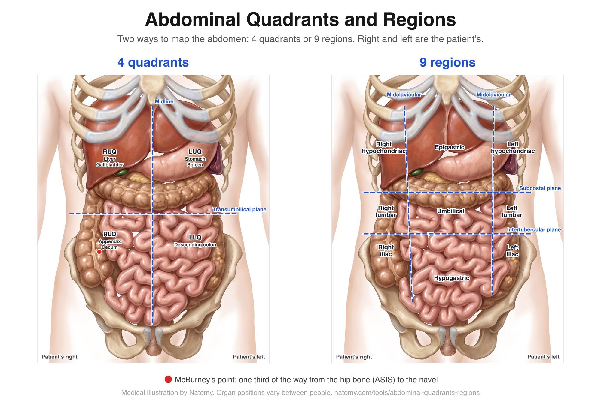
Download the chart (JPG) for teaching, charting, or case files.
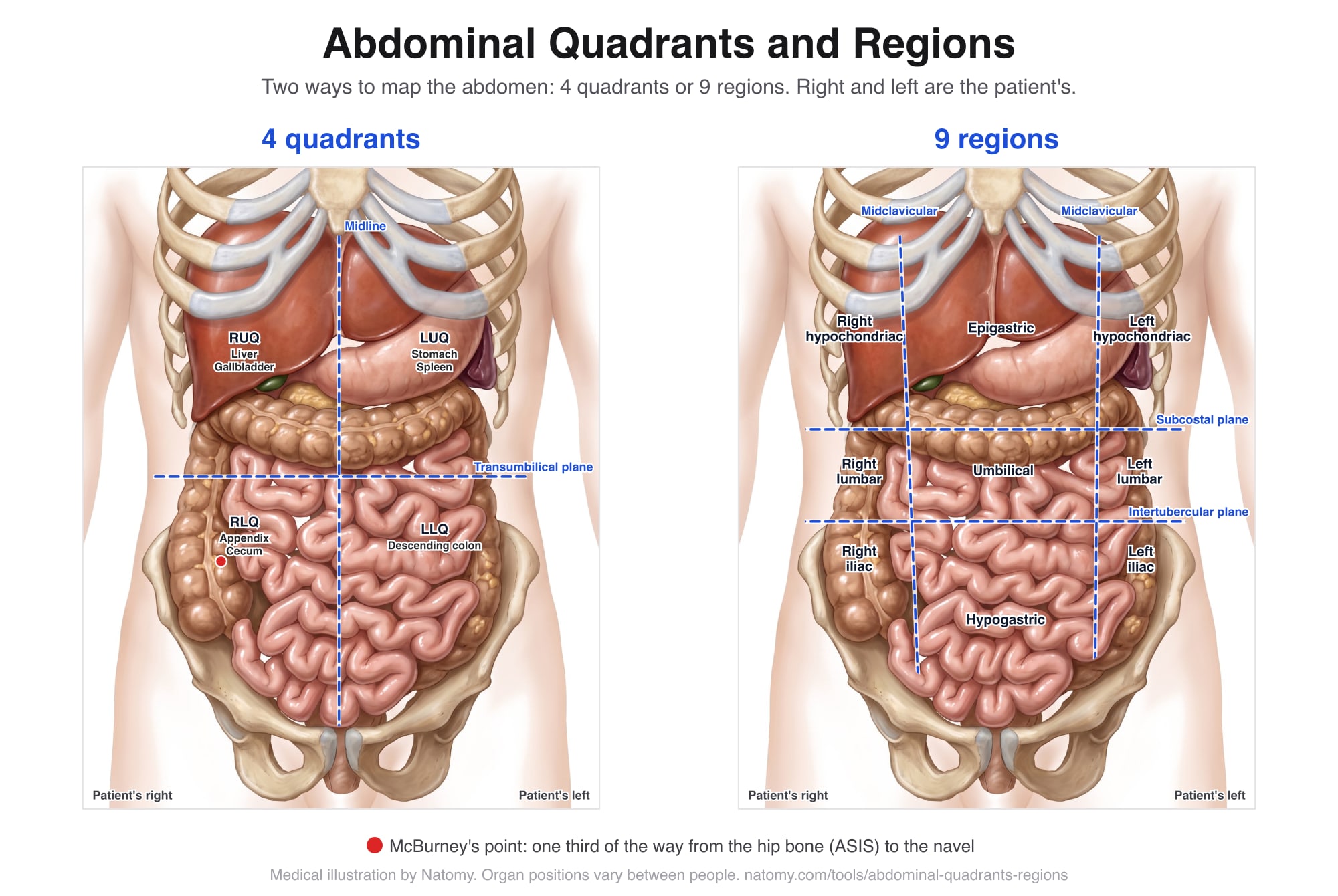
Interactive abdominal diagram
Switch between 4 quadrants and 9 regions, then tap an area to see the organs there and common causes of pain.
Tap an area. The body faces you, so the patient's right is on your left.
The 4 abdominal quadrants
Two lines cross at the navel: a vertical line down the middle of the body (the median plane) and a horizontal line through the navel (the transumbilical plane). Quadrants are the quickest way to describe a finding and are used every day in emergency rooms, clinic notes, and imaging requests.
| Quadrant | Organs | Common causes of pain |
|---|---|---|
| Right upper quadrant | Liver (most of it), Gallbladder, Duodenum, Head of the pancreas, Right kidney and adrenal gland, Hepatic flexure of the colon | Gallstones or an inflamed gallbladder (cholecystitis); Hepatitis or a liver abscess; Duodenal ulcer; Kidney stone or kidney infection; Pneumonia at the base of the right lung |
| Left upper quadrant | Stomach, Spleen, Body and tail of the pancreas, Left kidney and adrenal gland, Splenic flexure of the colon, Left lobe of the liver (part) | Gastritis or a stomach ulcer; Pancreatitis; Enlarged or injured spleen; Kidney stone or kidney infection; Pneumonia at the base of the left lung |
| Right lower quadrant | Cecum and appendix, End of the small intestine (terminal ileum), Ascending colon, Right ovary and fallopian tube, Right ureter | Appendicitis; Ovarian cyst or twisted ovary (torsion); Ectopic pregnancy; Inguinal hernia; Crohn's disease of the terminal ileum; Kidney stone passing down the ureter |
| Left lower quadrant | Descending colon, Sigmoid colon, Left ovary and fallopian tube, Left ureter | Diverticulitis (the most common cause in adults); Ovarian cyst or twisted ovary (torsion); Ectopic pregnancy; Inguinal hernia; Constipation; Kidney stone passing down the ureter |
The 9 abdominal regions
Four lines make a three by three grid:
- Two vertical midclavicular lines, one on each side, running down from the middle of each collarbone toward the middle of the groin (the midinguinal point).
- The subcostal plane, a horizontal line across the lowest edge of the rib cage (the 10th costal cartilages), at about the level of the third lumbar vertebra.
- The intertubercular (transtubercular) plane, a horizontal line across the tubercles of the iliac crests, the bony bumps you can feel about 5 cm behind the front point of each hip bone. It lies at about the level of the fifth lumbar vertebra.
Some textbooks use the transpyloric plane, halfway between the top of the breastbone and the pubic bone, as the upper line instead of the subcostal plane. The idea is the same.
| Region | Organs | Common causes of pain |
|---|---|---|
| Right hypochondriac region | Liver (right lobe), Gallbladder (at the costal margin, near the midclavicular line), Top of the right kidney, Hepatic flexure of the colon | Gallstones or cholecystitis; Hepatitis or a liver abscess; Pneumonia at the base of the right lung |
| Epigastric region | Stomach, Left lobe of the liver, Duodenum, Pancreas, Abdominal aorta | Indigestion, gastritis, or a stomach ulcer; Acid reflux (GERD); Pancreatitis; Heart attack, which can be felt here instead of the chest; Aortic dissection |
| Left hypochondriac region | Spleen, Upper stomach (fundus), Tail of the pancreas, Top of the left kidney, Splenic flexure of the colon | Enlarged or injured spleen; Gastritis; Pancreatitis; Pneumonia at the base of the left lung |
| Right lumbar (flank) region | Ascending colon, Right kidney, Small intestine | Kidney stone or kidney infection; Constipation; Colitis |
| Umbilical region | Small intestine, Transverse colon, Lower duodenum, Abdominal aorta | Early appendicitis (the pain often starts here, then moves to the lower right); Gastroenteritis; Small bowel obstruction; Umbilical hernia; Abdominal aortic aneurysm |
| Left lumbar (flank) region | Descending colon, Left kidney, Small intestine | Kidney stone or kidney infection; Constipation; Diverticulitis |
| Right iliac (inguinal) region | Cecum, Appendix, End of the small intestine (terminal ileum), Right ovary and fallopian tube | Appendicitis, typically sorest at McBurney's point; Ovarian cyst or torsion; Ectopic pregnancy; Inguinal hernia; Crohn's disease |
| Hypogastric (suprapubic) region | Urinary bladder, Uterus, Prostate (men), Sigmoid colon, Rectum, Loops of small intestine | Bladder infection (cystitis); Urinary retention; Period pain or endometriosis; Pelvic inflammatory disease; Prostatitis |
| Left iliac (inguinal) region | Sigmoid colon, Descending colon, Left ovary and fallopian tube | Diverticulitis; Ovarian cyst or torsion; Ectopic pregnancy; Inguinal hernia; Constipation |
Right and left are the patient's
Medical diagrams show the patient facing you, so the patient's right side is on your left. "RLQ" always means the patient's right lower quadrant, wherever the examiner is standing. Mixing up sides is a common source of errors in notes and diagrams.
Pain location: a clue, not a diagnosis
- Pain moves. Appendicitis often starts as a vague ache around the navel and moves to the right lower quadrant over several hours.
- Pain is referred. A heart attack can be felt in the epigastric region, gallbladder pain can spread to the right shoulder blade, and pneumonia at the base of a lung can cause upper abdominal pain.
- Organs sit differently. Pregnancy pushes the appendix and bowel upward, and organ positions vary between people.
- Some pain is everywhere. Widespread pain with a rigid belly can mean peritonitis or a perforation and needs emergency care.
Get emergency care for sudden severe belly pain, pain with fever and vomiting, a hard swollen belly, vomiting blood, black or bloody stool, or upper belly pain with chest pressure, sweating, or shortness of breath, or belly pain if you are or could be pregnant (possible ectopic pregnancy). For changes in bowel habit, see our Bristol stool chart.
Abdominal quadrants in injury and malpractice cases
The location of an abdominal injury or symptom matters in personal injury and medical malpractice cases, and records describe it in quadrants or regions. In car crashes, a lap belt can bruise the lower abdomen across both iliac regions and the hypogastric region (the seatbelt sign), which raises concern for bowel and spine injuries that are easy to miss at first. Left upper quadrant pain after a crash can point to a spleen injury, and right upper quadrant pain to a liver injury.
In missed diagnosis claims, reviewers compare where the pain was recorded with how the work-up was done: for example, right lower quadrant pain that was not followed up for appendicitis, or epigastric pain in a patient with heart risk factors that was treated as indigestion without an ECG. Clear, labeled diagrams help a jury see exactly where the findings were.
Illustrate an abdominal injury
Natomy turns records, imaging, and prompts into clear, labeled medical illustrations of abdominal injuries and conditions, such as spleen and liver lacerations, bowel injuries, and appendicitis, for patient education and medical-legal exhibits. See examples in the Natomy atlas.
Try Natomy TodayFAQ
What are the 4 quadrants of the abdomen?+
The right upper quadrant (RUQ), left upper quadrant (LUQ), right lower quadrant (RLQ), and left lower quadrant (LLQ). They are made by a vertical line down the middle of the body and a horizontal line through the navel. Right and left always mean the patient's right and left.
What are the 9 regions of the abdomen?+
Top row: right hypochondriac, epigastric, and left hypochondriac. Middle row: right lumbar, umbilical, and left lumbar. Bottom row: right iliac (inguinal), hypogastric (suprapubic), and left iliac (inguinal). They are made by two vertical midclavicular lines and two horizontal planes.
What organs are in the right upper quadrant?+
Most of the liver, the gallbladder, the duodenum, the head of the pancreas, the right kidney and adrenal gland, and the bend of the colon under the liver (hepatic flexure). Gallbladder problems are the most common cause of pain here.
What is in the right lower quadrant?+
The cecum and appendix, the end of the small intestine, the start of the ascending colon, and in women the right ovary and fallopian tube. Appendicitis is the classic cause of right lower quadrant pain.
What causes left lower quadrant pain?+
In adults, the most common cause is diverticulitis, inflammation of small pouches in the sigmoid colon. Other causes include constipation, ovarian cysts or torsion, ectopic pregnancy, kidney stones, and hernias.
What is McBurney's point?+
A spot in the right lower quadrant, about one third of the way from the bony point at the front of the right hip (the anterior superior iliac spine) to the navel. It roughly overlies the base of the appendix, and tenderness there is a classic sign of appendicitis.
Should I use quadrants or regions?+
Quadrants are quicker and are what most clinicians use day to day, for example 'RLQ tenderness'. The 9 regions give more detail and are common in anatomy teaching and in some exam and imaging reports. Both describe the same abdomen.
Why does a heart attack sometimes cause stomach pain?+
Pain from the heart can be felt in the upper abdomen (the epigastric region) because nerves from the heart and the upper gut enter the spinal cord at similar levels. This is called referred pain. Upper belly pain with sweating, shortness of breath, or nausea needs emergency care.
Where is the hypochondriac region?+
Just below the ribs on each side of the upper abdomen. The name means 'under the cartilage', meaning the rib cartilages. The right hypochondriac region holds most of the liver and the gallbladder; the left holds the spleen.
Sources: Moore KL, Dalley AF, Agur AMR. Clinically Oriented Anatomy, 8th ed. Wolters Kluwer; 2018 (abdomen chapter). Standring S, ed. Gray's Anatomy, 42nd ed. Elsevier; 2020. Cartwright SL, Knudson MP. Evaluation of acute abdominal pain in adults. Am Fam Physician. 2008;77(7):971-978. McBurney C. Experience with early operative interference in cases of disease of the vermiform appendix. N Y Med J. 1889;50:676-684. Illustration and descriptions are by Natomy. For education only; not a substitute for medical advice.
Related free tools
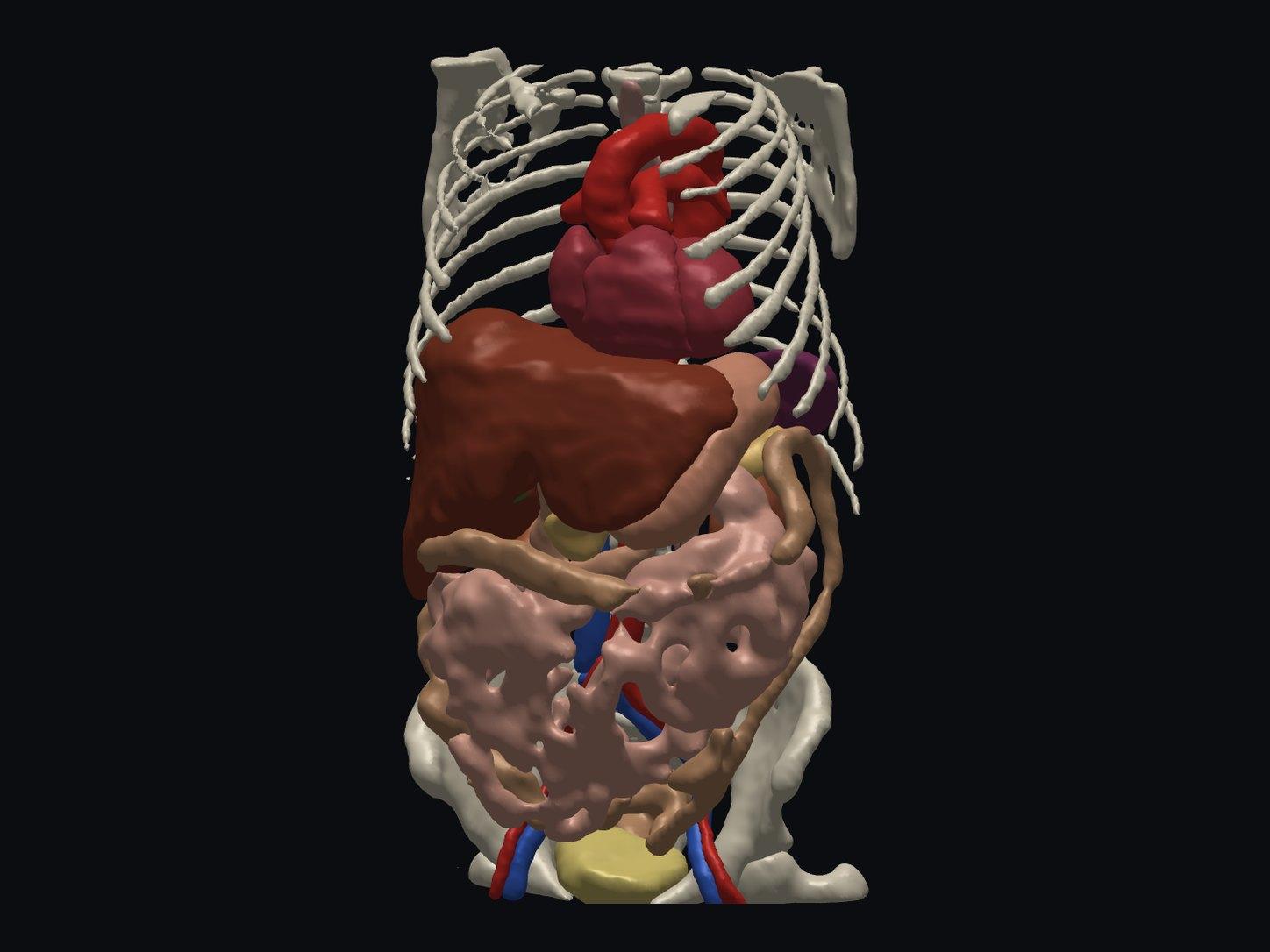
Free Online DICOM Viewer (3D)
Open a CT or MRI DICOM folder in your browser and view it in 3D, with AI anatomy labeling. Nothing is uploaded.
Try it free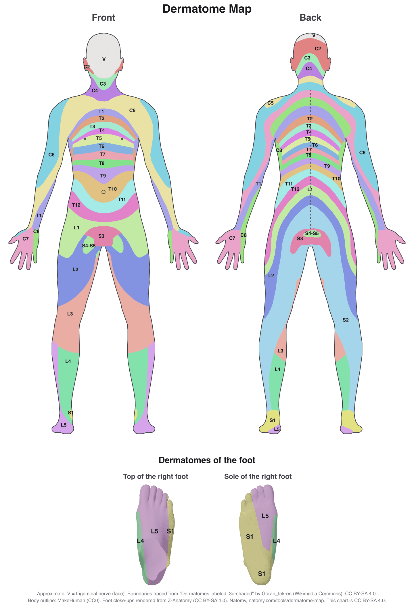
Interactive Dermatome Map
Tap the body to see each nerve root's skin area, test point, myotome, reflex, and disc level. Free printable chart.
Try it free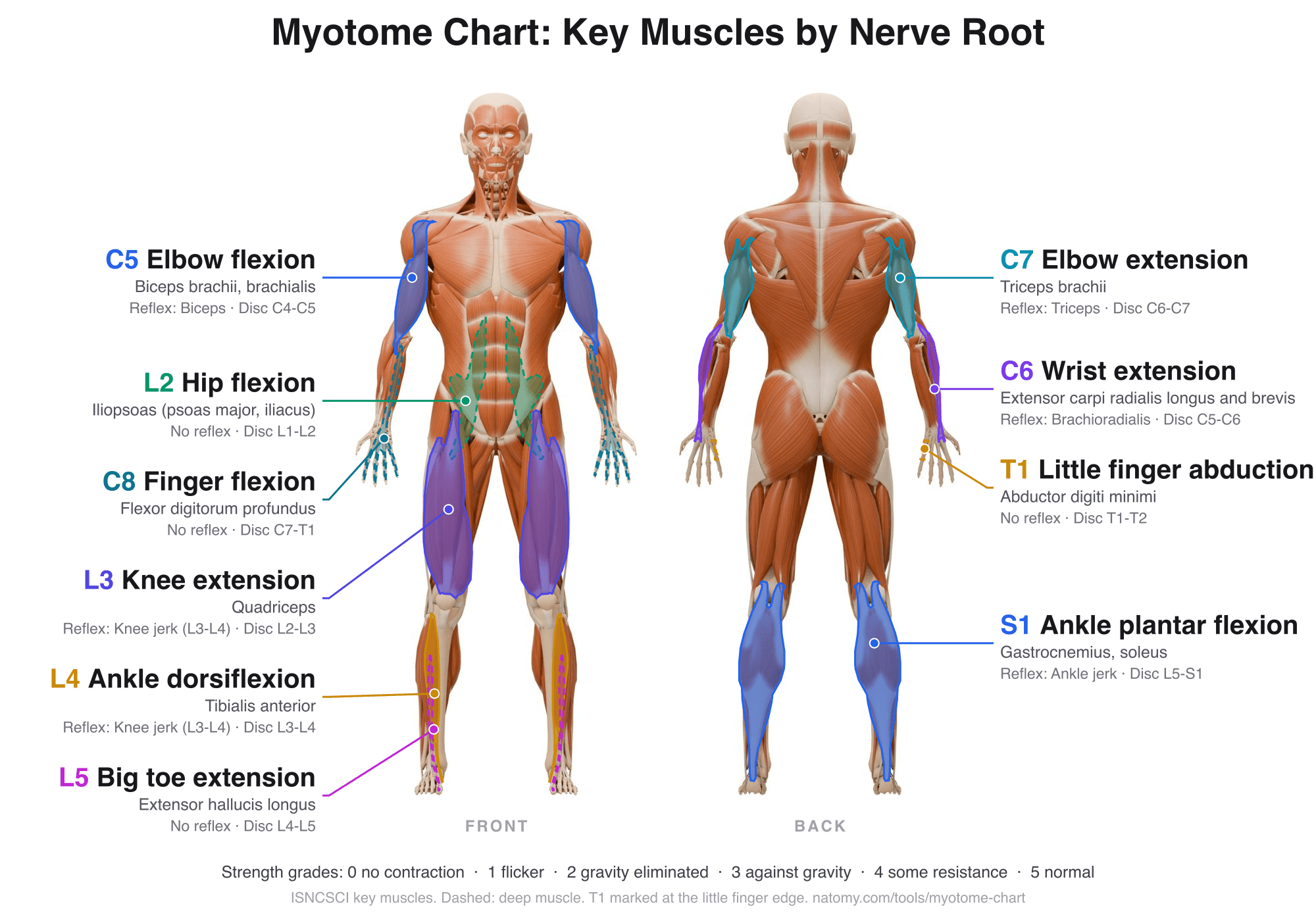
Myotome Chart and Motor Score
Tap a nerve root to see its key muscle, test position, and reflex. Grade strength 0 to 5 for the ISNCSCI motor score.
Try it free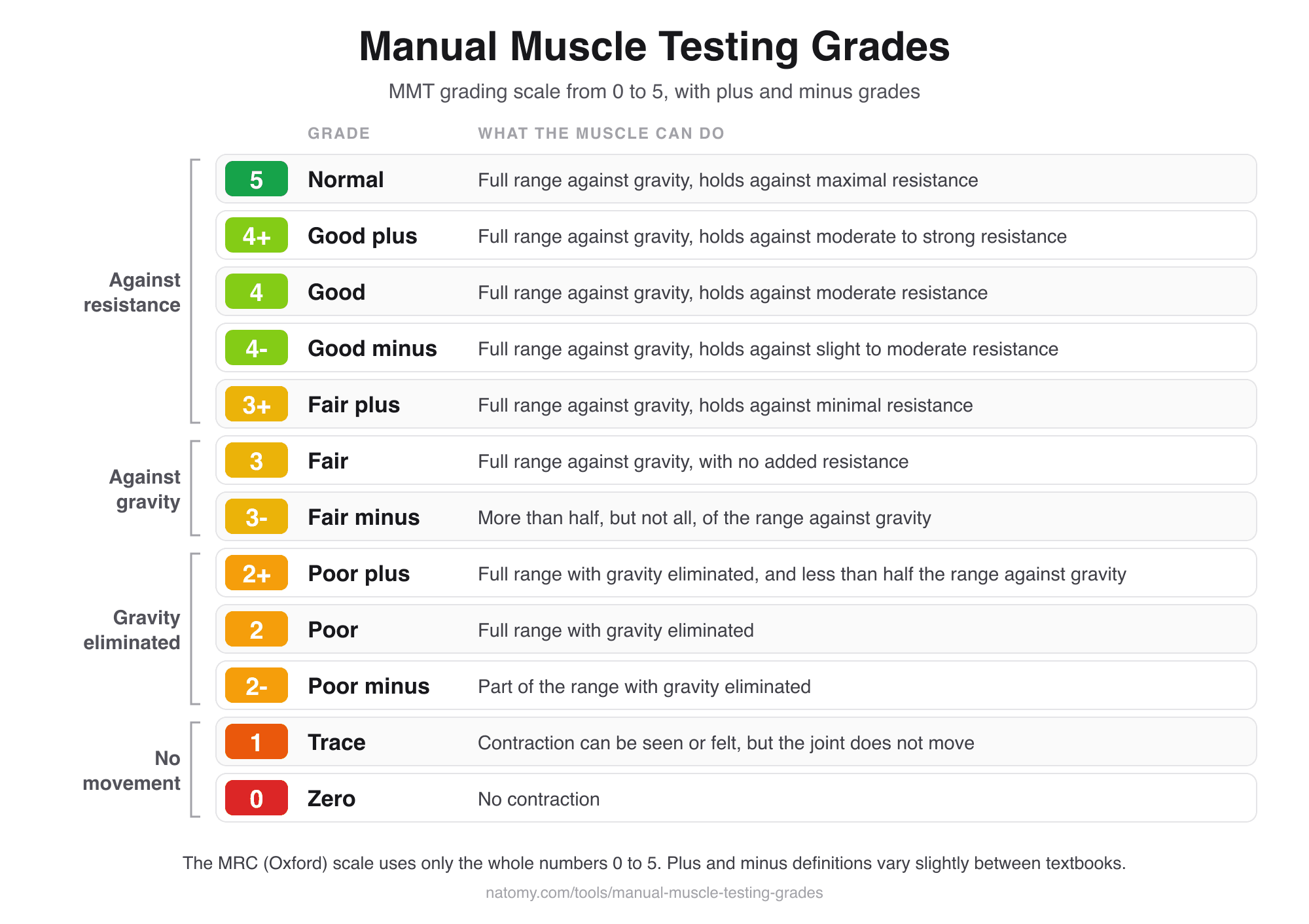
Manual Muscle Testing Grades
MMT grading scale chart from 0 to 5 with plus and minus grades, and a grade finder that writes the note for you.
Try it free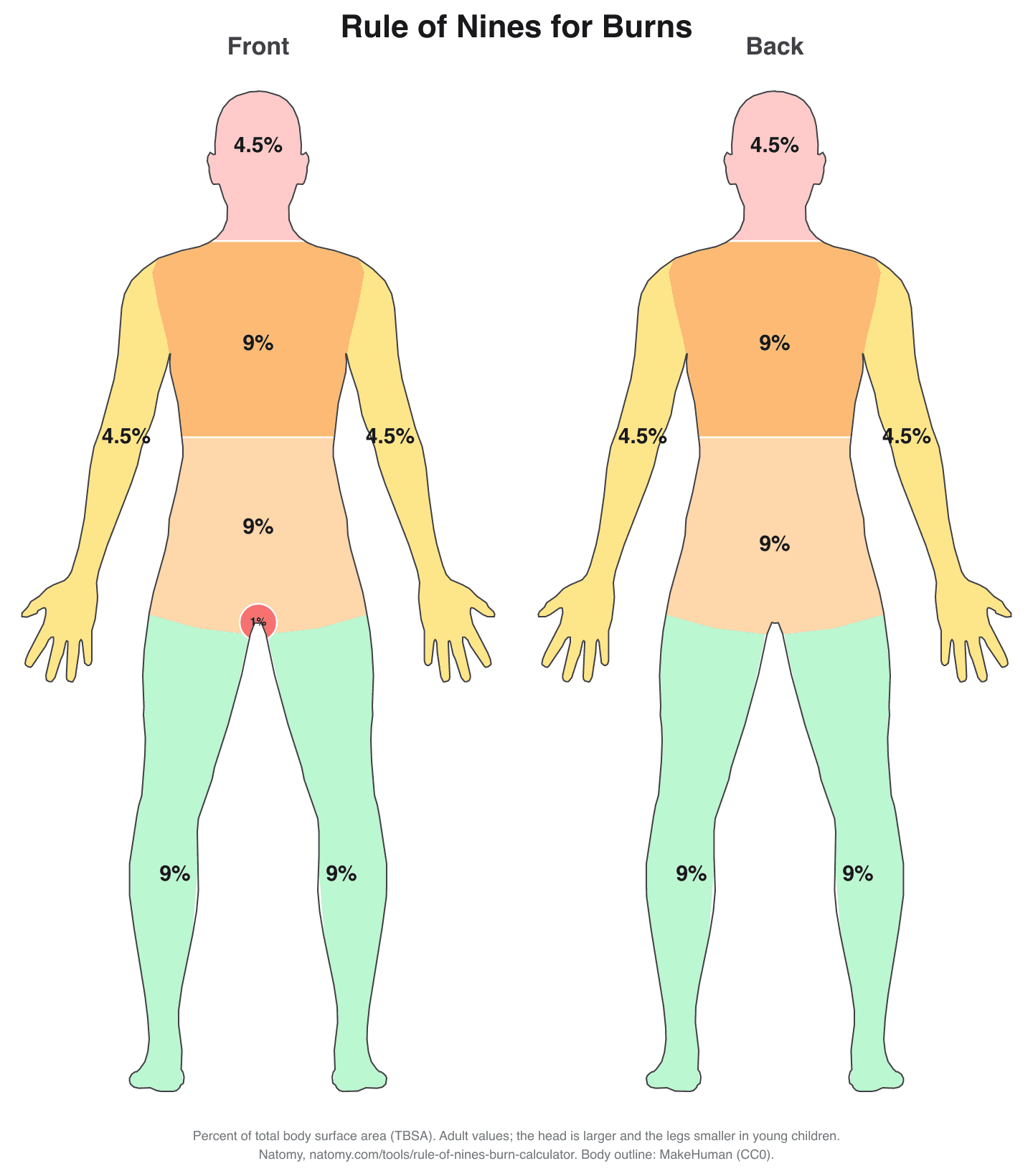
Rule of Nines Burn Calculator
Click the burned areas to get the total body surface area (TBSA), with the Parkland formula and a printable burn chart.
Try it free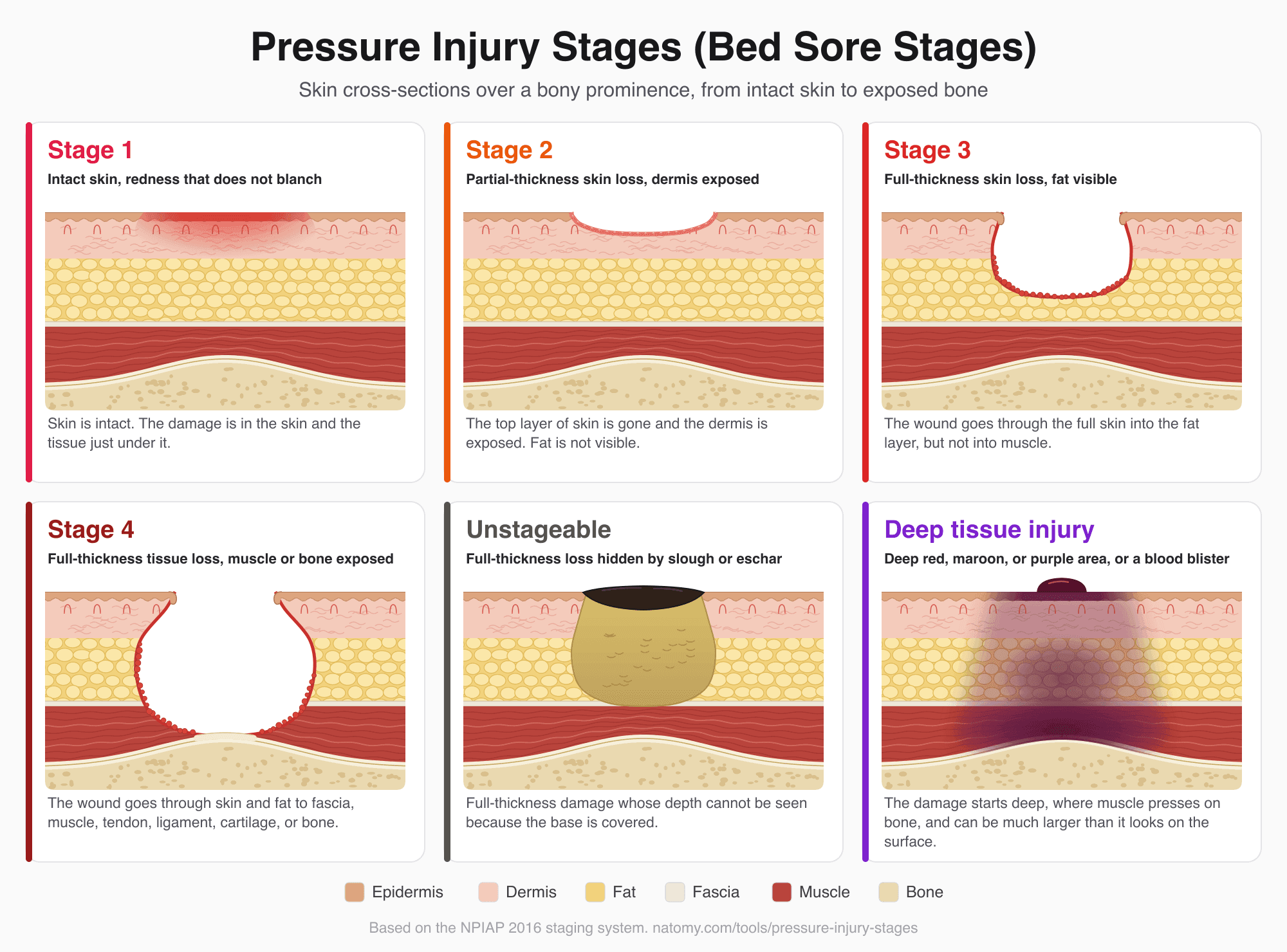
Pressure Injury (Bed Sore) Stages
Illustrated pressure ulcer stages chart, Stage 1 to 4, unstageable, and deep tissue injury, with a stage finder and wound note.
Try it free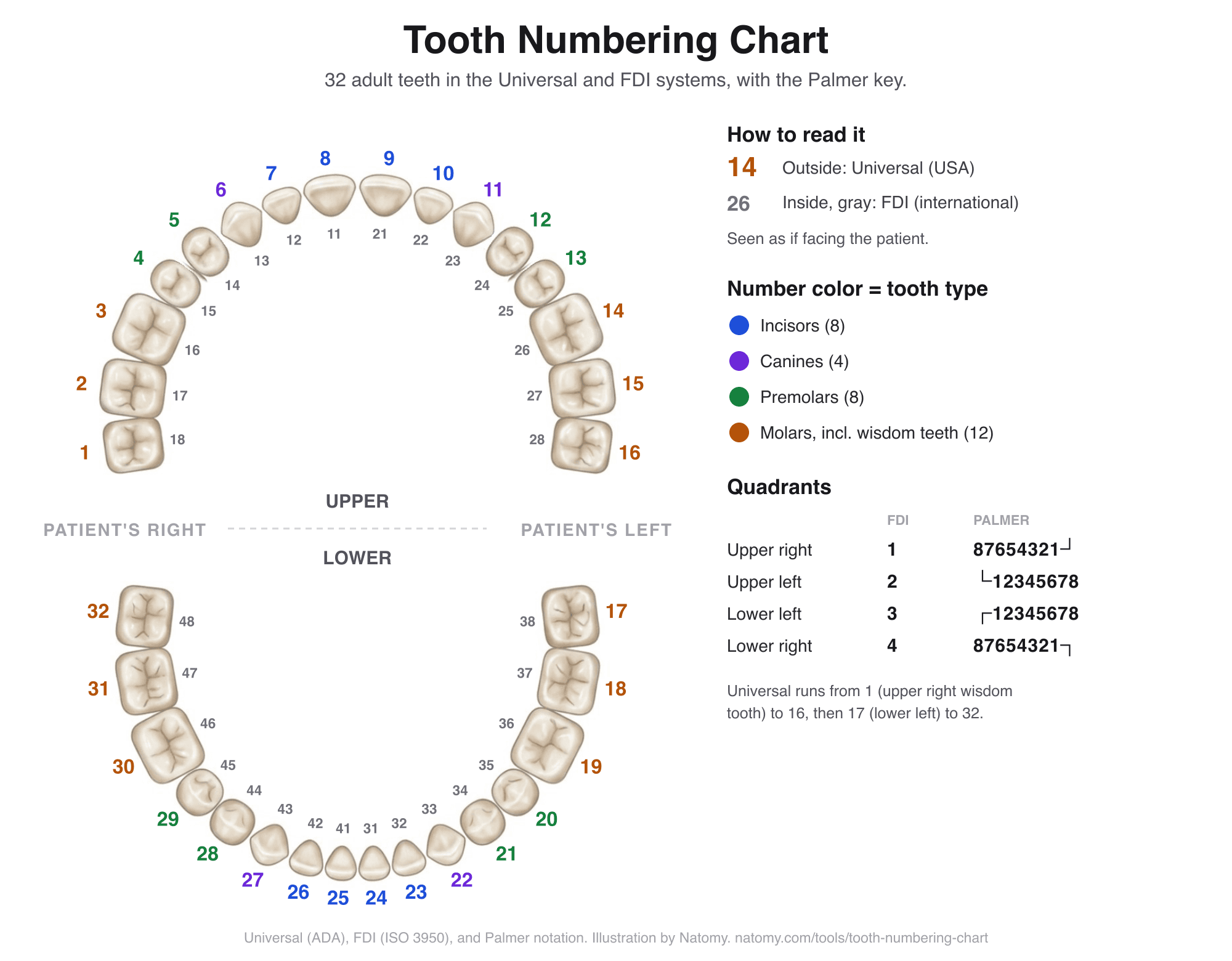
Tooth Numbering Chart
Tap any adult or baby tooth to see its Universal, FDI, and Palmer number and name, and convert numbers between systems.
Try it free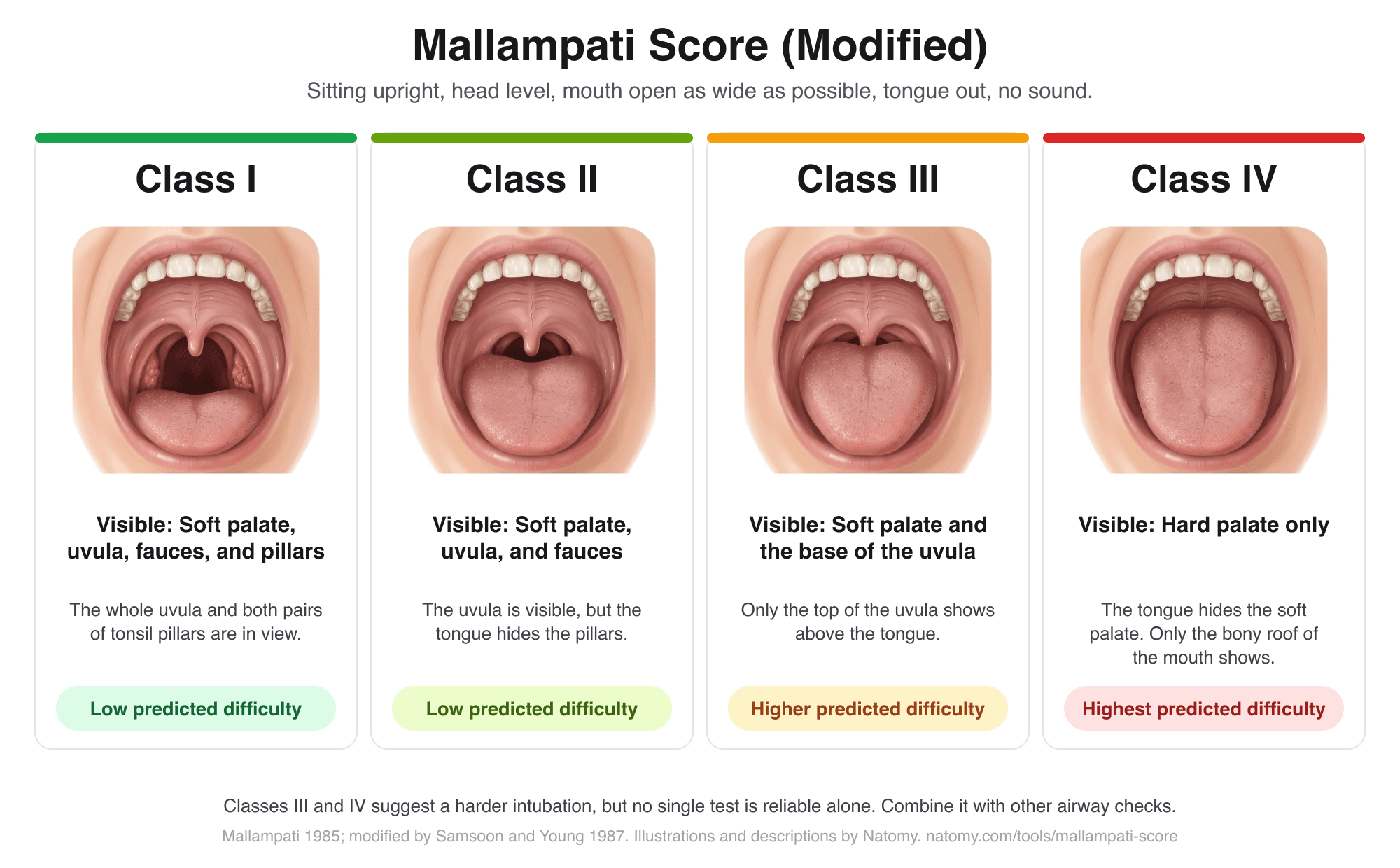
Mallampati Score Classifier
Match an open-mouth view to Mallampati classes I to IV with illustrated pictures, add other airway findings, and copy an airway note.
Try it free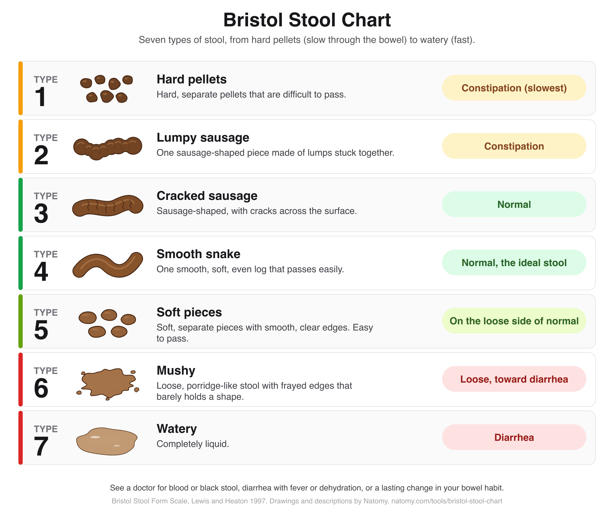
Bristol Stool Chart
Find your stool type from 1 to 7 on an illustrated chart, see what it means, and keep a simple bowel diary for your doctor.
Try it free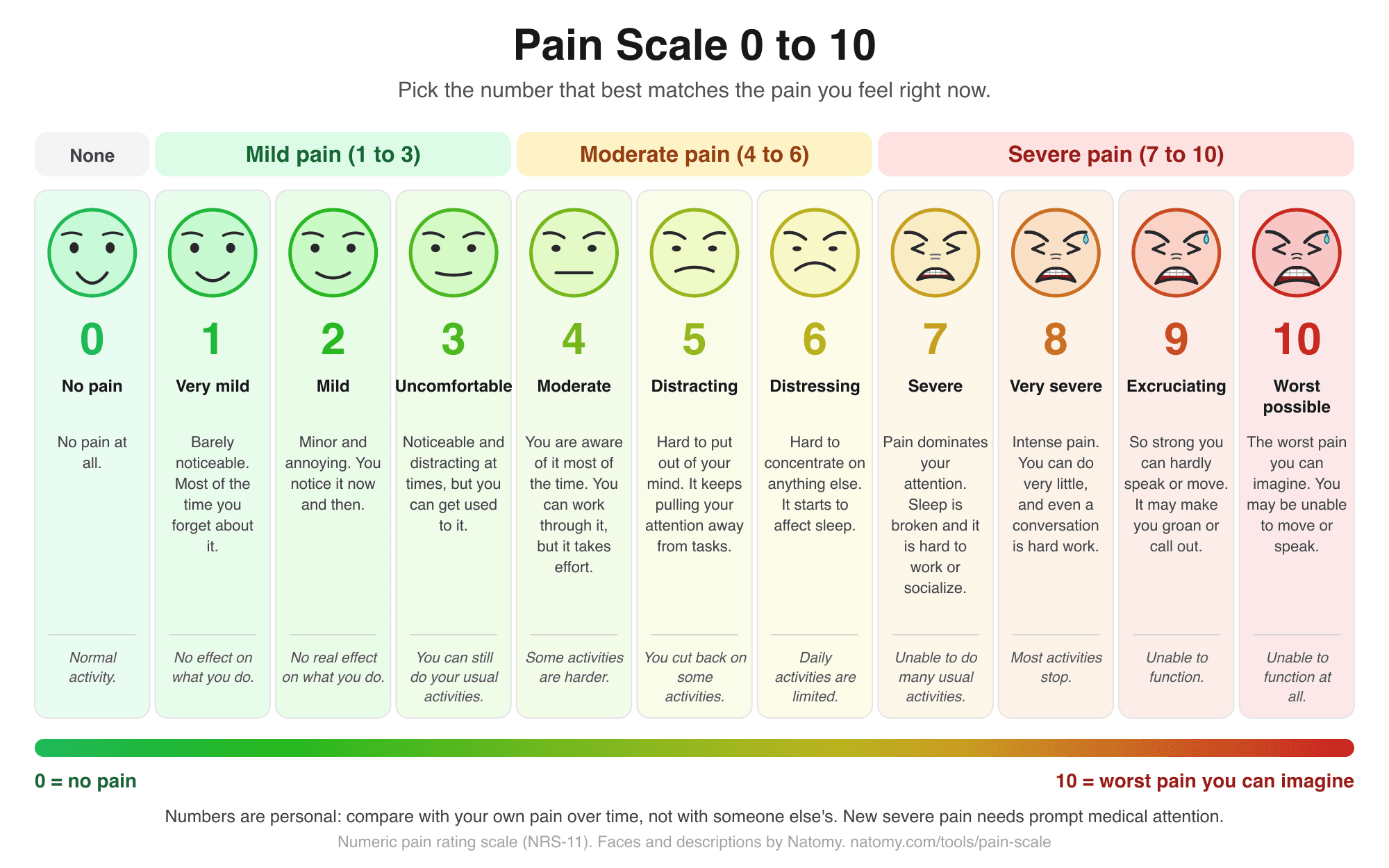
Pain Scale 0 to 10 Chart
Rate pain from 0 to 10 with drawn faces, see what mild, moderate, and severe mean, and copy a pain journal entry.
Try it free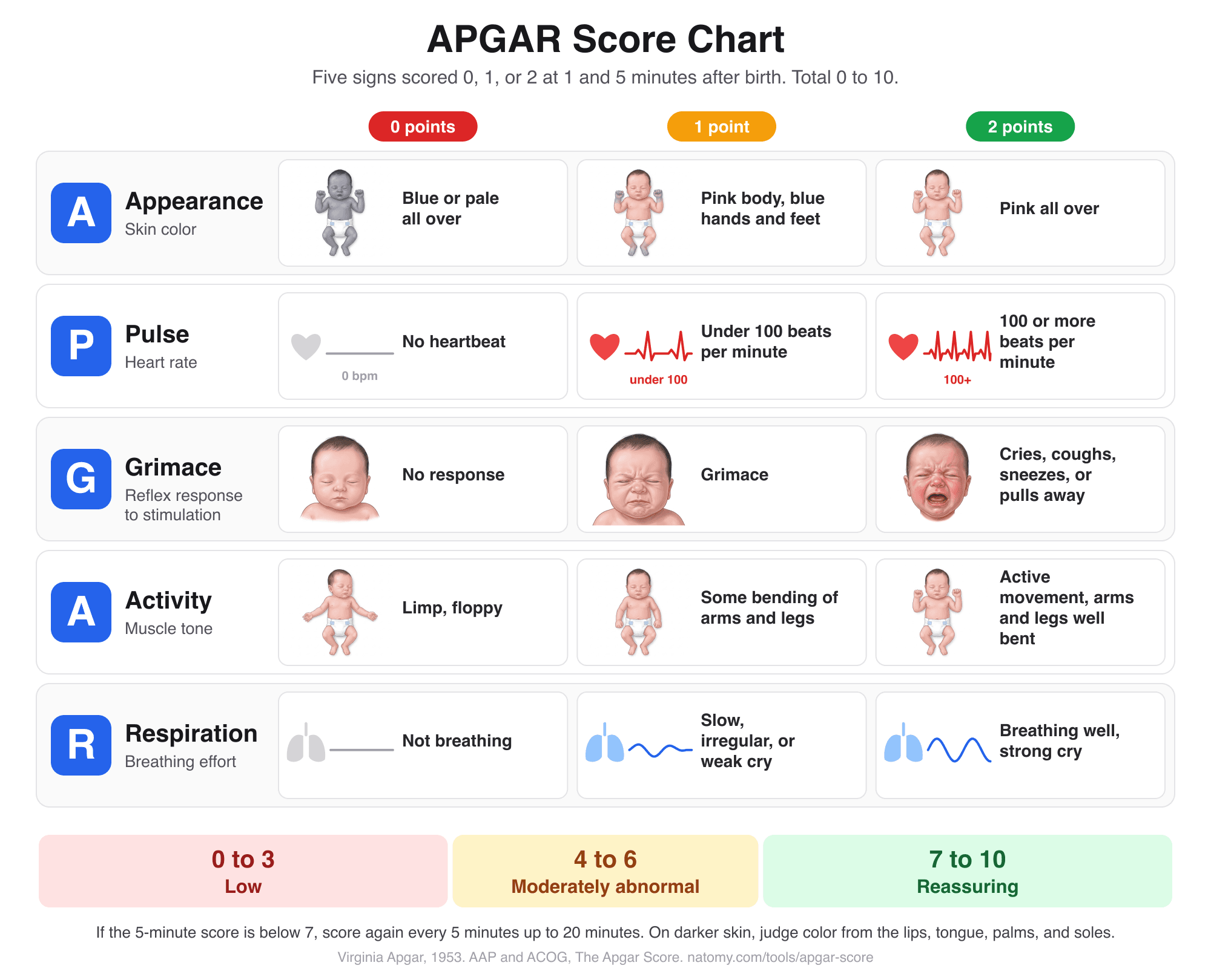
APGAR Score Chart and Calculator
Score the 5 newborn signs at 1, 5, and 10 minutes with an illustrated chart, what normal and low scores mean, and a note for the record.
Try it free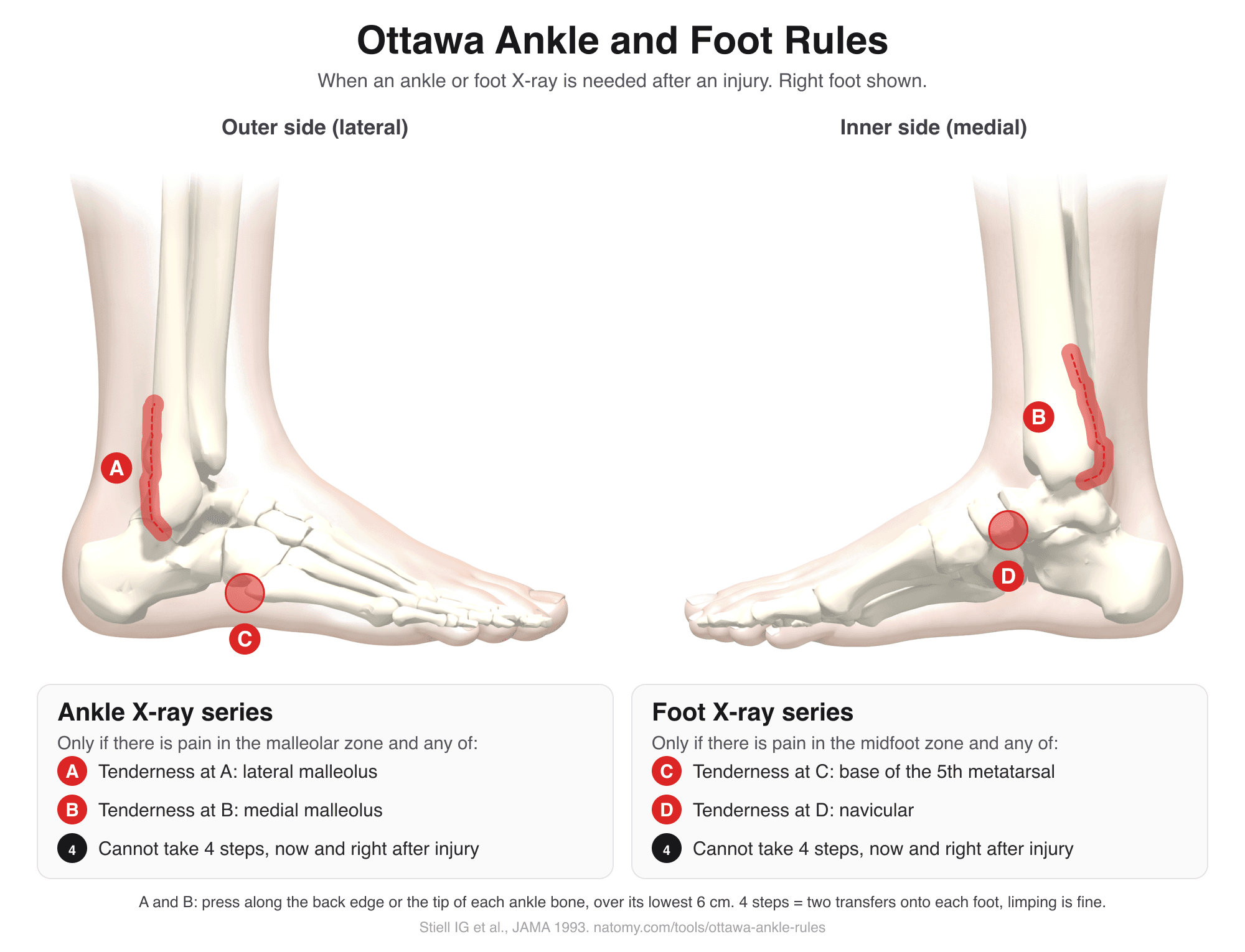
Ottawa Ankle Rules
Tap the tender spots on an illustrated ankle and foot to see if the Ottawa rules call for an X-ray. Includes the Ottawa knee rule.
Try it free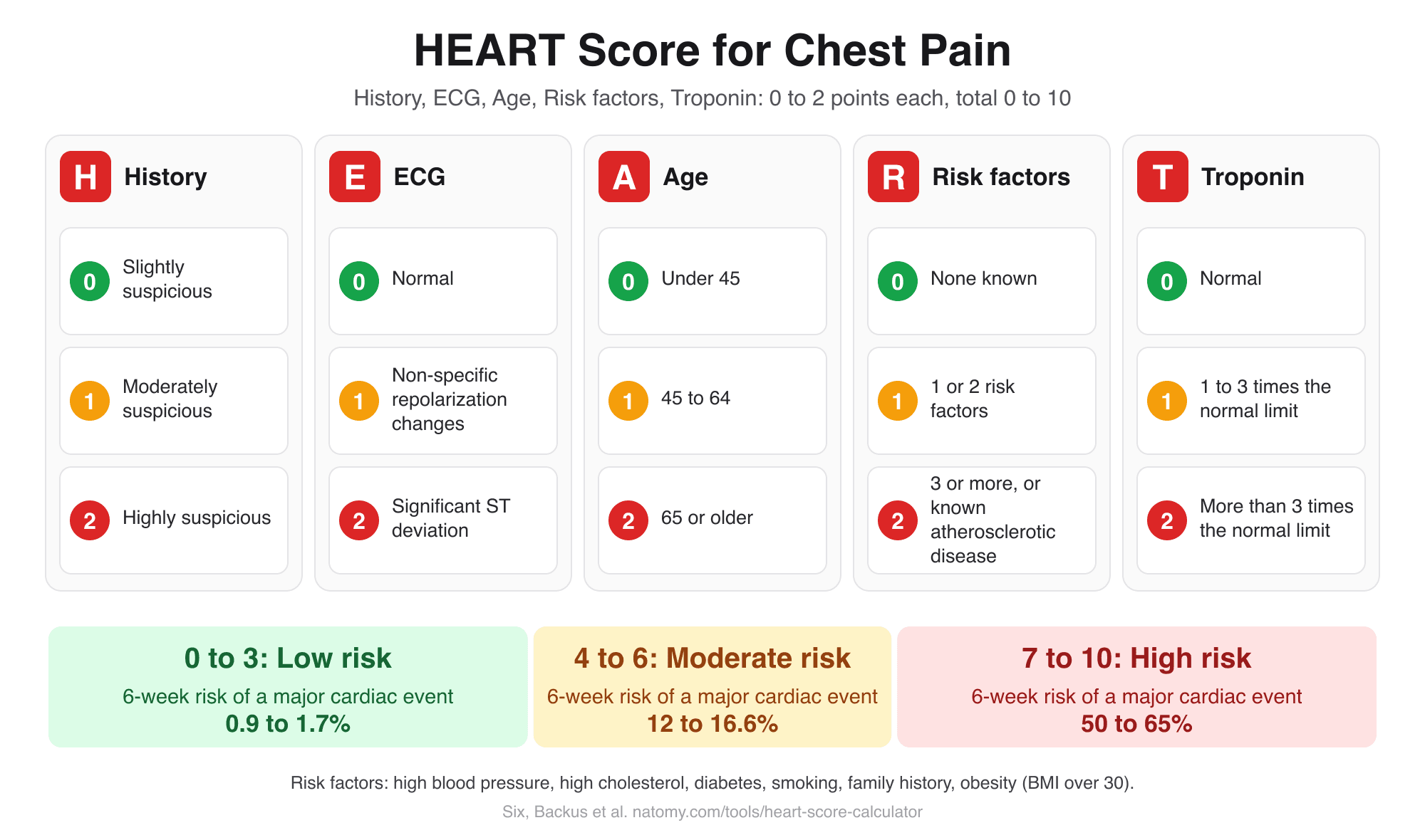
HEART Score Calculator
Score history, ECG, age, risk factors, and troponin for chest pain to get the HEART score, risk group, and 6-week MACE risk.
Try it free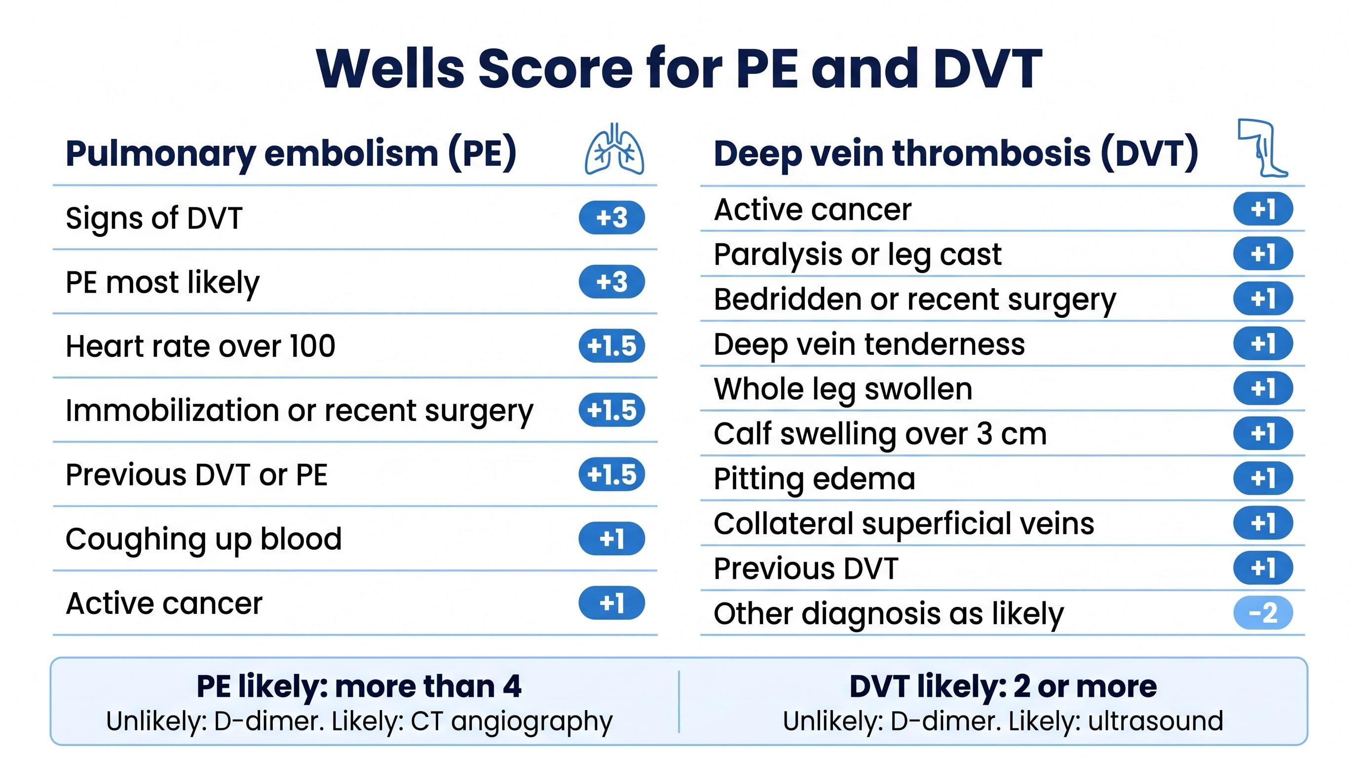
Wells Score Calculator (PE and DVT)
Check the Wells criteria for pulmonary embolism or deep vein thrombosis to get the score, the risk group, and the next test.
Try it free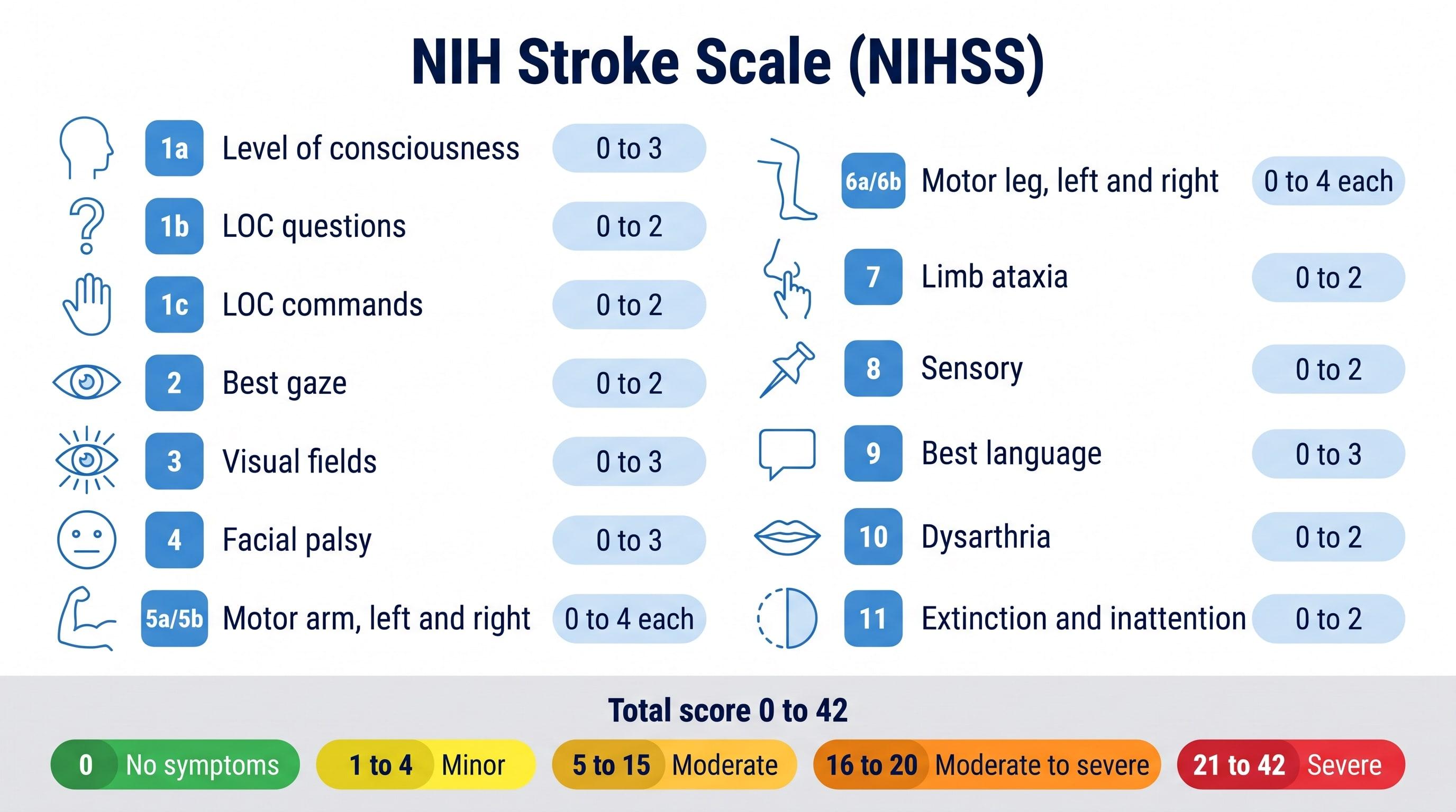
NIHSS Calculator (NIH Stroke Scale)
Score all 15 NIH Stroke Scale items to get the NIHSS total from 0 to 42 and the stroke severity range.
Try it free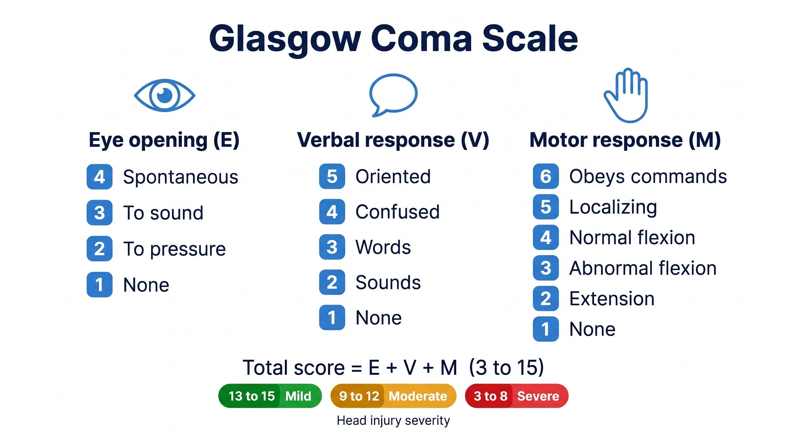
Glasgow Coma Scale Calculator
Score eye, verbal, and motor responses to get the GCS total, E V M code, severity range, and GCS-P.
Try it free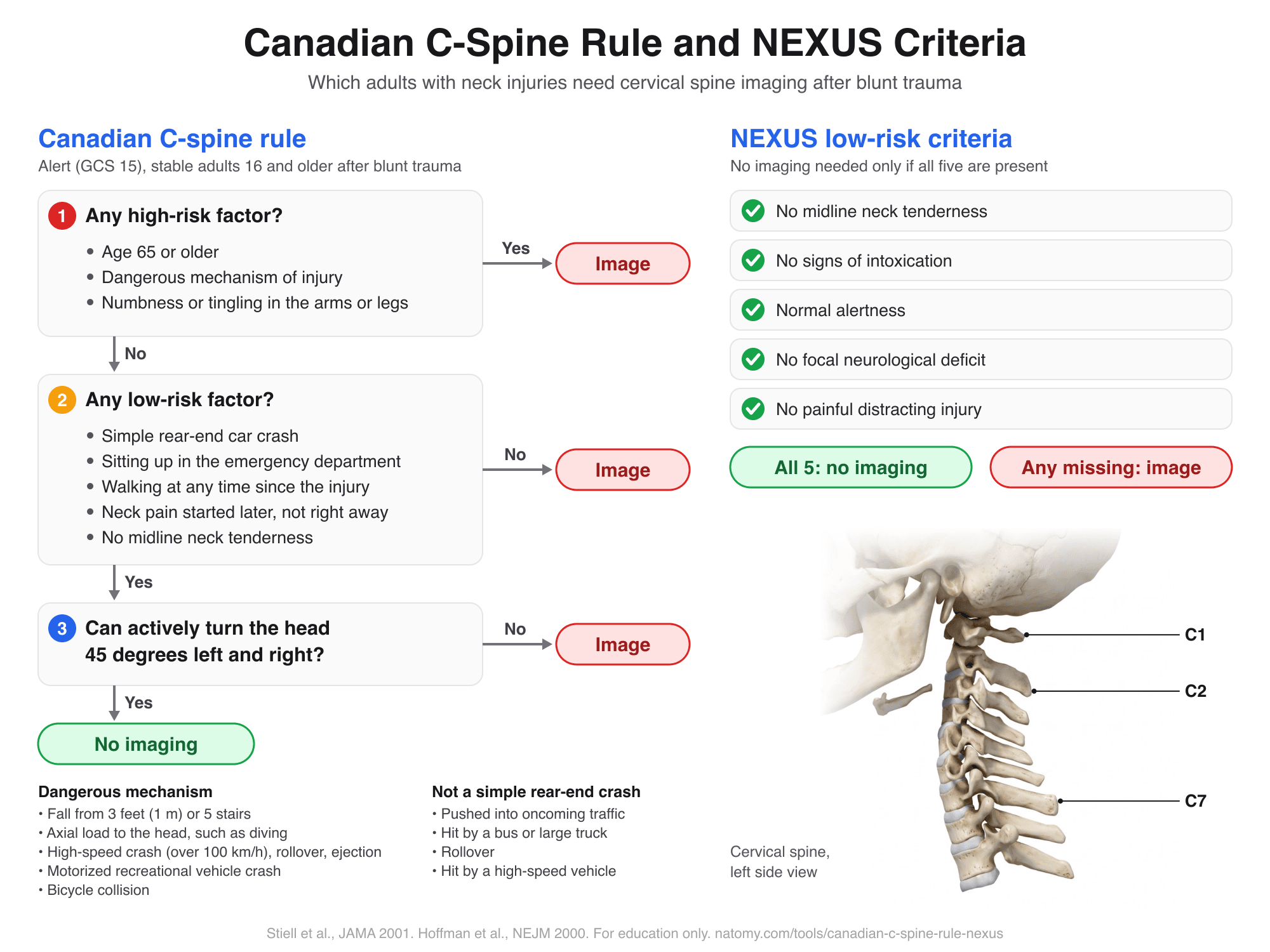
Canadian C-Spine Rule and NEXUS
Run the Canadian C-spine rule or the NEXUS criteria step by step to see which adults need neck imaging after trauma.
Try it free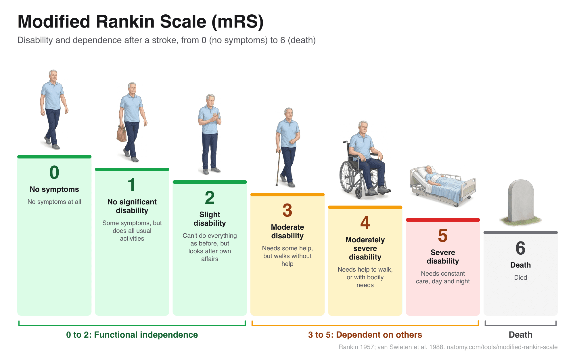
Modified Rankin Scale (mRS)
Illustrated mRS chart from 0 to 6 with a question-based score finder, what each grade means after a stroke, and a copyable note.
Try it free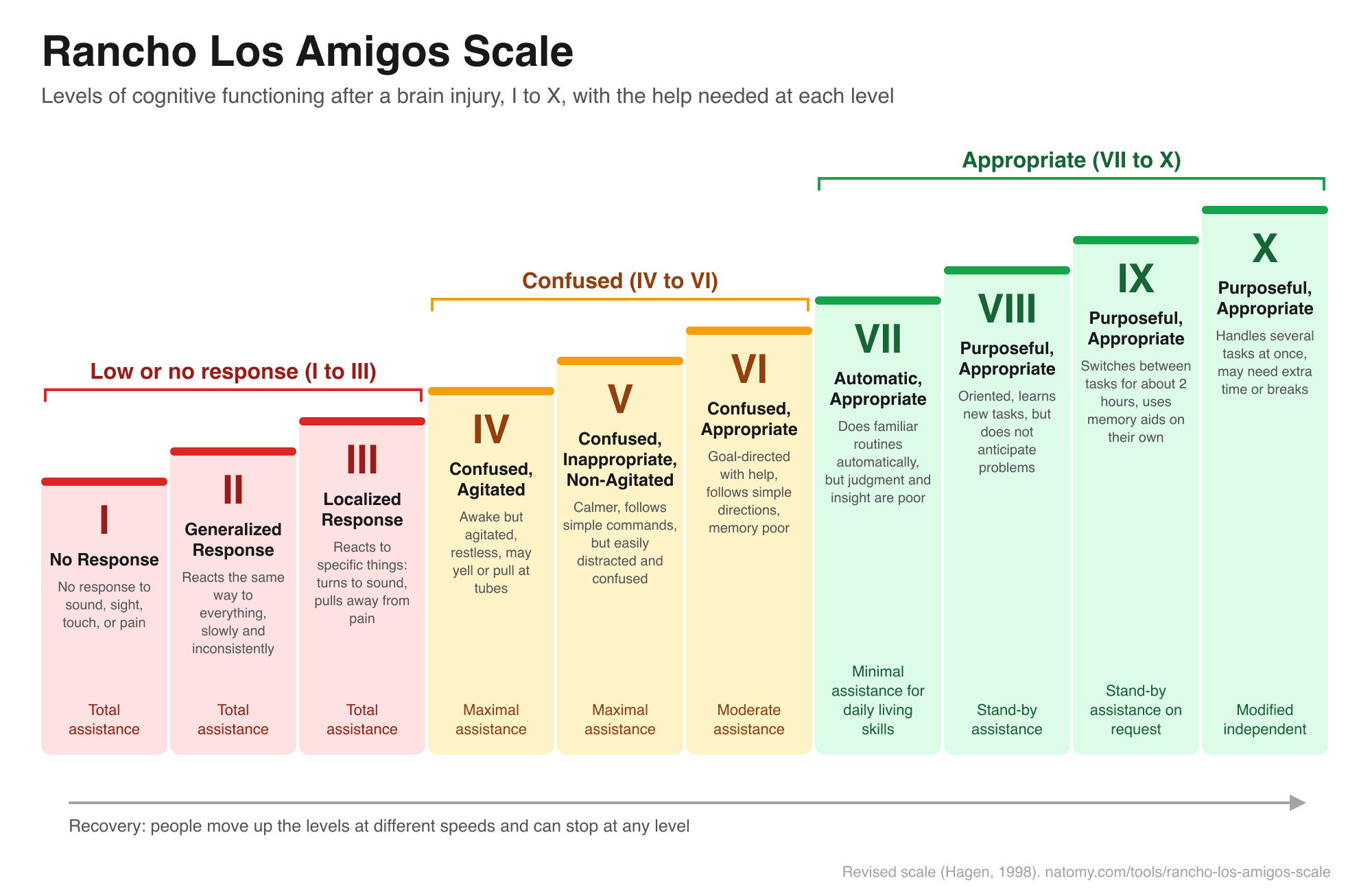
Rancho Los Amigos Scale
The 10 levels of cognitive functioning after a brain injury, with a level finder, what to expect, and how to help.
Try it free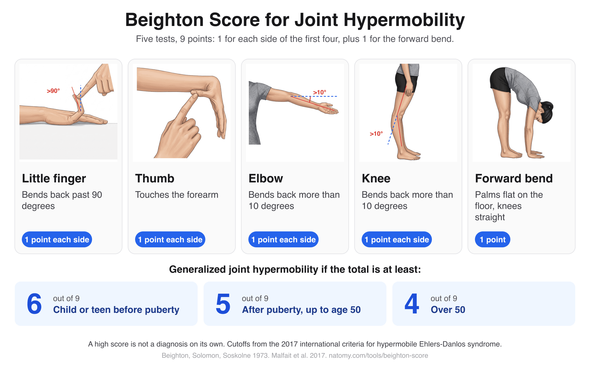
Beighton Score Calculator
Illustrated hypermobility test: score the 5 Beighton movements out of 9, compare with the age cutoff, and copy a note.
Try it free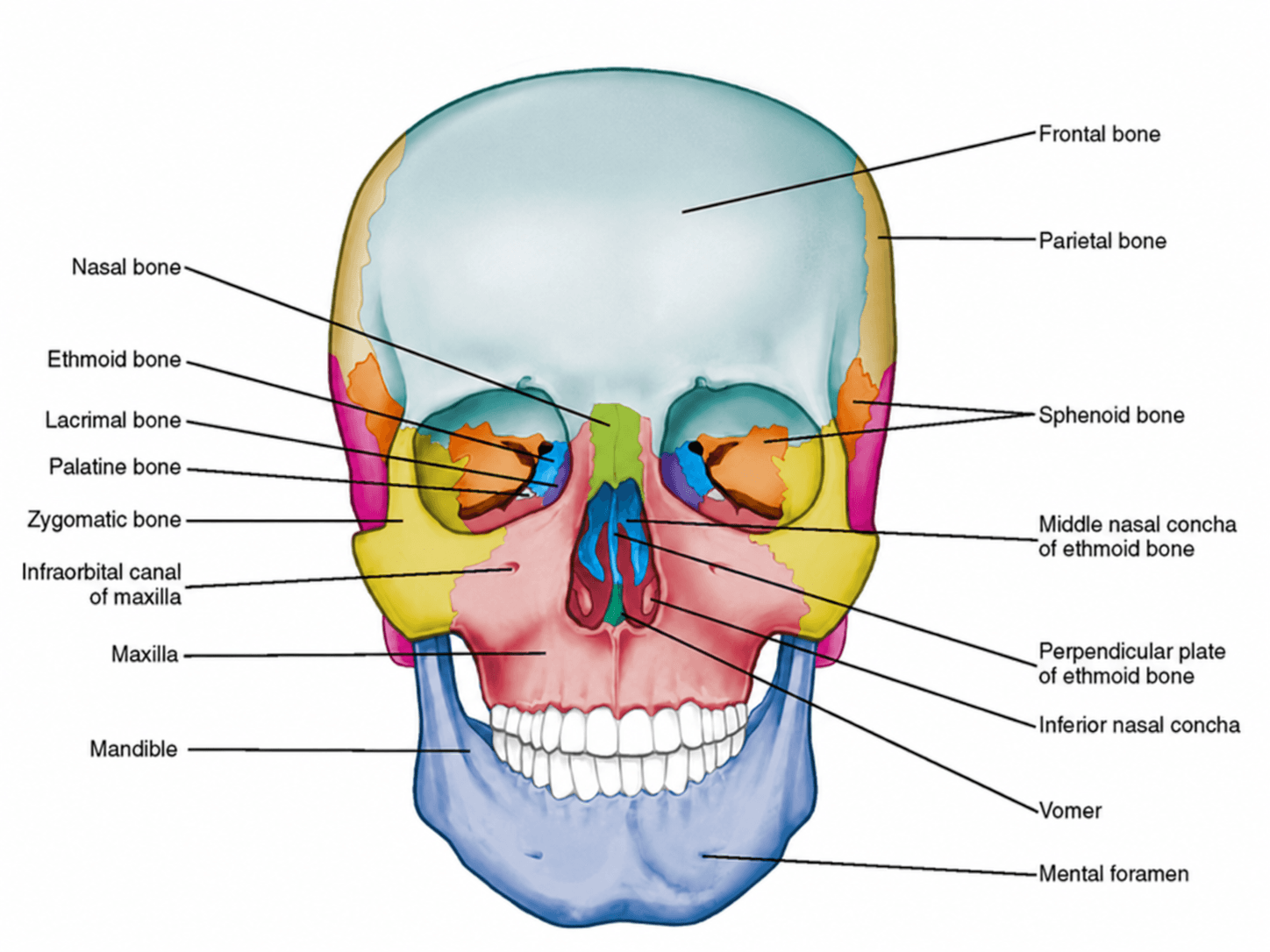
Anterior View of Skull Labeled
Free labeled and unlabeled anterior view of the skull, downloadable as PNG.
Try it free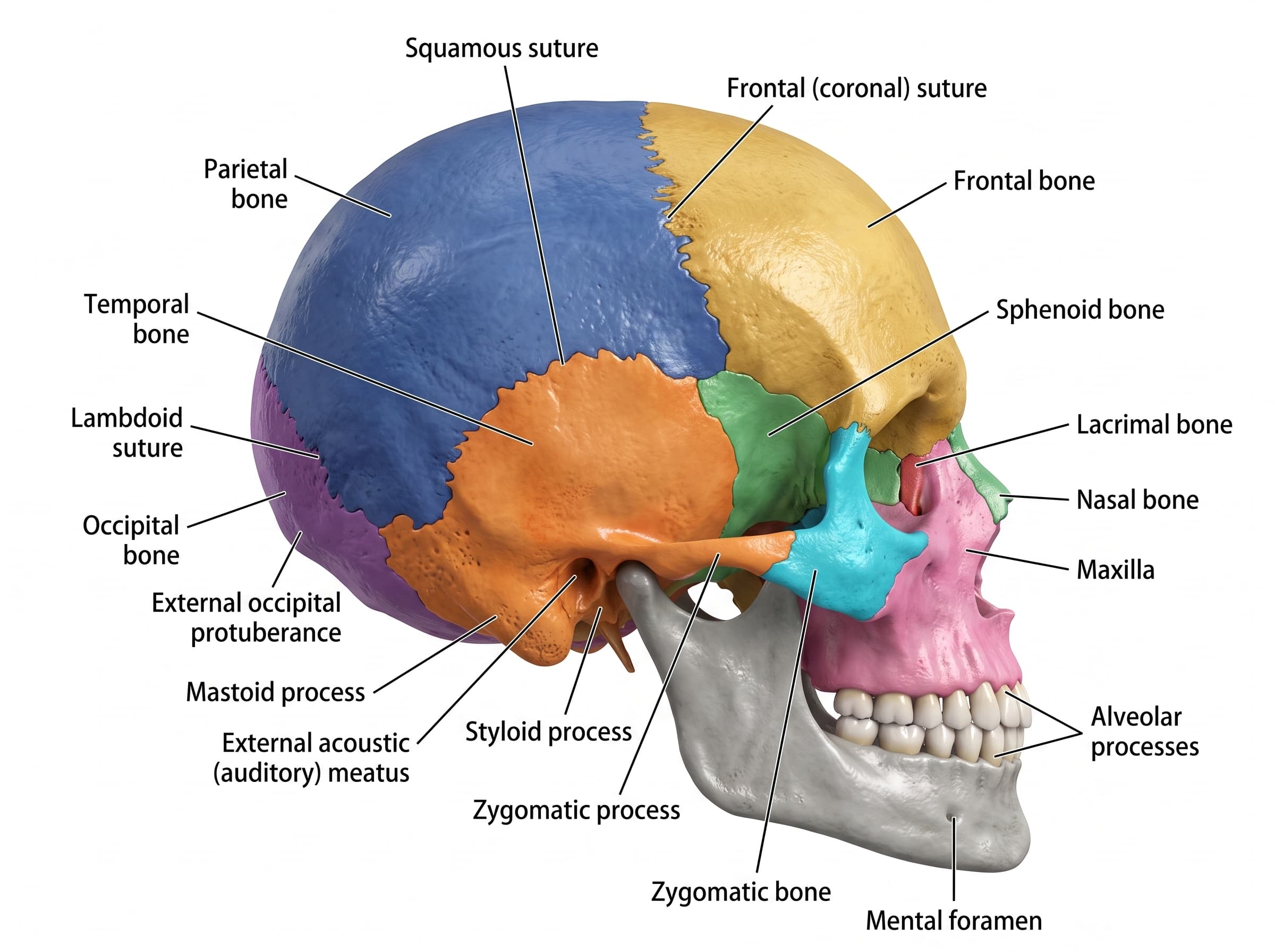
Lateral View of Skull Labeled
Free labeled and unlabeled lateral view of the skull, downloadable as PNG.
Try it free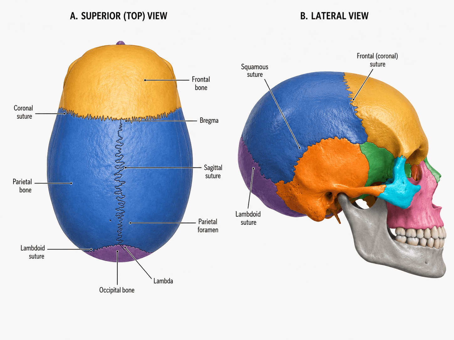
Skull Sutures Labeled
Free labeled and unlabeled skull sutures diagram, superior and lateral views, downloadable as PNG.
Try it free