Free Tools
Ottawa Ankle Rules
The Ottawa ankle rules tell you when an ankle or foot injury needs an X-ray. Press four spots on the ankle and foot and check whether the patient can take 4 steps. If none of these are positive, a fracture is very unlikely. Use the free checker below to tap the tender spots and get a note for the record.
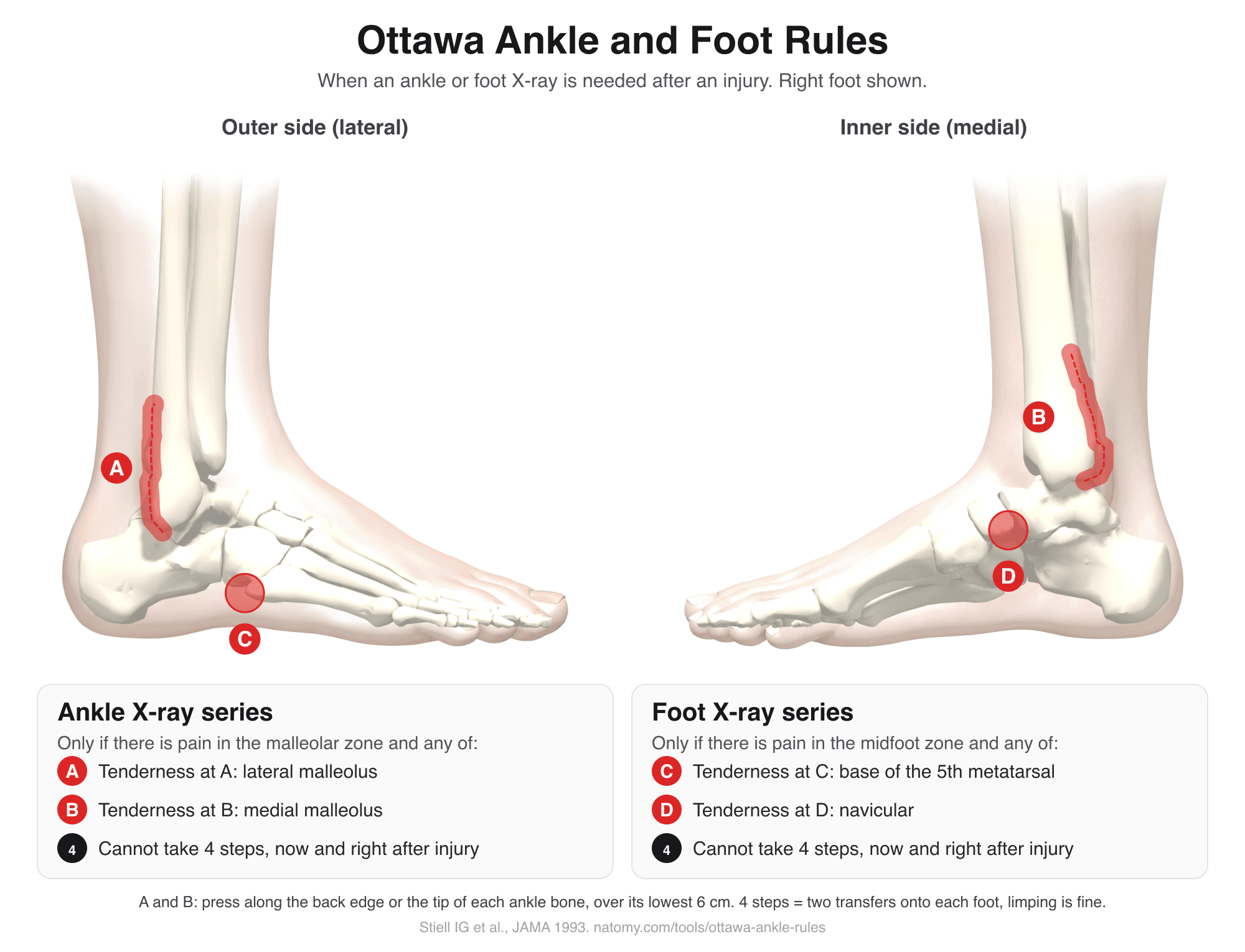
Download the chart (PNG) for teaching, the emergency department, or case files.
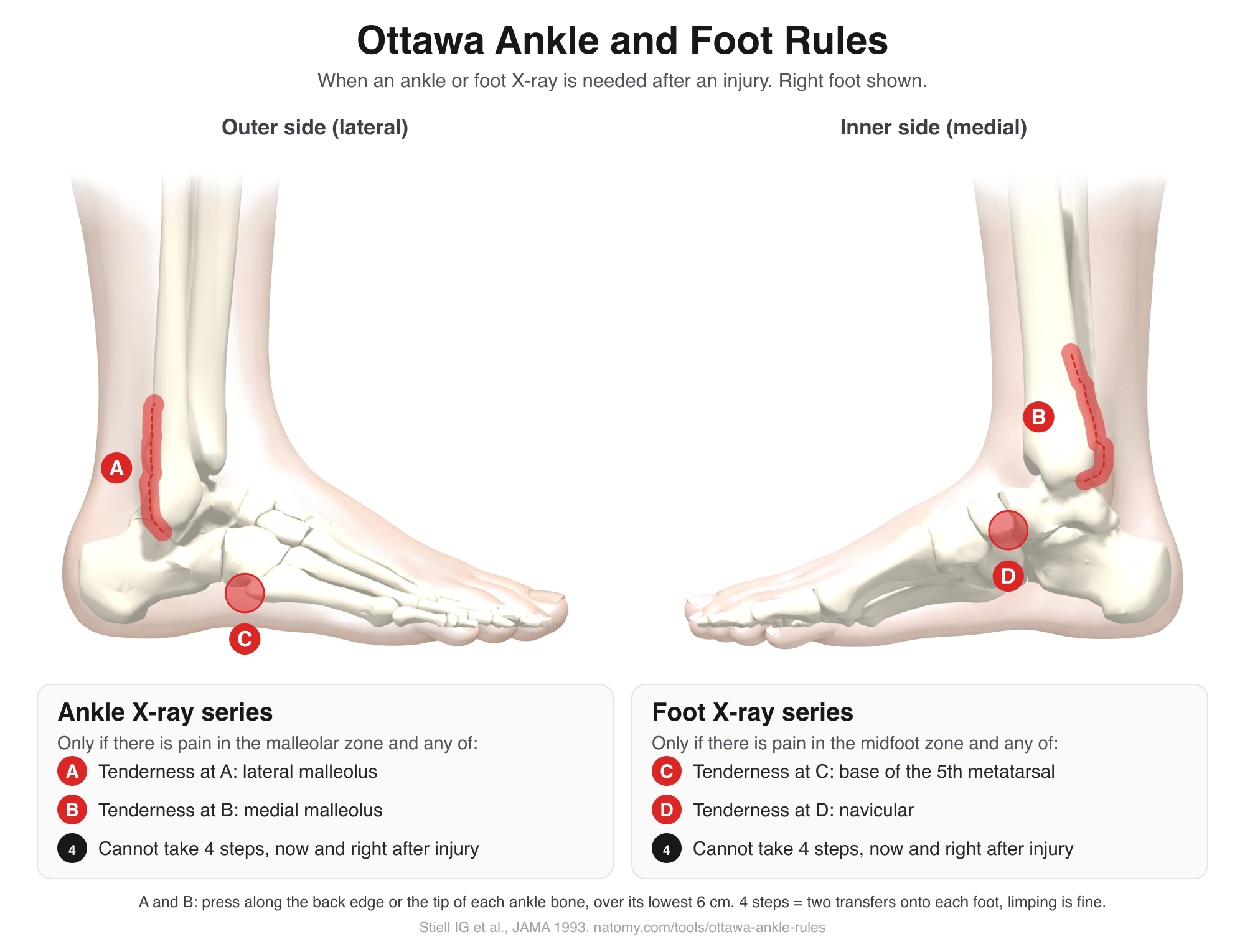
Ottawa ankle rules checker
Tap the zones that are tender to firm pressure, then answer the weight-bearing question.
1. Where is it painful?
2. Tap every spot that is tender to firm pressure
- A: Posterior edge or tip of the lateral malleolus (fibula), lower 6 cm
- B: Posterior edge or tip of the medial malleolus (tibia), lower 6 cm
- C: The bony bump halfway along the outer edge of the foot
- D: The bony bump on the inner side of the midfoot, in front of the ankle
3. Weight bearing
Unable to bear weight both right after the injury and in the emergency department: cannot take 4 steps (two transfers onto each foot), even with a limp.
When the rule may not apply (optional)
The Ottawa ankle and foot rules
Ankle X-ray series
Needed only if there is pain in the malleolar zone (around the ankle bones) and any of these:
- Bone tenderness at A: posterior edge or tip of the lateral malleolus (fibula), lower 6 cm
- Bone tenderness at B: posterior edge or tip of the medial malleolus (tibia), lower 6 cm
- Unable to take 4 steps both right after the injury and in the emergency department
Foot X-ray series (Ottawa foot rules)
Needed only if there is pain in the midfoot zone and any of these:
- Bone tenderness at C: the base of the 5th metatarsal
- Bone tenderness at D: the navicular
- Unable to take 4 steps both right after the injury and in the emergency department
How to examine the ankle and foot
- Press the back edge, not the front. Start at the tip of each malleolus and work up its back edge for 6 cm. The front of the lateral malleolus is where the ankle ligaments attach, so it is tender in an ordinary sprain and does not count.
- Find the bumps on the foot. The base of the 5th metatarsal is the bony bump halfway along the outer edge of the foot, at and just beyond which avulsion and Jones fractures happen. The navicular is the bump on the inner side, about a thumb's width in front of and below the medial malleolus.
- Count 4 steps. The patient must transfer weight onto each foot twice. Limping is fine. It has to be impossible both right after the injury and at the exam to count.
How accurate are the rules?
The rules were designed to miss almost nothing. A 2003 BMJ review of 27 studies and more than 15,000 patients found a pooled sensitivity of about 98% for the ankle and 99% for the midfoot, so when the rules are negative the chance of a fracture is very low. Specificity varies widely between studies, with a median of about 40%, which is the trade-off for that safety. The rules are estimated to cut ankle X-rays by about 30 to 40%; when they were put into practice in Ottawa, ankle X-rays fell by 28% and foot X-rays by 14%, with no missed fractures.
When the rules may not apply
Formally excluded from the 1993 validation study:
- Under 18 (excluded from the original studies; later studies support use in children over 5)
- Pregnancy
- Injury more than 10 days old, or a return visit for the same injury
- Referred in with X-rays already taken elsewhere
- Isolated skin injuries such as cuts or burns
Clinicians also use caution when the exam is unreliable:
- Intoxication, a head injury, or other painful injuries that make the exam unreliable
- Reduced sensation in the leg, for example from diabetic neuropathy or a spinal injury
The Ottawa knee rule
A companion rule from the same Ottawa group, studied in adults. After an acute knee injury, a knee X-ray is needed if any one of these applies:
- Age 55 or older
- Tenderness at the head of the fibula
- Isolated tenderness of the kneecap (patella), with no other bone tenderness
- Unable to bend the knee to 90 degrees
- Unable to bear weight both right after the injury and in the emergency department (4 steps)
Like the ankle rules, it is very sensitive for fractures that matter, such as patella fractures and tibial plateau fractures, and cuts unnecessary knee X-rays.
Ottawa ankle rules in missed fracture cases
Ankle and foot injuries are among the most common reasons for an emergency visit, and a missed fracture is a frequent source of malpractice claims. When an X-ray was not ordered, the question is usually whether the Ottawa rules were applied and documented: which spots were pressed, whether the patient could take 4 steps, and whether anything made the rules unreliable. When an X-ray was taken, the question shifts to how it was read. A documented negative exam, with clear return advice, supports a finding of reasonable care; a chart that says only "ankle sprain" does not. The rules are a widely accepted decision aid, not a legal requirement.
For attorneys, an illustration that marks the exact zones on the patient's ankle, next to the ankle fracture that was later found, makes the standard of care easy for a jury to follow.
Illustrate an ankle or foot injury
Natomy turns X-rays, records, and prompts into clear, labeled medical illustrations of fractures and ligament injuries for patient education and medical-legal exhibits. See examples in the Natomy atlas.
Try Natomy TodayFAQ
What are the Ottawa ankle rules?+
The Ottawa ankle rules are a quick bedside check that tells a clinician whether an ankle or foot X-ray is needed after an injury. An ankle X-ray is needed only if there is pain around the ankle bones and either bone tenderness along the back edge or tip of either malleolus, or the patient cannot take 4 steps. A foot X-ray is needed only if there is midfoot pain and either tenderness at the base of the 5th metatarsal or the navicular, or the patient cannot take 4 steps.
How accurate are the Ottawa ankle rules?+
Very good at ruling fractures out. A 2003 BMJ review of 27 studies with more than 15,000 patients found a sensitivity of about 98% for the ankle and 99% for the midfoot, so when the rules are negative the chance of a fracture is very low (under about 1.5% when roughly 15% of patients have a fracture). Specificity varies widely between studies, with a median of about 40%, so many patients who meet the rules turn out not to have a fracture. The rules are estimated to cut ankle X-rays by about 30 to 40%; when they were put into practice in Ottawa, ankle X-rays fell by 28% and foot X-rays by 14%, with no missed fractures.
What counts as unable to bear weight?+
Not being able to take 4 steps, which means transferring weight onto each foot twice, both right after the injury and when examined. Limping still counts as bearing weight. If the patient can walk 4 steps with a limp, that criterion is not met.
Where exactly do you press?+
Along the back (posterior) edge and the tip of the lateral malleolus (the outer ankle bone, the end of the fibula) and the medial malleolus (the inner ankle bone, the end of the tibia), over the lowest 6 cm of each. On the foot, press the base of the 5th metatarsal, the bump halfway along the outer edge, and the navicular, the bump on the inner side in front of the ankle. Pressing the back edge rather than the front matters, because the front of the lateral malleolus is where ligament sprains hurt.
Do the Ottawa ankle rules work in children?+
The rules were developed in adults. A 2009 meta-analysis of 12 studies (3,130 children) found a sensitivity of 98.5% in children over 5, with X-rays cut by about 25%. A few Salter-Harris type I fractures were missed. The rules are less reliable in younger children.
When should the Ottawa ankle rules not be used?+
The 1993 validation study formally excluded patients under 18, pregnant patients, isolated skin injuries, patients referred with X-rays from elsewhere, injuries more than 10 days old, and return visits for the same injury. Clinicians also use caution when the exam is unreliable, such as with intoxication, a head injury, multiple painful injuries, or reduced sensation in the leg.
What is the Ottawa knee rule?+
A companion rule for knee injuries, studied in adults. A knee X-ray is needed if any of these apply: age 55 or older, tenderness at the head of the fibula, tenderness of the kneecap alone, inability to bend the knee to 90 degrees, or inability to take 4 steps both right after the injury and when examined.
Can I get an X-ray anyway if the rules are negative?+
Yes. The rules support clinical judgment and do not replace it. If pain or walking does not improve within about a week, or something else is concerning, an X-ray or follow-up is reasonable.
Sources: Stiell IG, et al. A study to develop clinical decision rules for the use of radiography in acute ankle injuries. Ann Emerg Med. 1992;21(4):384-390. Stiell IG, et al. Decision rules for the use of radiography in acute ankle injuries: refinement and prospective validation. JAMA. 1993;269(9):1127-1132. Bachmann LM, et al. Accuracy of Ottawa ankle rules to exclude fractures of the ankle and mid-foot: systematic review. BMJ. 2003;326(7386):417. Stiell IG, et al. Implementation of the Ottawa ankle rules. JAMA. 1994;271(11):827-832. Dowling S, et al. Accuracy of Ottawa Ankle Rules to exclude fractures of the ankle and midfoot in children: a meta-analysis. Acad Emerg Med. 2009;16(4):277-287. Stiell IG, et al. Derivation of a decision rule for the use of radiography in acute knee injuries. Ann Emerg Med. 1995;26(4):405-413. Stiell IG, et al. Prospective validation of a decision rule for the use of radiography in acute knee injuries. JAMA. 1996;275(8):611-615. Descriptions are written in our own words. For education and documentation only; not a substitute for clinical judgment.
Related free tools
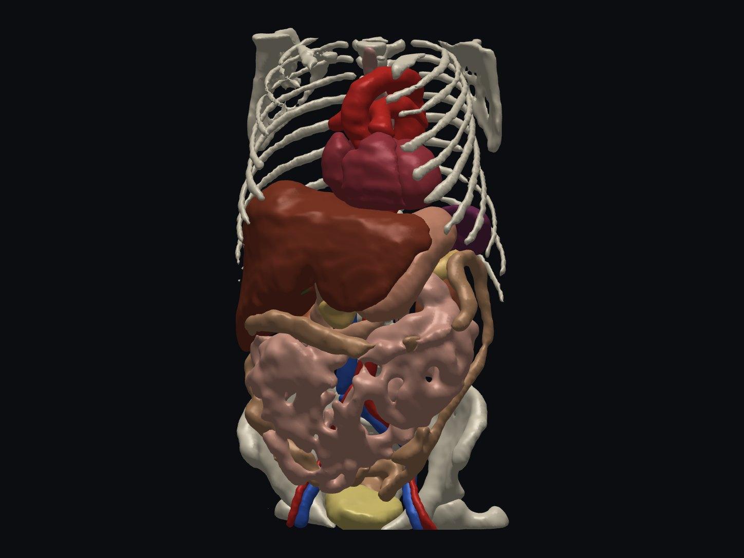
Free Online DICOM Viewer (3D)
Open a CT or MRI DICOM folder in your browser and view it in 3D, with AI anatomy labeling. Nothing is uploaded.
Try it free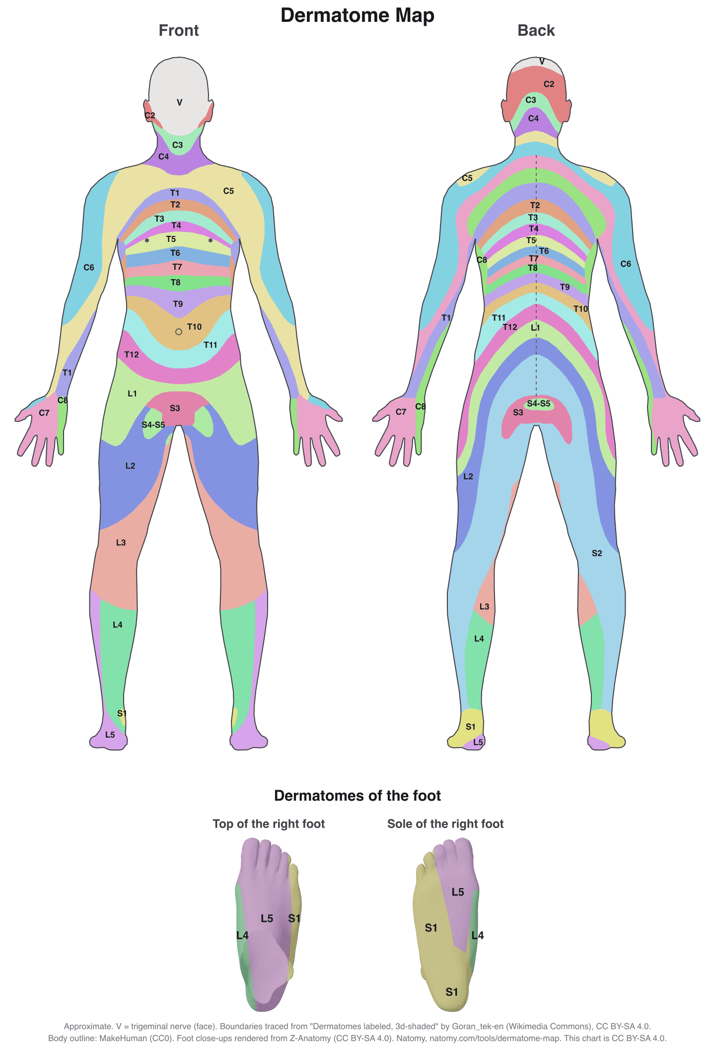
Interactive Dermatome Map
Tap the body to see each nerve root's skin area, test point, myotome, reflex, and disc level. Free printable chart.
Try it free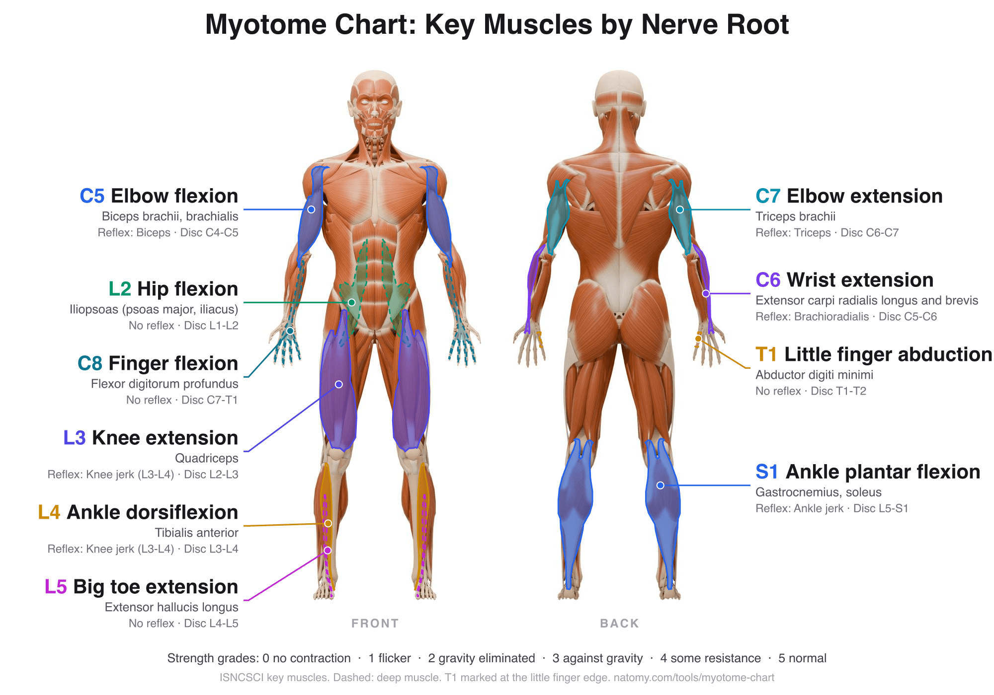
Myotome Chart and Motor Score
Tap a nerve root to see its key muscle, test position, and reflex. Grade strength 0 to 5 for the ISNCSCI motor score.
Try it free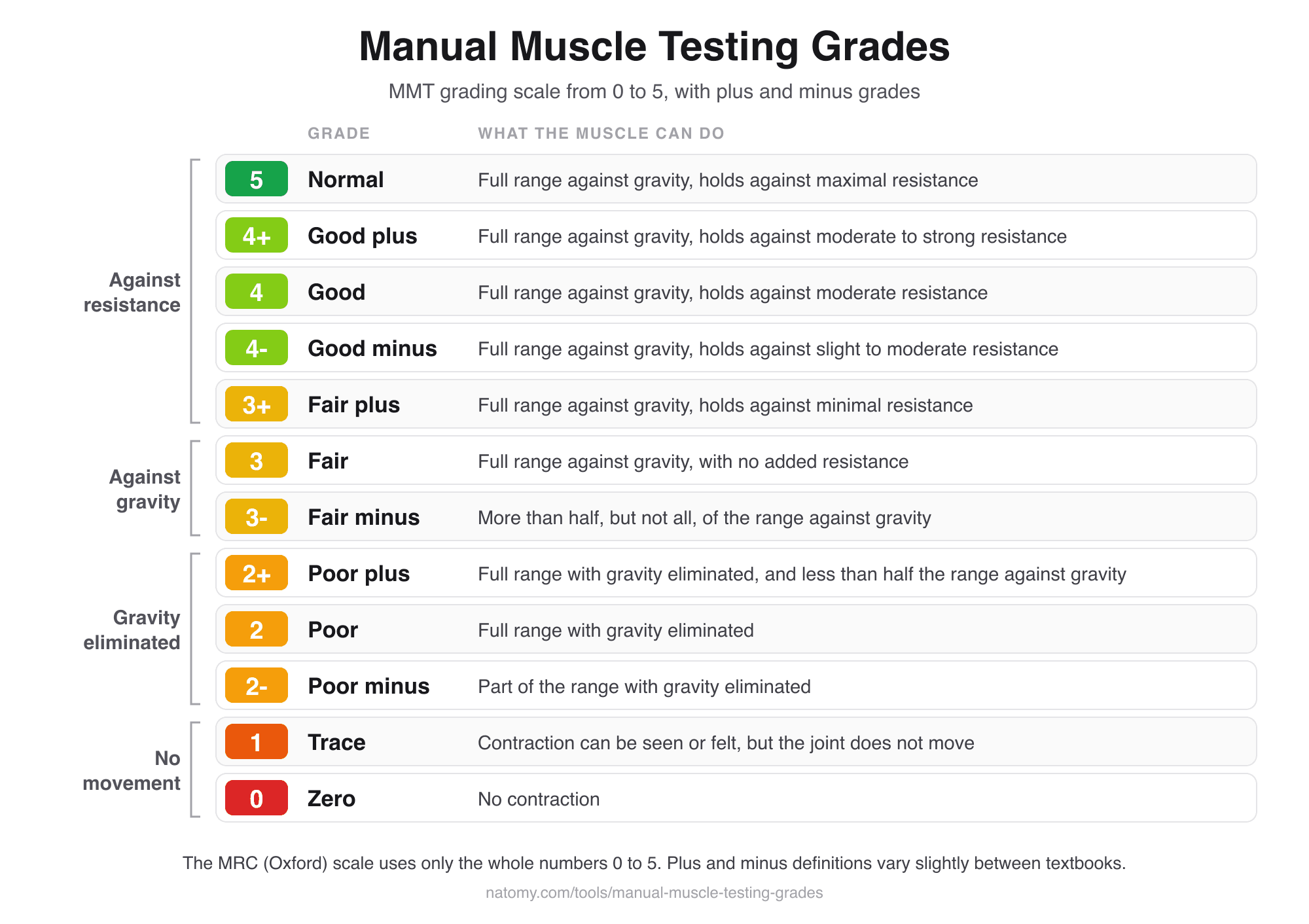
Manual Muscle Testing Grades
MMT grading scale chart from 0 to 5 with plus and minus grades, and a grade finder that writes the note for you.
Try it free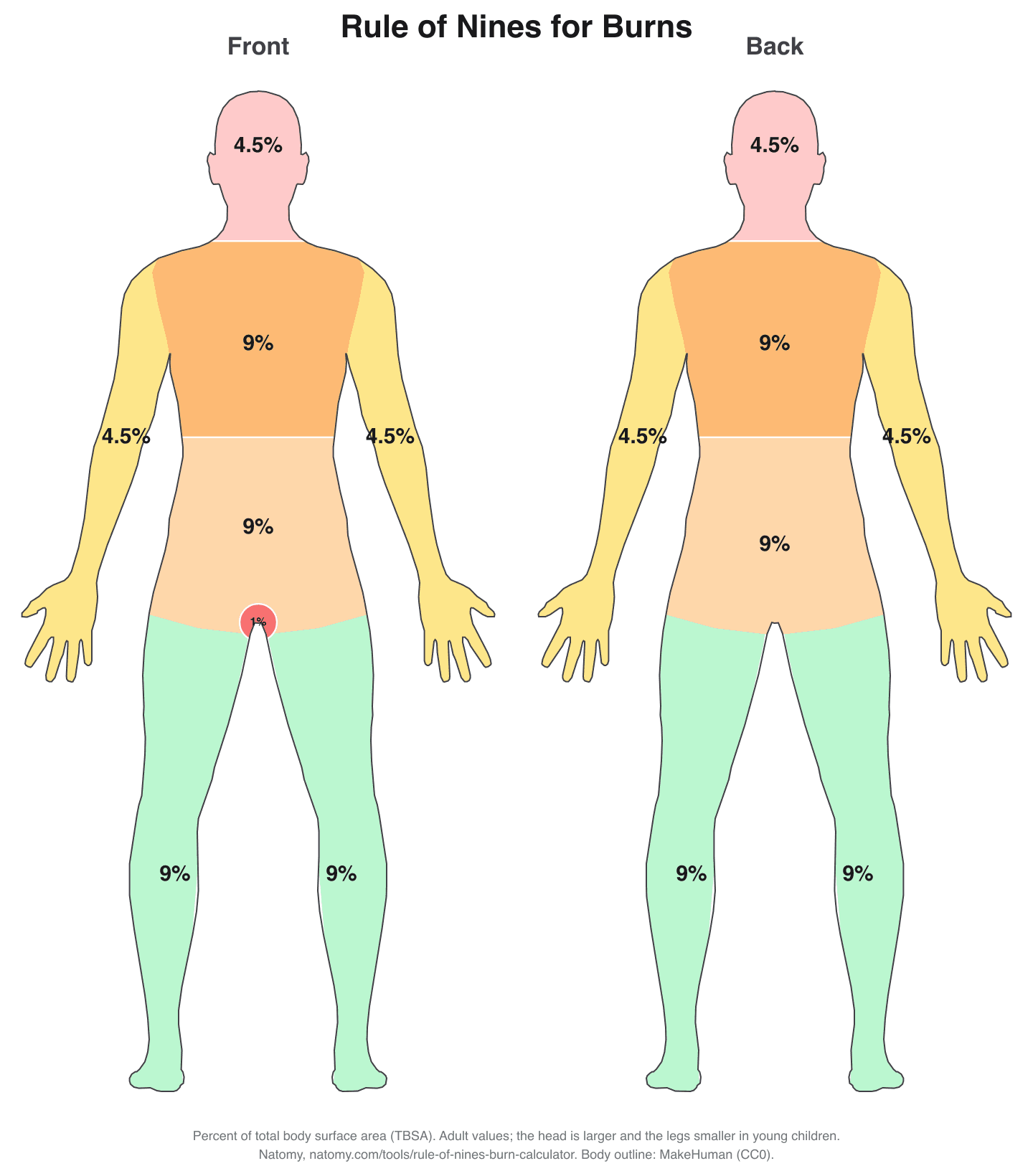
Rule of Nines Burn Calculator
Click the burned areas to get the total body surface area (TBSA), with the Parkland formula and a printable burn chart.
Try it free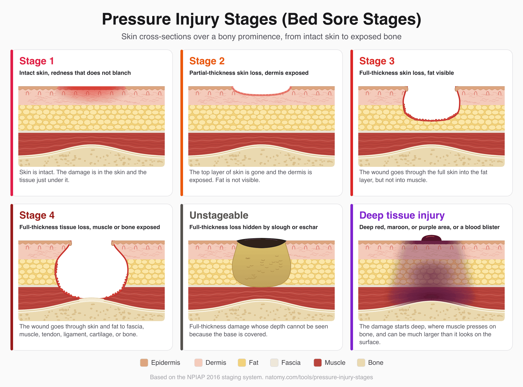
Pressure Injury (Bed Sore) Stages
Illustrated pressure ulcer stages chart, Stage 1 to 4, unstageable, and deep tissue injury, with a stage finder and wound note.
Try it free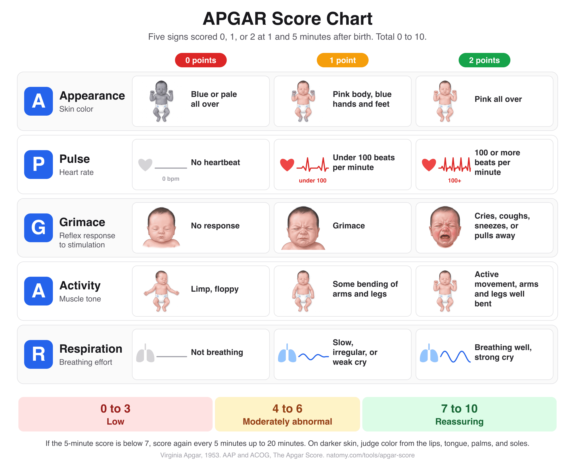
APGAR Score Chart and Calculator
Score the 5 newborn signs at 1, 5, and 10 minutes with an illustrated chart, what normal and low scores mean, and a note for the record.
Try it free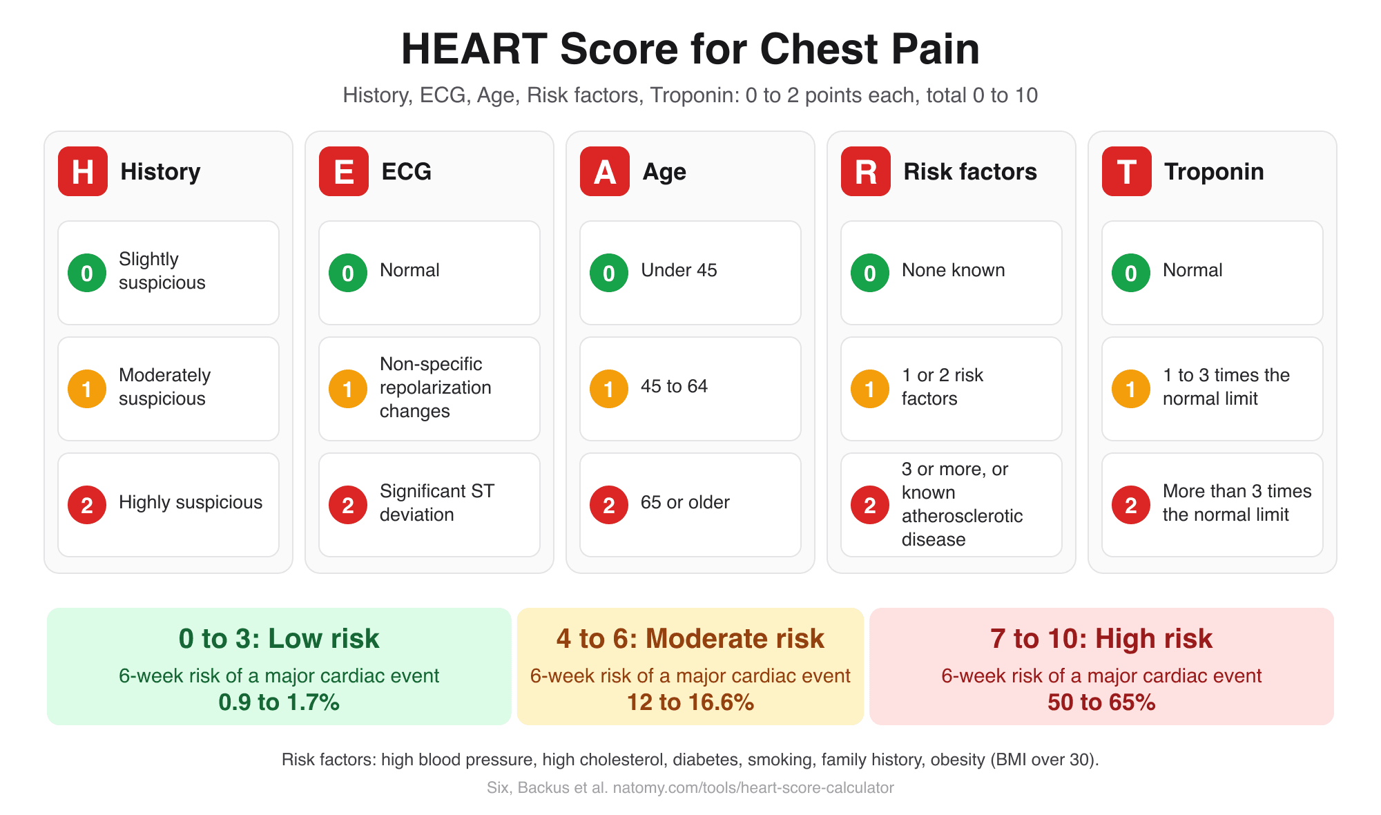
HEART Score Calculator
Score history, ECG, age, risk factors, and troponin for chest pain to get the HEART score, risk group, and 6-week MACE risk.
Try it free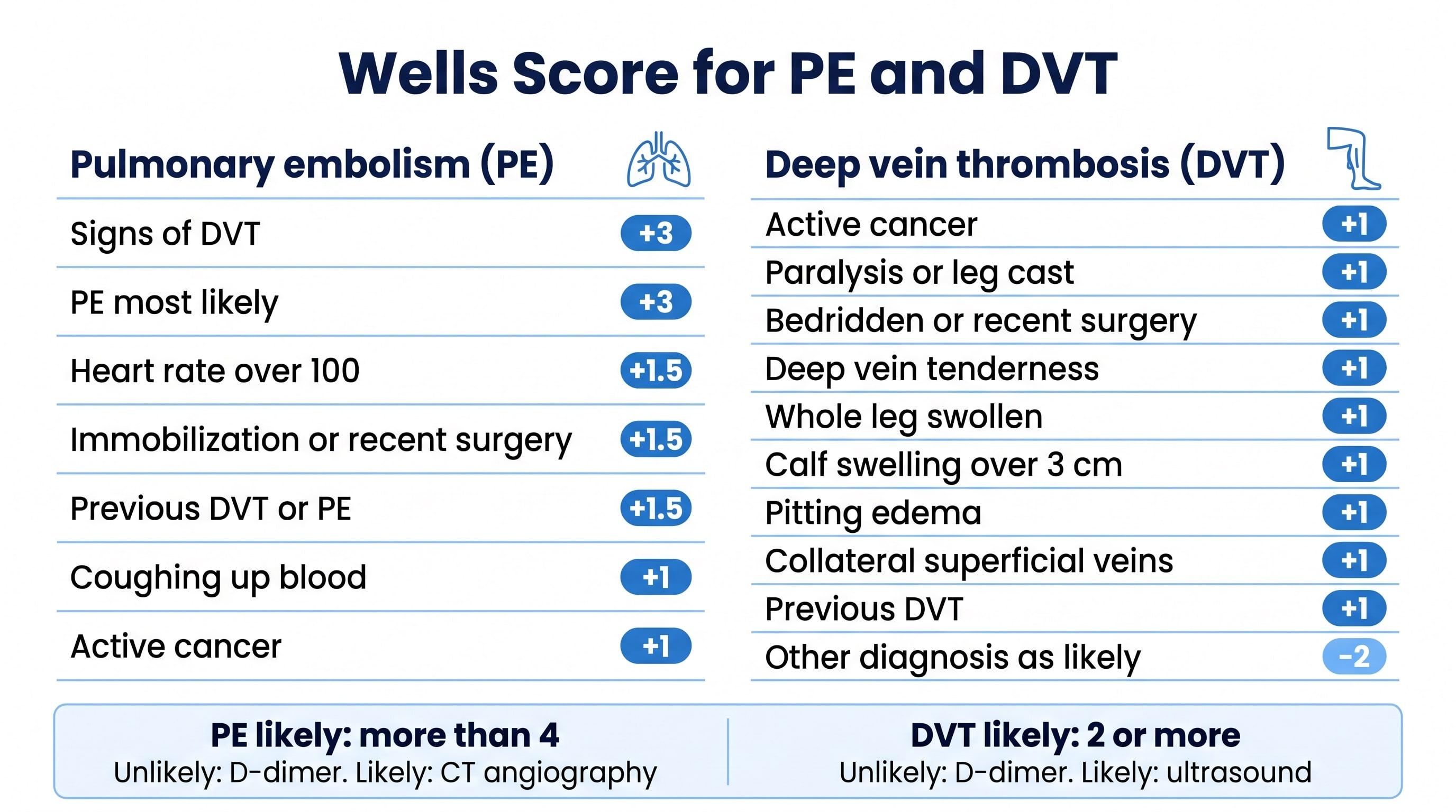
Wells Score Calculator (PE and DVT)
Check the Wells criteria for pulmonary embolism or deep vein thrombosis to get the score, the risk group, and the next test.
Try it free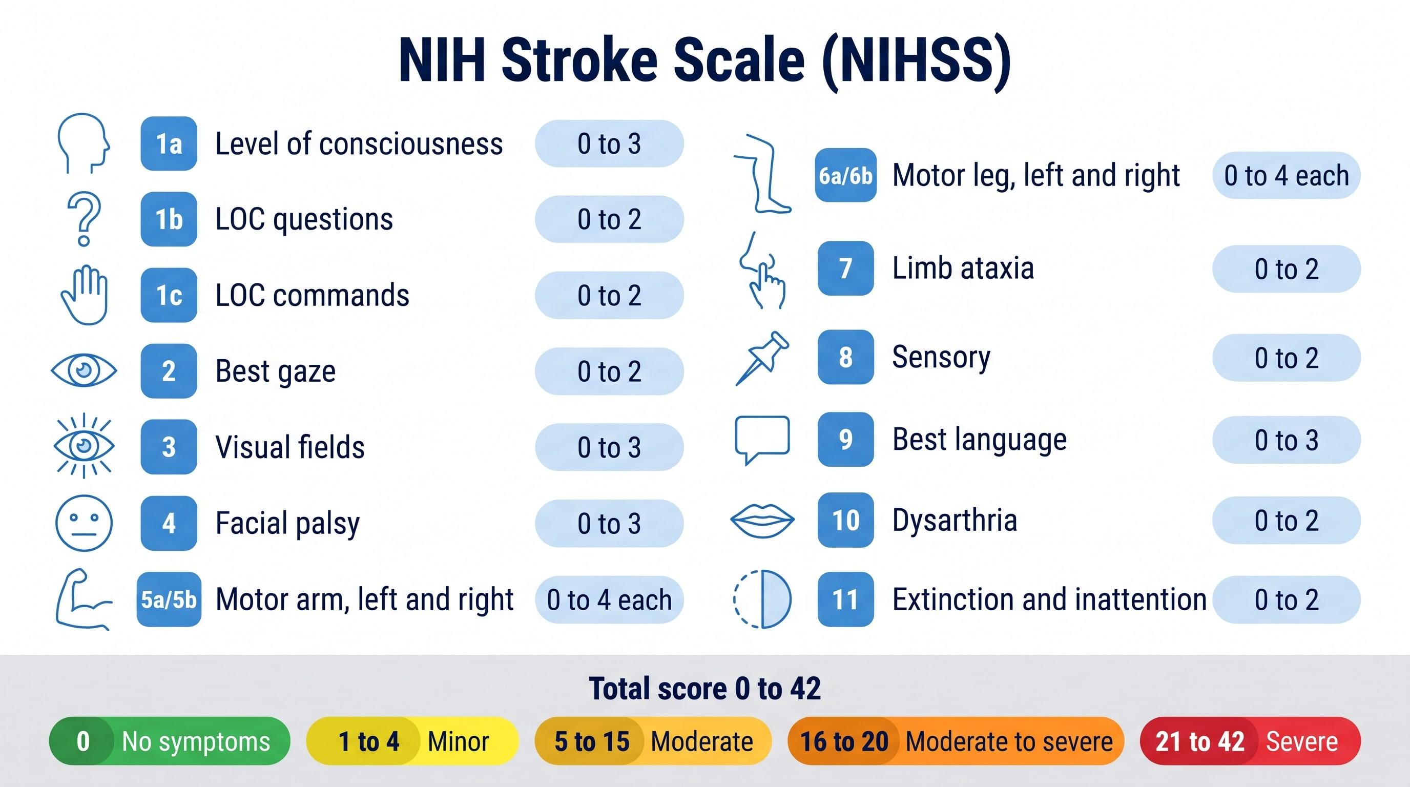
NIHSS Calculator (NIH Stroke Scale)
Score all 15 NIH Stroke Scale items to get the NIHSS total from 0 to 42 and the stroke severity range.
Try it free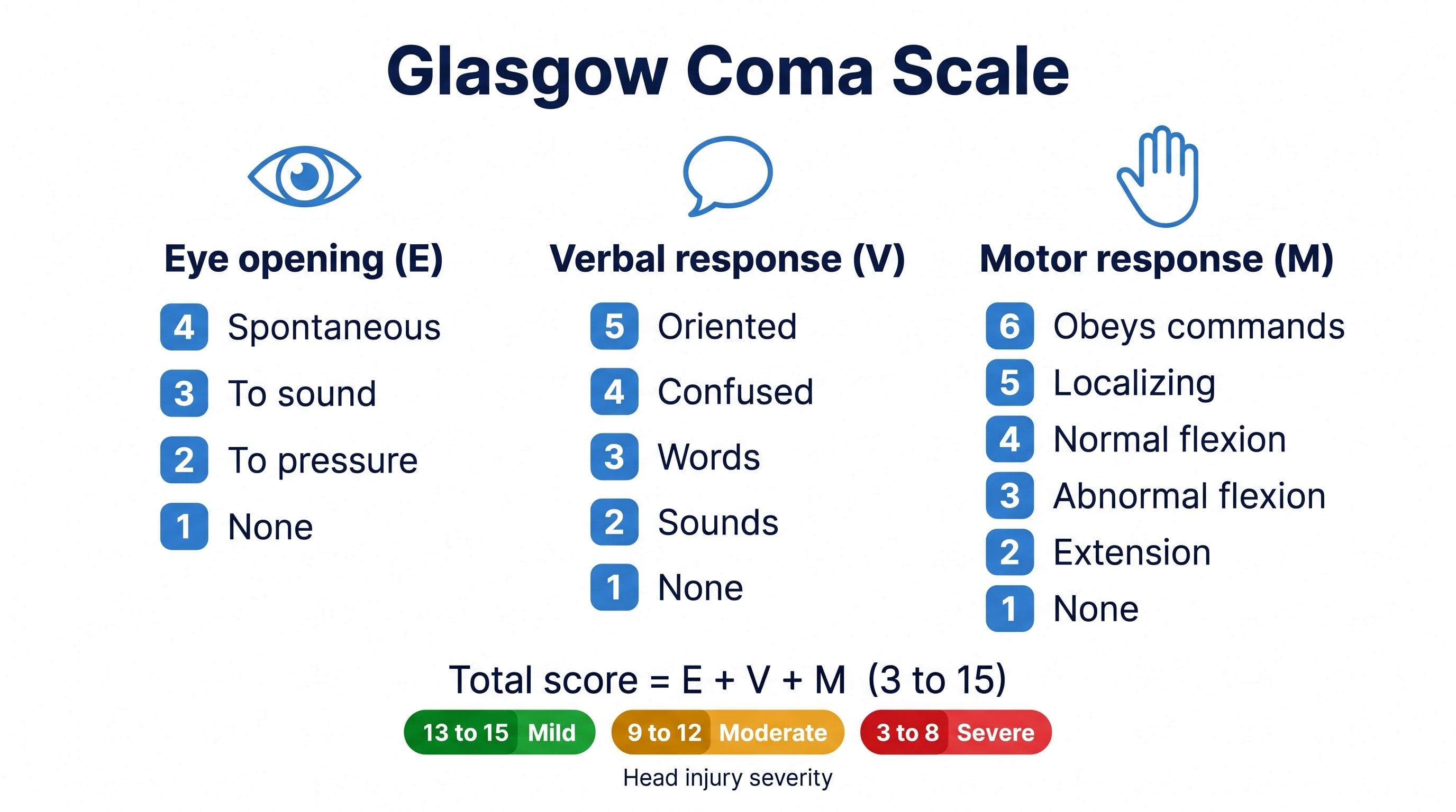
Glasgow Coma Scale Calculator
Score eye, verbal, and motor responses to get the GCS total, E V M code, severity range, and GCS-P.
Try it free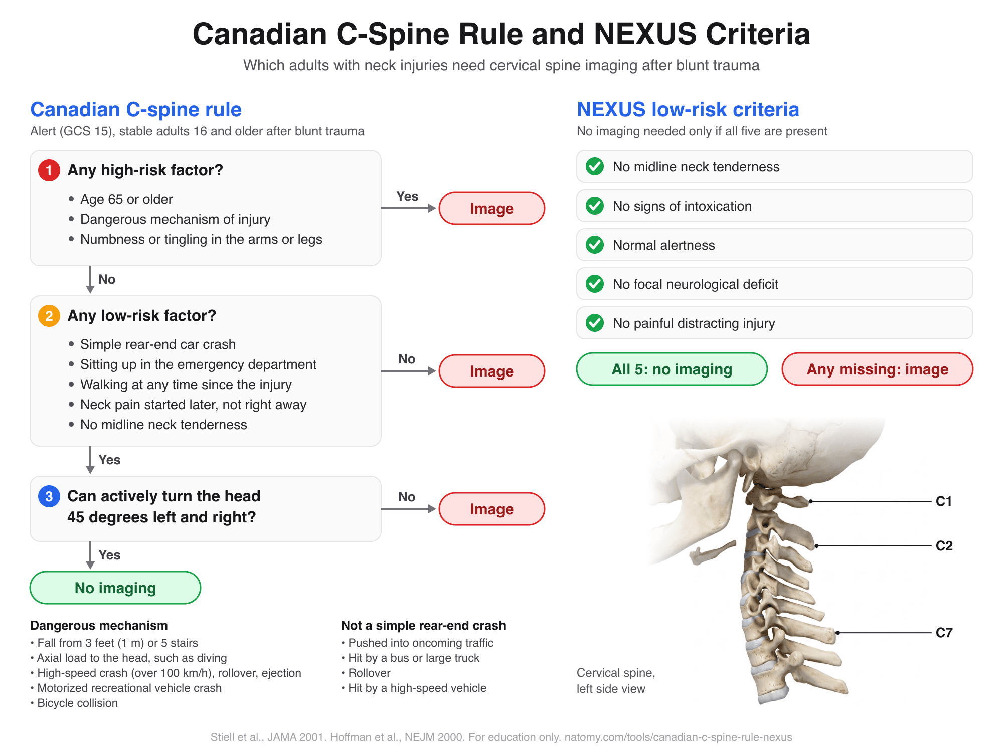
Canadian C-Spine Rule and NEXUS
Run the Canadian C-spine rule or the NEXUS criteria step by step to see which adults need neck imaging after trauma.
Try it free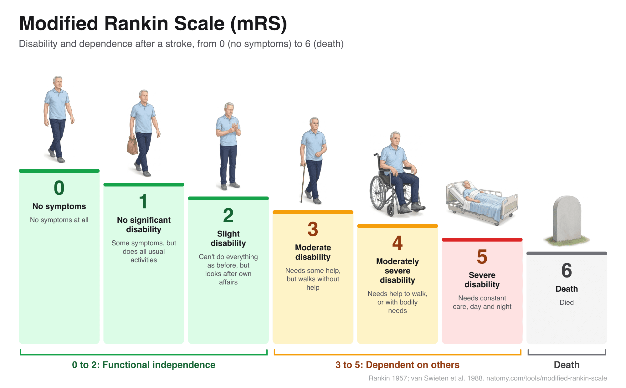
Modified Rankin Scale (mRS)
Illustrated mRS chart from 0 to 6 with a question-based score finder, what each grade means after a stroke, and a copyable note.
Try it free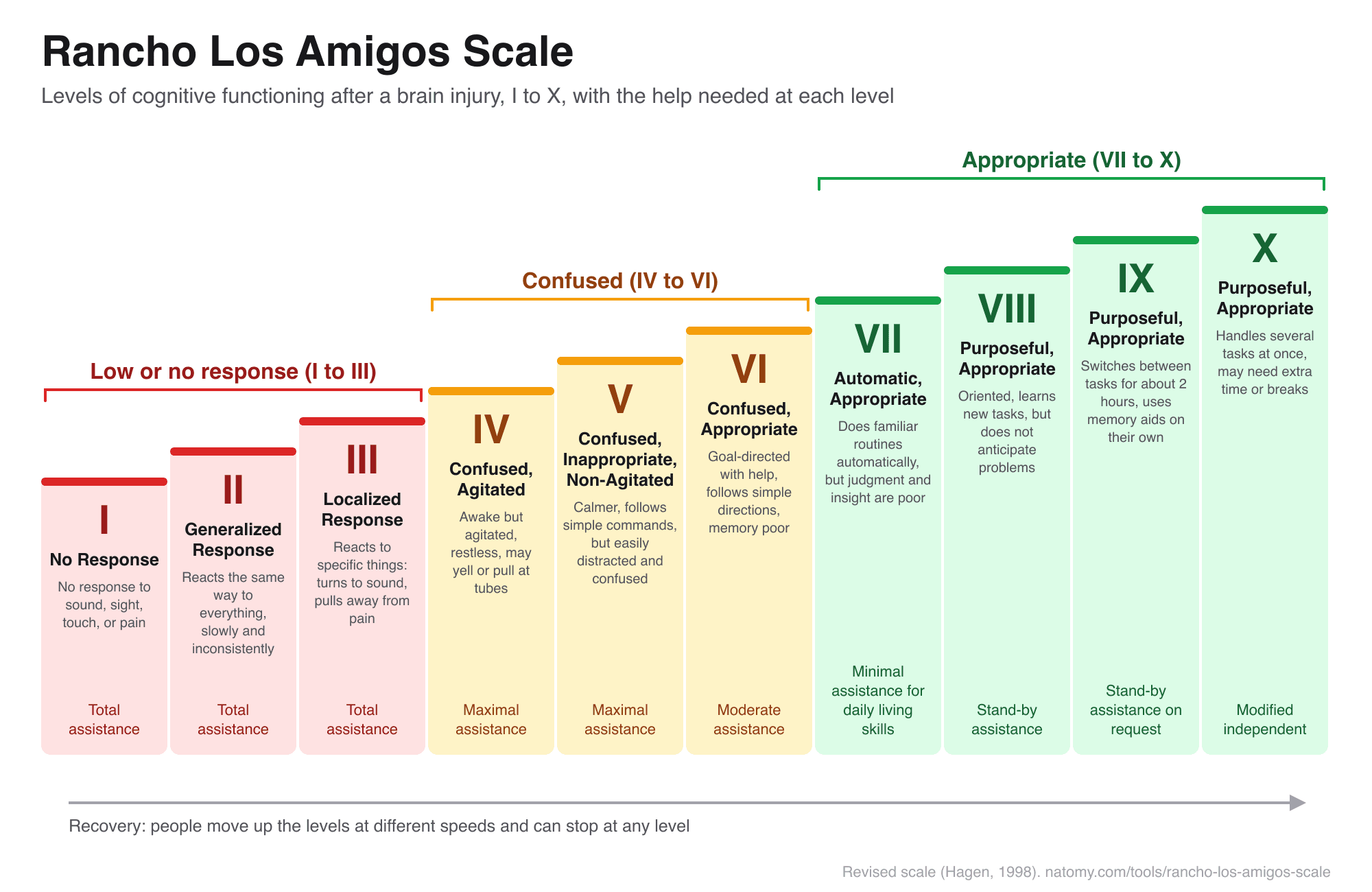
Rancho Los Amigos Scale
The 10 levels of cognitive functioning after a brain injury, with a level finder, what to expect, and how to help.
Try it free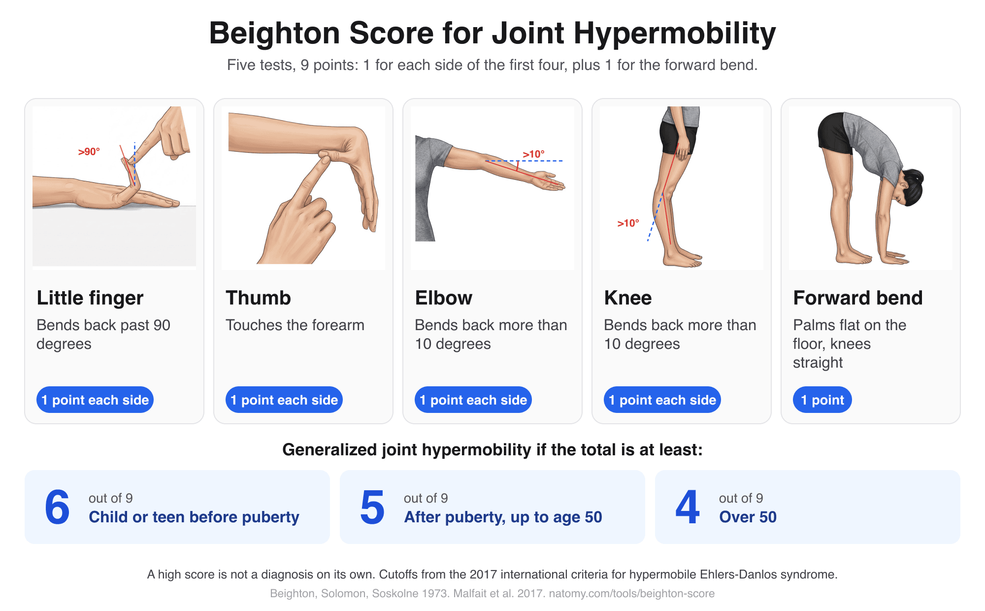
Beighton Score Calculator
Illustrated hypermobility test: score the 5 Beighton movements out of 9, compare with the age cutoff, and copy a note.
Try it free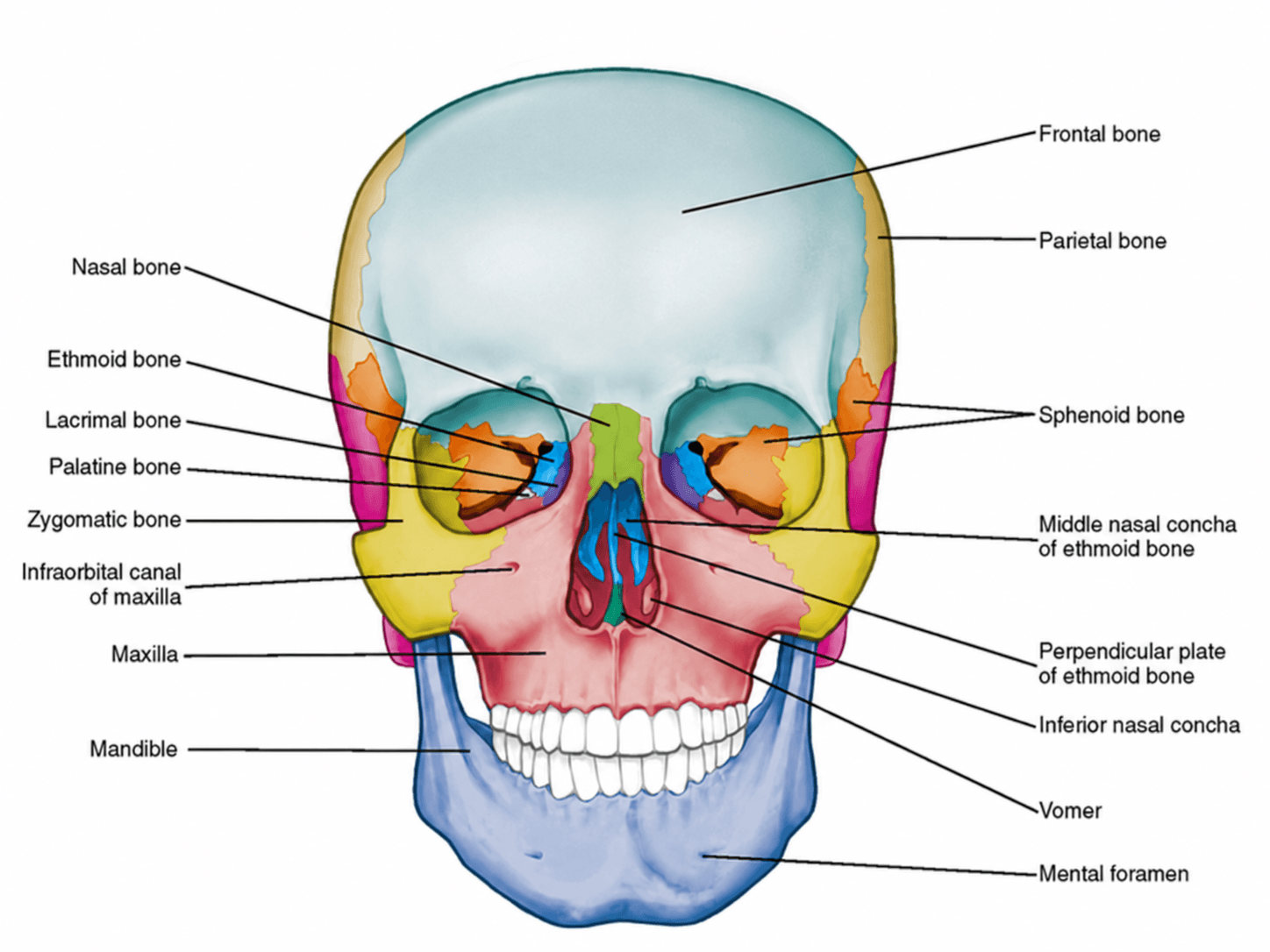
Anterior View of Skull Labeled
Free labeled and unlabeled anterior view of the skull, downloadable as PNG.
Try it free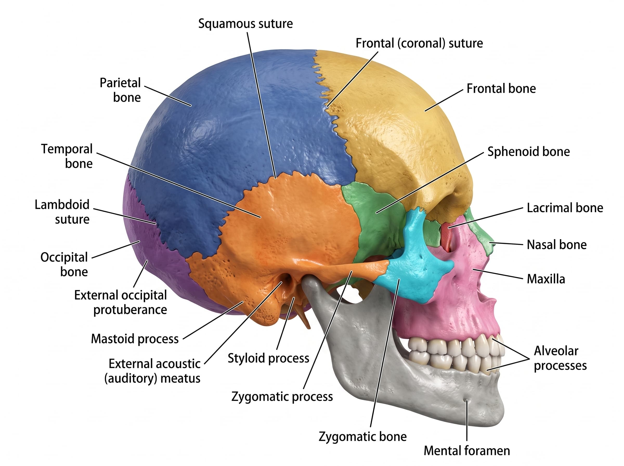
Lateral View of Skull Labeled
Free labeled and unlabeled lateral view of the skull, downloadable as PNG.
Try it free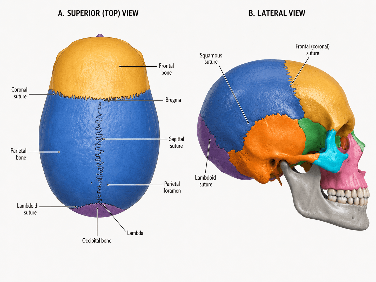
Skull Sutures Labeled
Free labeled and unlabeled skull sutures diagram, superior and lateral views, downloadable as PNG.
Try it free