Free Tools
Skull Sutures Labelled
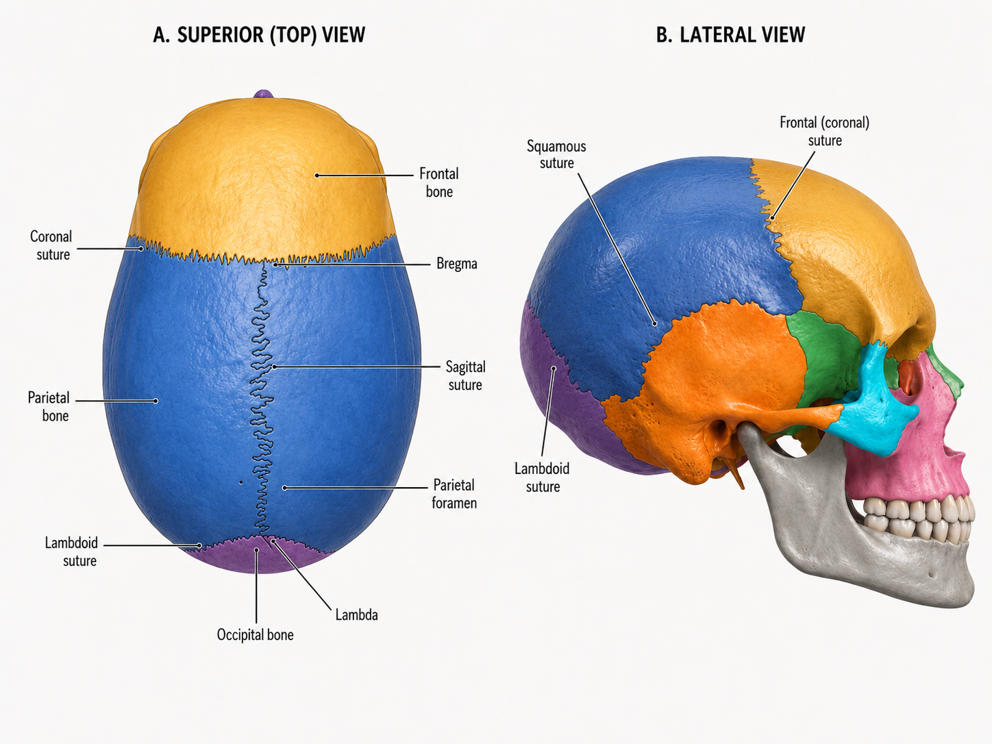
A free, downloadable diagram of the skull sutures from superior and lateral views, labelled with the coronal, sagittal, lambdoid, and squamous sutures plus key landmarks. Switch to the unlabelled version to quiz yourself before checking your answers.
What are skull sutures?
Sutures are the fibrous joints that hold the flat bones of the skull together. In adults they're essentially immobile, but in infants they remain flexible, letting the skull compress slightly during birth and expand as the brain grows. This diagram shows the four major sutures from two angles: a superior (top-down) view and a lateral view.
The major skull sutures
- Coronal suture
- Runs side to side across the top of the skull, joining the frontal bone to the two parietal bones. Visible from both the superior and lateral views.
- Sagittal suture
- Runs front to back along the midline of the skull roof, joining the two parietal bones to each other. Only visible from the superior view.
- Lambdoid suture
- Runs across the back of the skull, joining the parietal bones to the occipital bone. Visible from both views.
- Squamous suture
- A curved suture on each side of the skull joining the temporal bone to the parietal bone. Only visible from the lateral view.
Key landmarks
A few named points mark where sutures intersect or where small openings occur along them. These are commonly tested alongside the sutures themselves.
- Bregma
- The point where the coronal and sagittal sutures meet. In infants, this location corresponds to the anterior fontanelle, the soft spot that allows the skull to flex during birth and expand as the brain grows.
- Lambda
- The point where the sagittal and lambdoid sutures meet, at the back of the skull. It corresponds to the posterior fontanelle in infants, which closes earlier than the anterior fontanelle.
- Parietal foramen
- A small opening near the back of the parietal bone, close to the sagittal suture, that transmits a small emissary vein connecting scalp veins to the venous sinuses inside the skull.
Bones bordering these sutures
- Frontal bone
- Forms the forehead and meets the parietal bones at the coronal suture.
- Parietal bone
- A pair of bones forming the roof and sides of the skull, bordered by the coronal, sagittal, lambdoid, and squamous sutures.
- Occipital bone
- Forms the back and base of the skull, meeting the parietal bones at the lambdoid suture.
- Temporal bone
- Forms the lower side wall of the skull around the ear, meeting the parietal bone at the squamous suture. Only visible from the lateral view.
Why sutures matter
Sutures aren't just landmarks — they're clinically relevant. Their flexibility in infancy is why newborns have soft spots (fontanelles) at bregma and lambda, and premature fusion of a suture, known as craniosynostosis, can restrict skull growth and require surgical correction. Later in life, the gradual fusion of sutures is one of the features used to estimate age from skeletal remains.
Tips for studying with this diagram
- Learn the sutures by the bones they connect — coronal (frontal–parietal), sagittal (parietal–parietal), lambdoid (parietal–occipital), and squamous (temporal–parietal).
- Use bregma and lambda as anchor points: bregma sits where the coronal and sagittal sutures cross, lambda where the sagittal and lambdoid sutures cross.
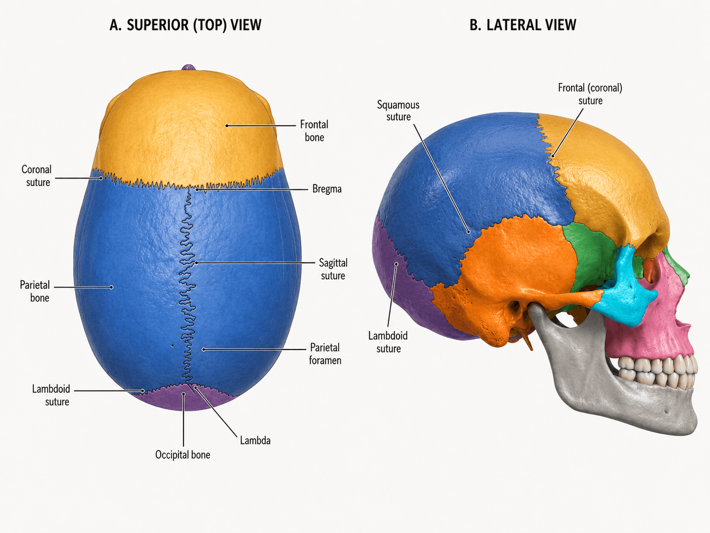
- Switch to the unlabelled version and try to sketch the suture lines from memory before checking the labelled diagram.
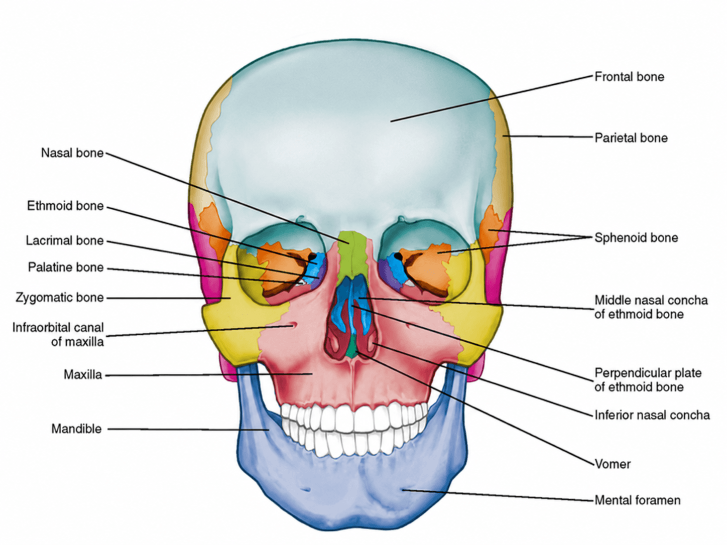
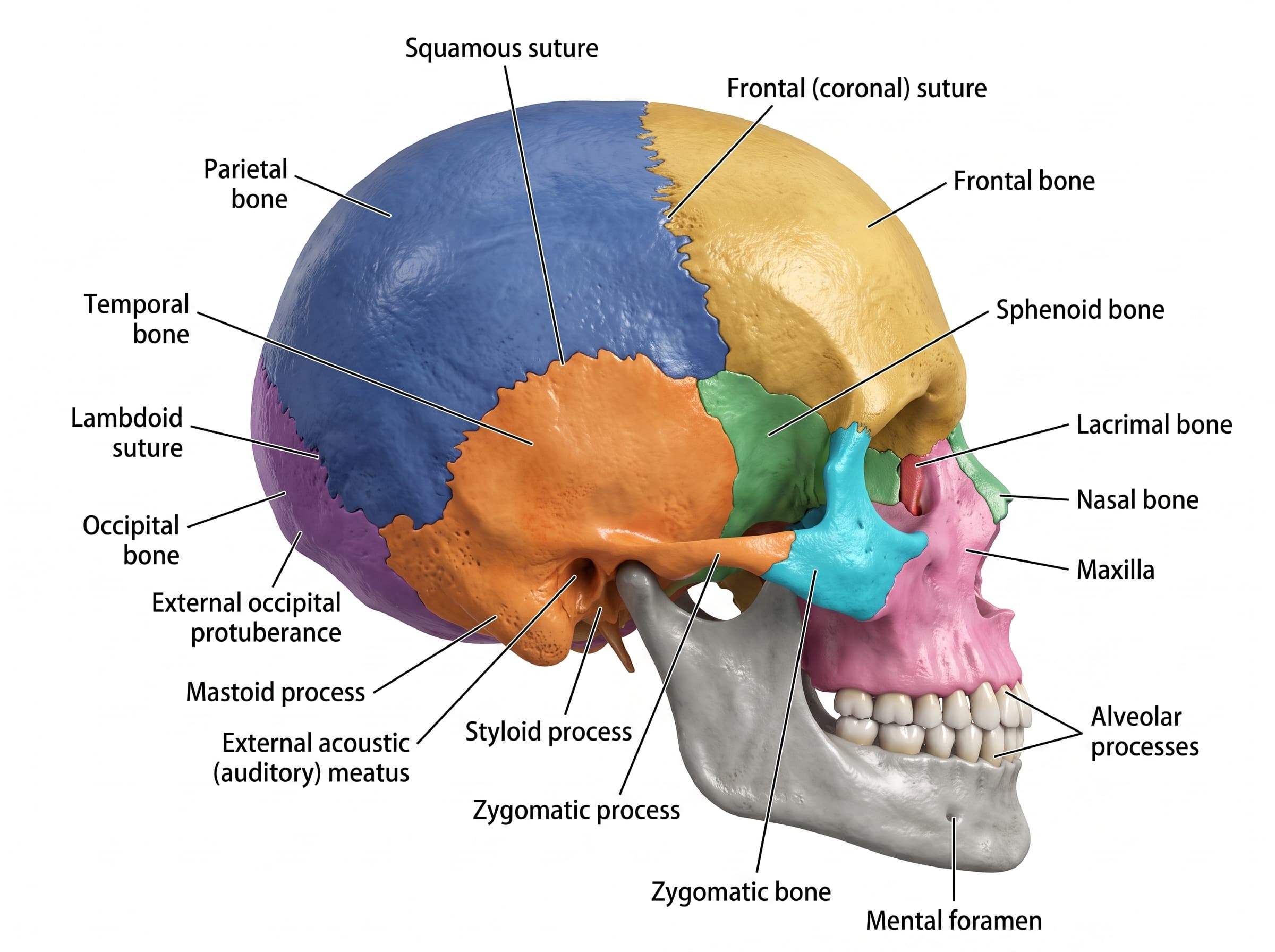
- Pair this with the anterior view and lateral view diagrams for a complete picture of the skull before an exam.
Need a custom illustration?
Natomy turns clinical photos and prompts into publication-ready medical illustrations in seconds — including custom anatomy diagrams beyond the skull sutures.
Try Natomy FreeFAQ
Is the skull sutures labelled diagram free to use?+
Yes. Both the labelled and unlabelled skull suture diagrams are free to download as PNG files for personal study, teaching, and educational use.
What are skull sutures?+
Sutures are fibrous joints that connect the flat bones of the skull. Unlike most joints, they're immobile in adults, though they allow some flexibility in infancy while the skull is still growing.
What's the difference between the superior and lateral views in this diagram?+
The superior (top-down) view shows the coronal, sagittal, and lambdoid sutures along with bregma, lambda, and the parietal foramen. The lateral (side) view adds the squamous suture, which isn't visible from directly above.
Do skull sutures ever fuse?+
Yes. Sutures gradually ossify (fuse) with age, a process that typically continues over decades. Premature fusion of one or more sutures in infancy is a condition called craniosynostosis.
Can I use the unlabelled version to quiz myself?+
Yes. The unlabelled diagram keeps the same color-coded bones and both views without text labels, so you can test your recall of each suture and landmark before checking the labelled version.
Can I generate diagrams of other skull views or structures?+
Natomy can generate custom medical illustrations from clinical photos or prompts, including other skull views and anatomical structures beyond the sutures.