Free Tools
Wells Score Calculator (PE and DVT)
Choose pulmonary embolism or deep vein thrombosis, then check every criterion that applies. The score updates as you go, with the two-level and three-level risk groups and the usual next test.
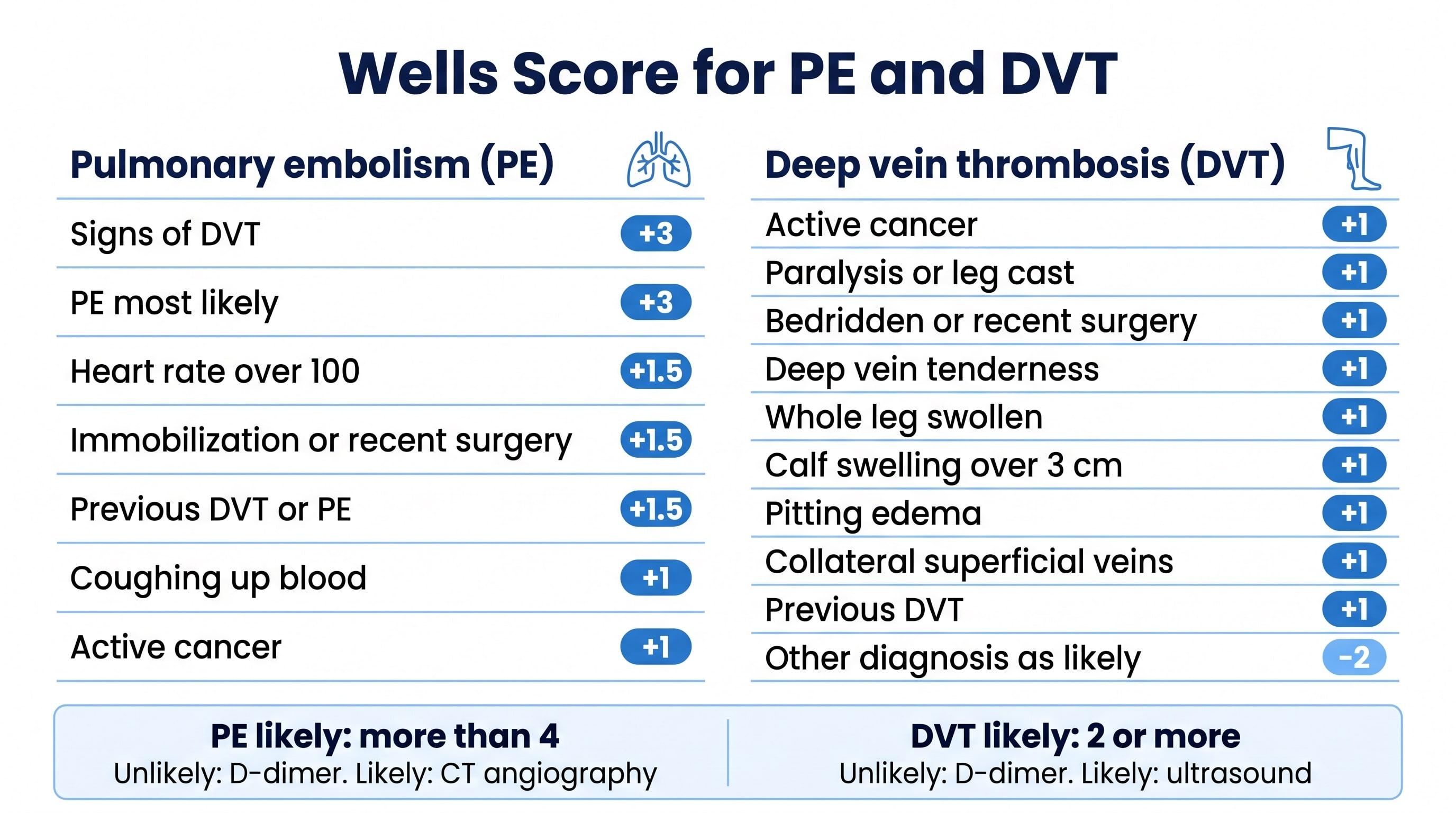
The Wells score in 60 seconds
Wells criteria for pulmonary embolism
The Wells score for PE (Wells et al., 2000) is the most widely used clinical prediction rule for pulmonary embolism. Scores range from 0 to 12.5.
| Criterion | Points |
|---|---|
| Clinical signs and symptoms of DVT | 3 |
| PE is the most likely diagnosis | 3 |
| Heart rate over 100 | 1.5 |
| Immobilization 3 days or more, or surgery in the last 4 weeks | 1.5 |
| Previous DVT or PE | 1.5 |
| Hemoptysis | 1 |
| Active cancer | 1 |
Two-level: more than 4 is PE likely. Three-level: under 2 low, 2 to 6 moderate, over 6 high.
Wells criteria for deep vein thrombosis
The Wells score for DVT (Wells et al., 2003) estimates the chance of a clot in the deep veins of the leg. Scores range from -2 to 9; the only negative item is a likely alternative diagnosis.
| Criterion | Points |
|---|---|
| Active cancer | 1 |
| Paralysis, paresis, or recent plaster cast on the leg | 1 |
| Bedridden more than 3 days, or major surgery in the last 12 weeks | 1 |
| Localized tenderness along the deep veins | 1 |
| Entire leg swollen | 1 |
| Calf swelling more than 3 cm compared with the other leg (10 cm below the tibial tuberosity) | 1 |
| Pitting edema confined to the symptomatic leg | 1 |
| Collateral superficial (non-varicose) veins | 1 |
| Previously documented DVT | 1 |
| Alternative diagnosis at least as likely as DVT | -2 |
Two-level: 2 or more is DVT likely. Three-level: 0 or less low, 1 to 2 moderate, 3 or more high.
What to do with the score
- Clot unlikely: check a D-dimer. A normal result effectively rules out PE or DVT in most patients.
- Clot likely, or a raised D-dimer: go to imaging. CT pulmonary angiography for PE, compression ultrasound for DVT.
- The score supports clinical judgment; it does not replace it, and it is less reliable in pregnancy and in hospital inpatients.
Who uses it
- Emergency physicians, hospitalists, and nurses
- Score a suspected PE or DVT quickly and copy a documented result with the criteria into the record.
- Medical malpractice attorneys
- Check whether a missed or delayed clot diagnosis followed the standard pathway: was the risk scored, and did the D-dimer or imaging that the score called for happen?
Illustrate a PE or DVT case
Natomy turns CT and ultrasound images, reports, and prompts into clear, labeled medical illustrations of deep vein thrombosis and pulmonary embolism, from the clot in the leg to the blocked lung artery, for patient education and medical-legal exhibits. Related scores: NIHSS and Glasgow Coma Scale.
Try Natomy TodayFAQ
What is the Wells score?+
The Wells score is a set of bedside criteria, developed by Philip Wells and colleagues, that estimates how likely a patient is to have a blood clot before any testing. There are two versions: one for pulmonary embolism (PE), a clot in the lungs, and one for deep vein thrombosis (DVT), a clot in the leg veins. Each criterion adds points, and the total sorts the patient into a risk group.
What is a high Wells score for PE?+
With the two-level rule, more than 4 points means PE is likely and 4 or fewer means PE is unlikely. With the older three-level rule, under 2 points is low probability, 2 to 6 is moderate, and over 6 is high.
What is a high Wells score for DVT?+
With the two-level rule, 2 or more points means DVT is likely and under 2 means DVT is unlikely. With the three-level rule, 0 or less is low probability, 1 to 2 is moderate, and 3 or more is high.
How is the Wells score used with a D-dimer test?+
When the Wells score says a clot is unlikely, a normal D-dimer blood test makes a clot very unlikely and imaging can usually be avoided. When the score says a clot is likely, or the D-dimer is raised, the next step is imaging: CT pulmonary angiography for PE or compression ultrasound for DVT.
Why does PE score 1.5 for some criteria and DVT only 1?+
The two scores were built separately from different patient groups, and each criterion's weight reflects how strongly it predicted a clot in that study. In the PE score, signs of DVT and PE being the most likely diagnosis carry the most weight, at 3 points each.
What is the PERC rule?+
The PERC rule (pulmonary embolism rule-out criteria) is a separate checklist for patients a clinician already judges to be at low risk of PE. If a low-risk patient meets all eight PERC criteria, PE can usually be ruled out without a D-dimer. It is used alongside, not instead of, clinical judgment and the Wells score.
Why do lawyers look at Wells scores?+
Missed or delayed diagnosis of pulmonary embolism and DVT is a common medical malpractice claim. Whether a Wells score was documented, what it was, and whether the recommended D-dimer or imaging followed are often central questions when reviewing the emergency department records.
Sources: Wells PS, et al. Derivation of a simple clinical model to categorize patients probability of pulmonary embolism. Thromb Haemost 2000;83:416-20. Wells PS, et al. Evaluation of D-dimer in the diagnosis of suspected deep-vein thrombosis. N Engl J Med 2003;349:1227-35. Criterion wording on this page is a plain-language summary. For education and documentation only; not a substitute for clinical judgment.
Related free tools
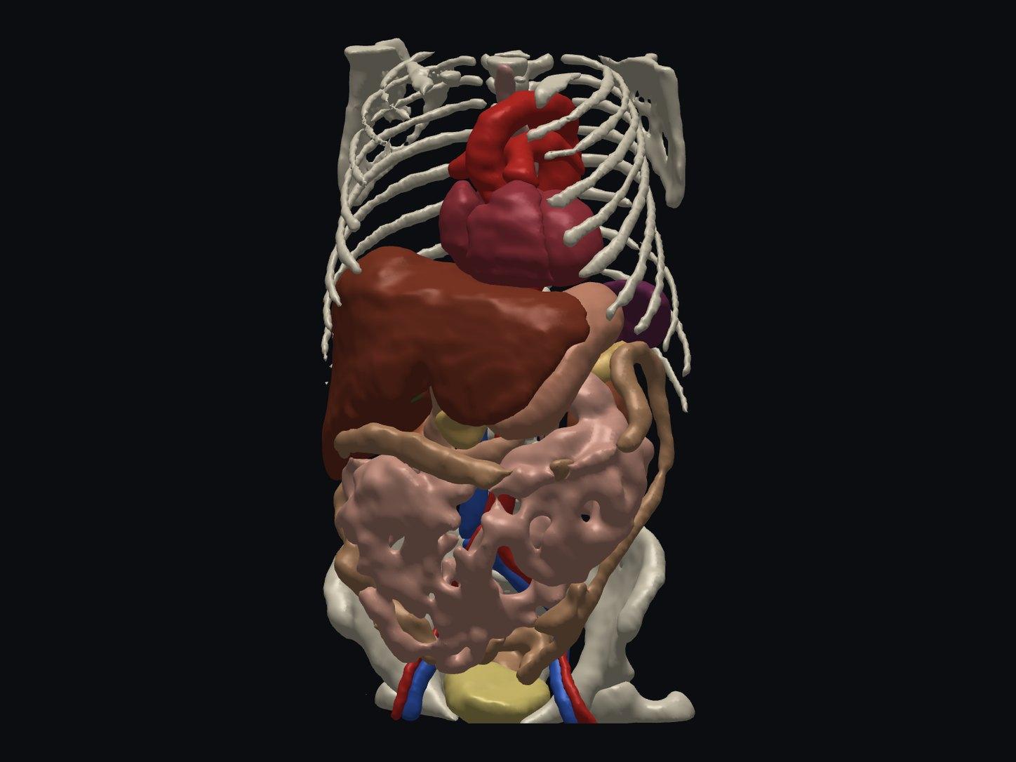
Free Online DICOM Viewer (3D)
Open a CT or MRI DICOM folder in your browser and view it in 3D, with AI anatomy labeling. Nothing is uploaded.
Try it free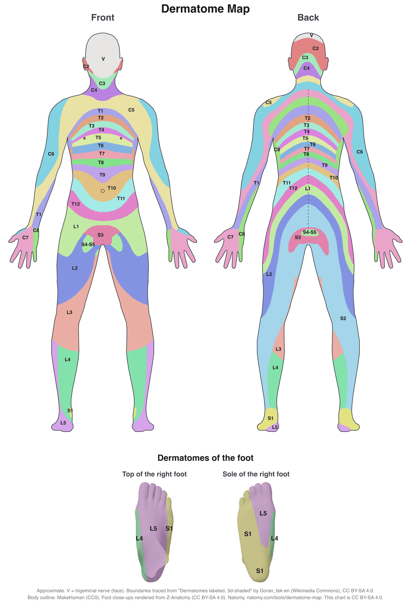
Interactive Dermatome Map
Tap the body to see each nerve root's skin area, test point, myotome, reflex, and disc level. Free printable chart.
Try it free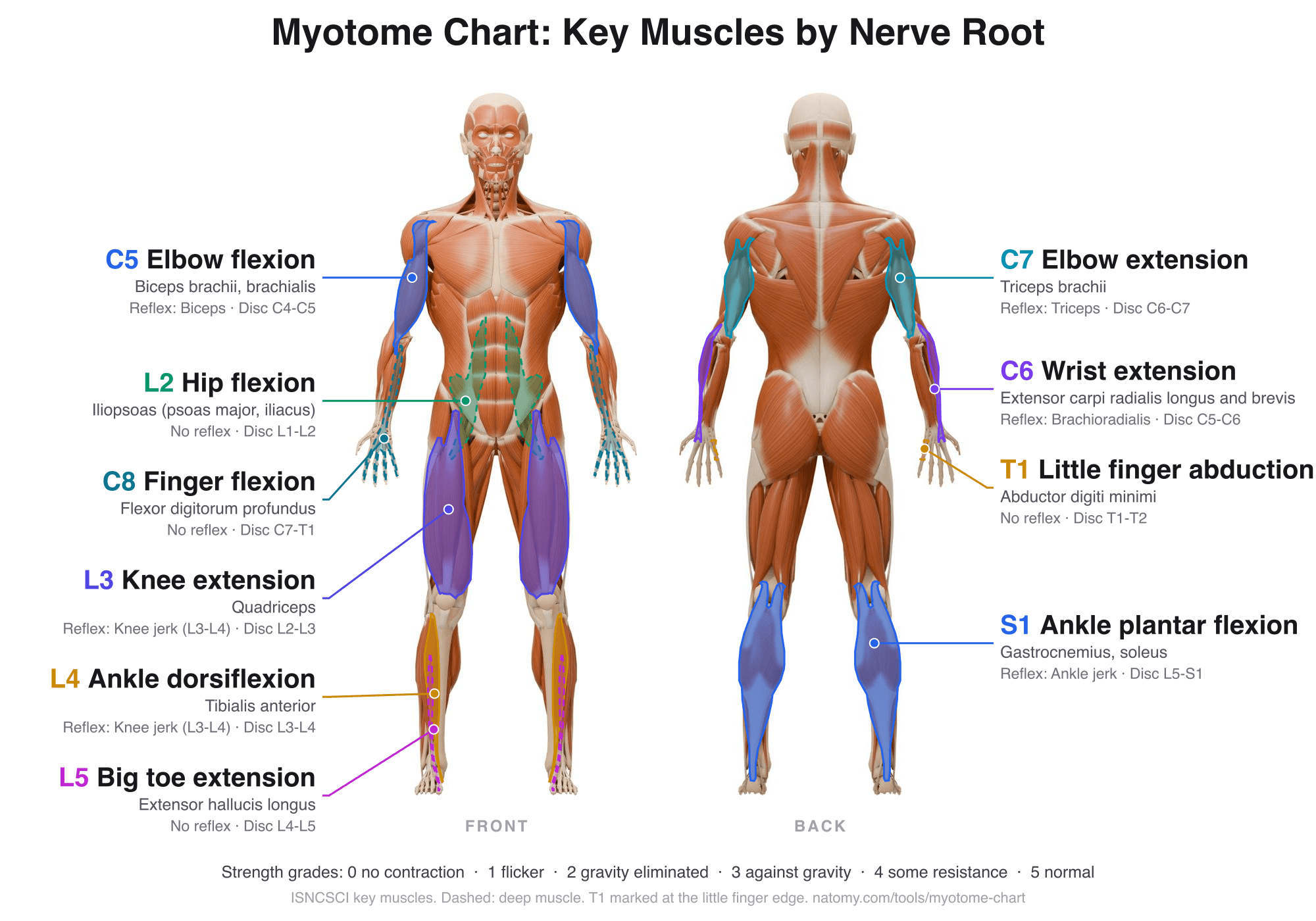
Myotome Chart and Motor Score
Tap a nerve root to see its key muscle, test position, and reflex. Grade strength 0 to 5 for the ISNCSCI motor score.
Try it free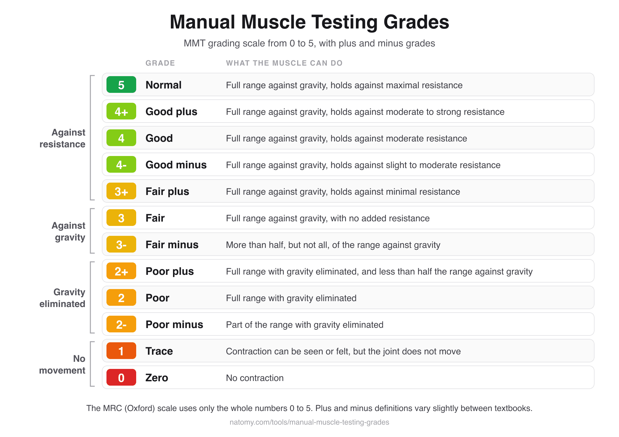
Manual Muscle Testing Grades
MMT grading scale chart from 0 to 5 with plus and minus grades, and a grade finder that writes the note for you.
Try it free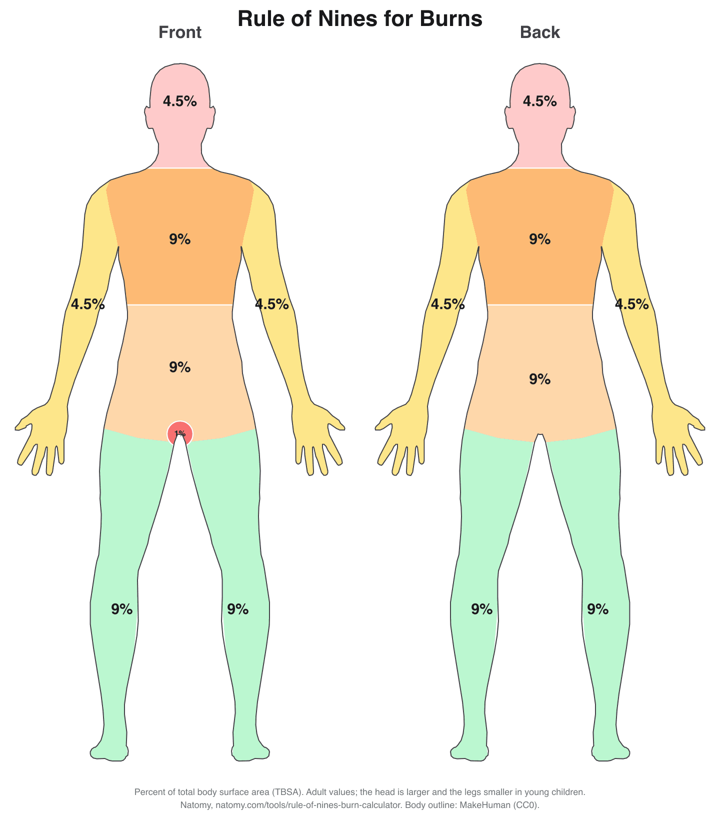
Rule of Nines Burn Calculator
Click the burned areas to get the total body surface area (TBSA), with the Parkland formula and a printable burn chart.
Try it free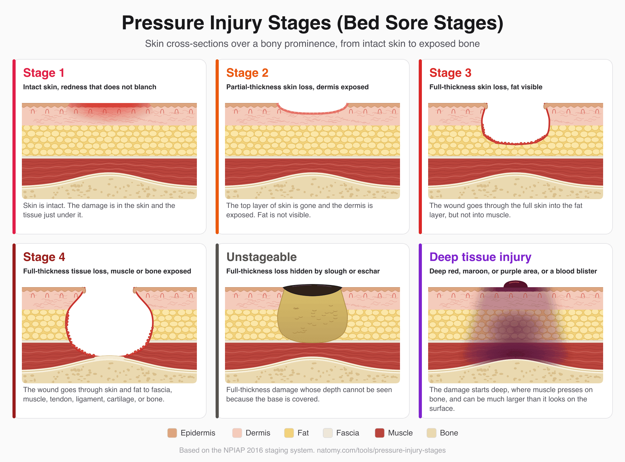
Pressure Injury (Bed Sore) Stages
Illustrated pressure ulcer stages chart, Stage 1 to 4, unstageable, and deep tissue injury, with a stage finder and wound note.
Try it free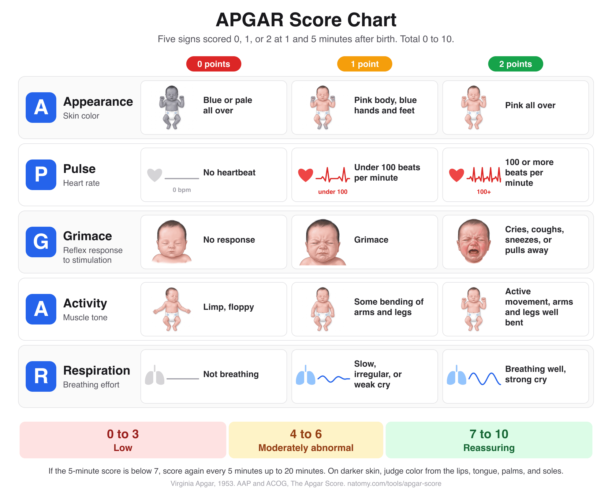
APGAR Score Chart and Calculator
Score the 5 newborn signs at 1, 5, and 10 minutes with an illustrated chart, what normal and low scores mean, and a note for the record.
Try it free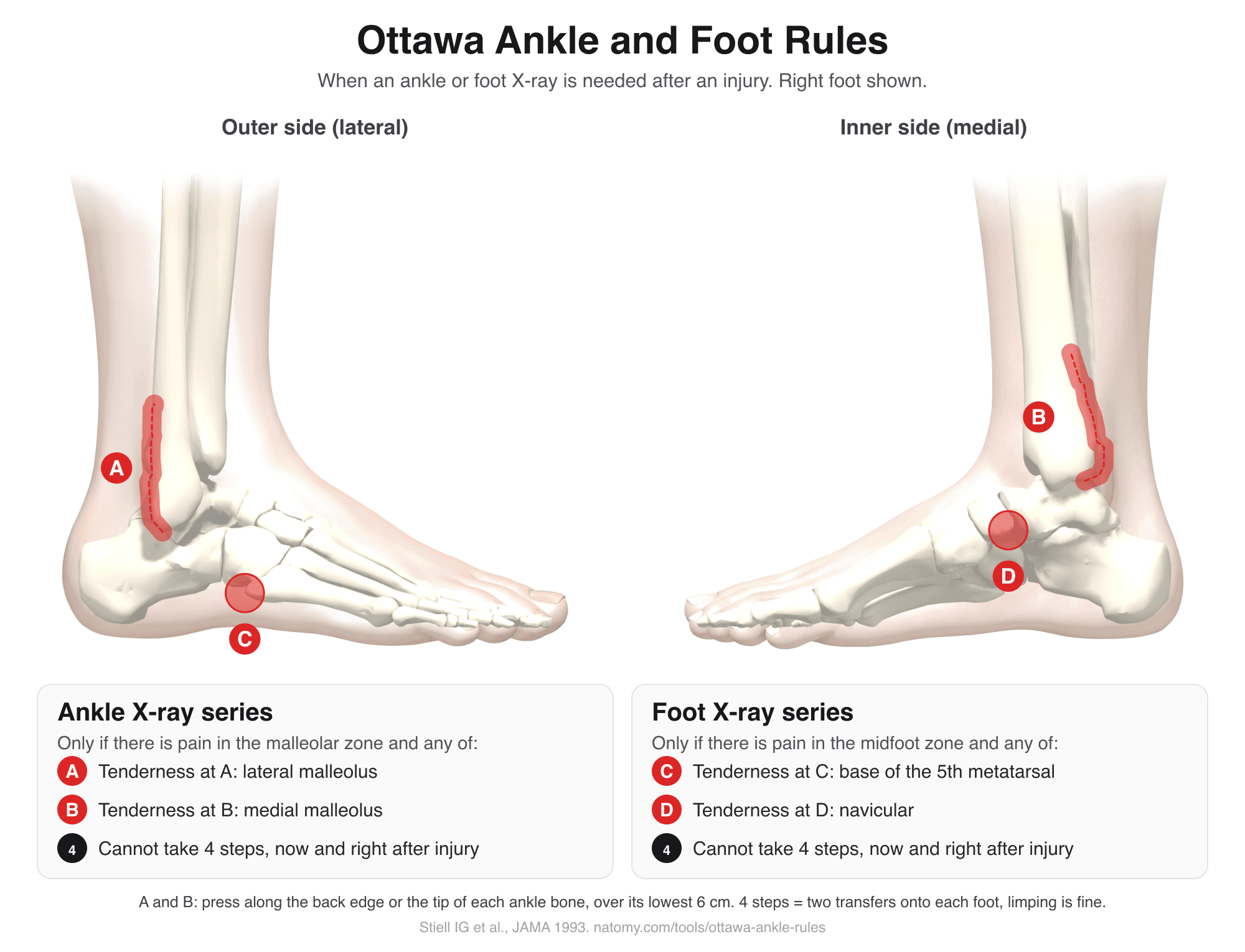
Ottawa Ankle Rules
Tap the tender spots on an illustrated ankle and foot to see if the Ottawa rules call for an X-ray. Includes the Ottawa knee rule.
Try it free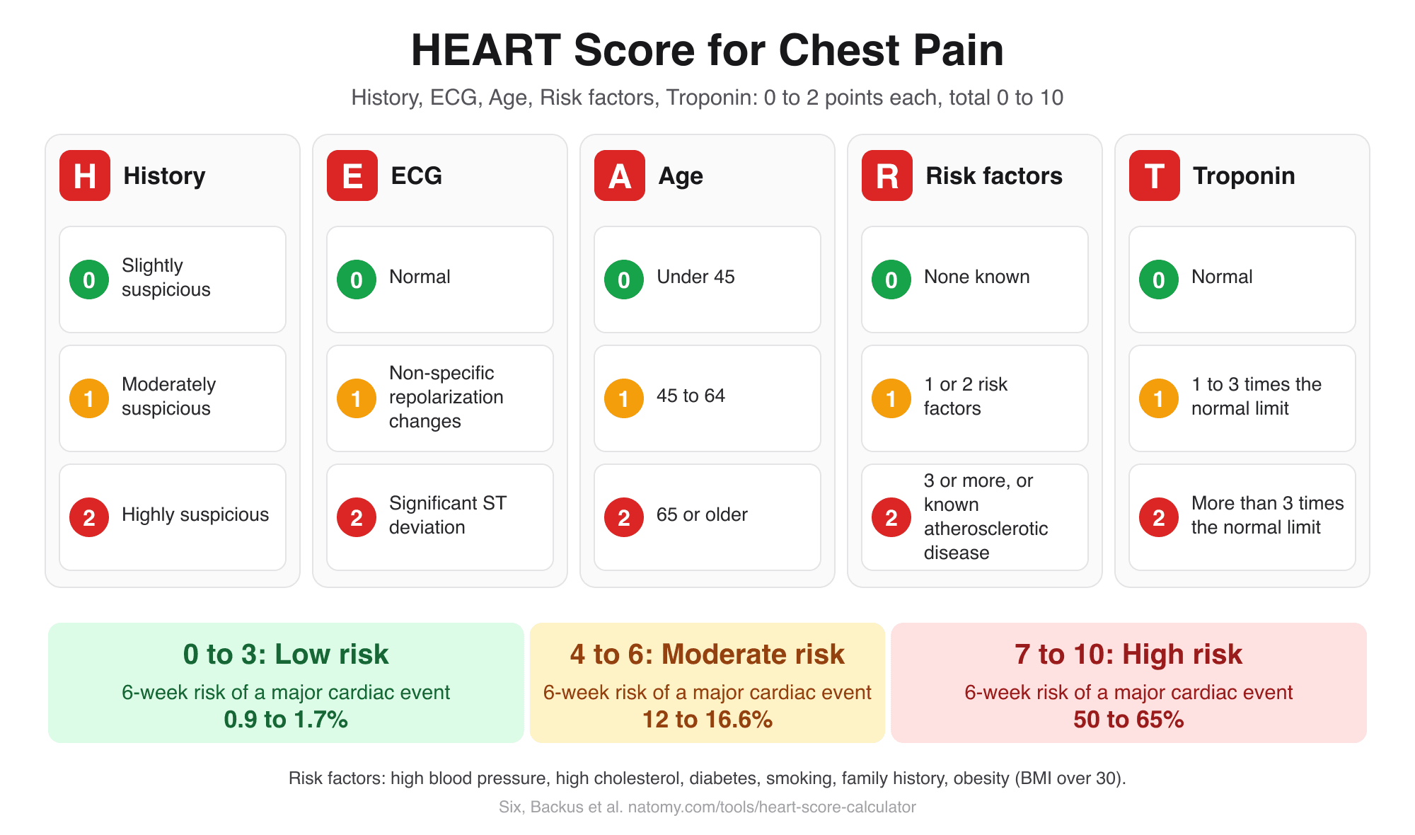
HEART Score Calculator
Score history, ECG, age, risk factors, and troponin for chest pain to get the HEART score, risk group, and 6-week MACE risk.
Try it free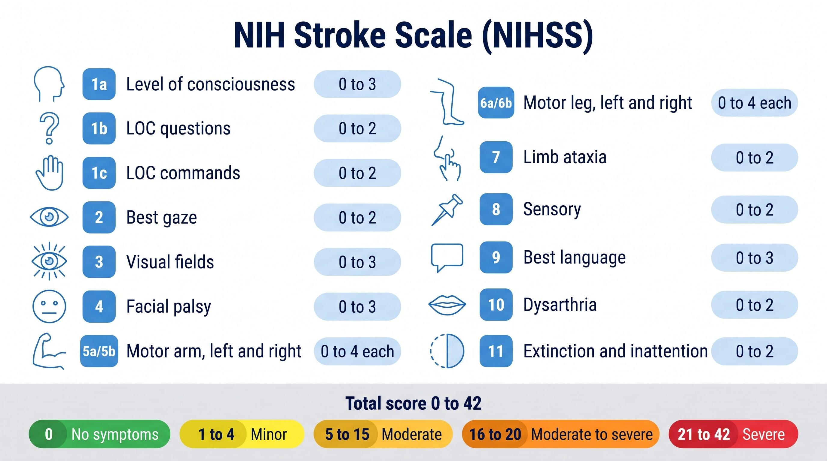
NIHSS Calculator (NIH Stroke Scale)
Score all 15 NIH Stroke Scale items to get the NIHSS total from 0 to 42 and the stroke severity range.
Try it free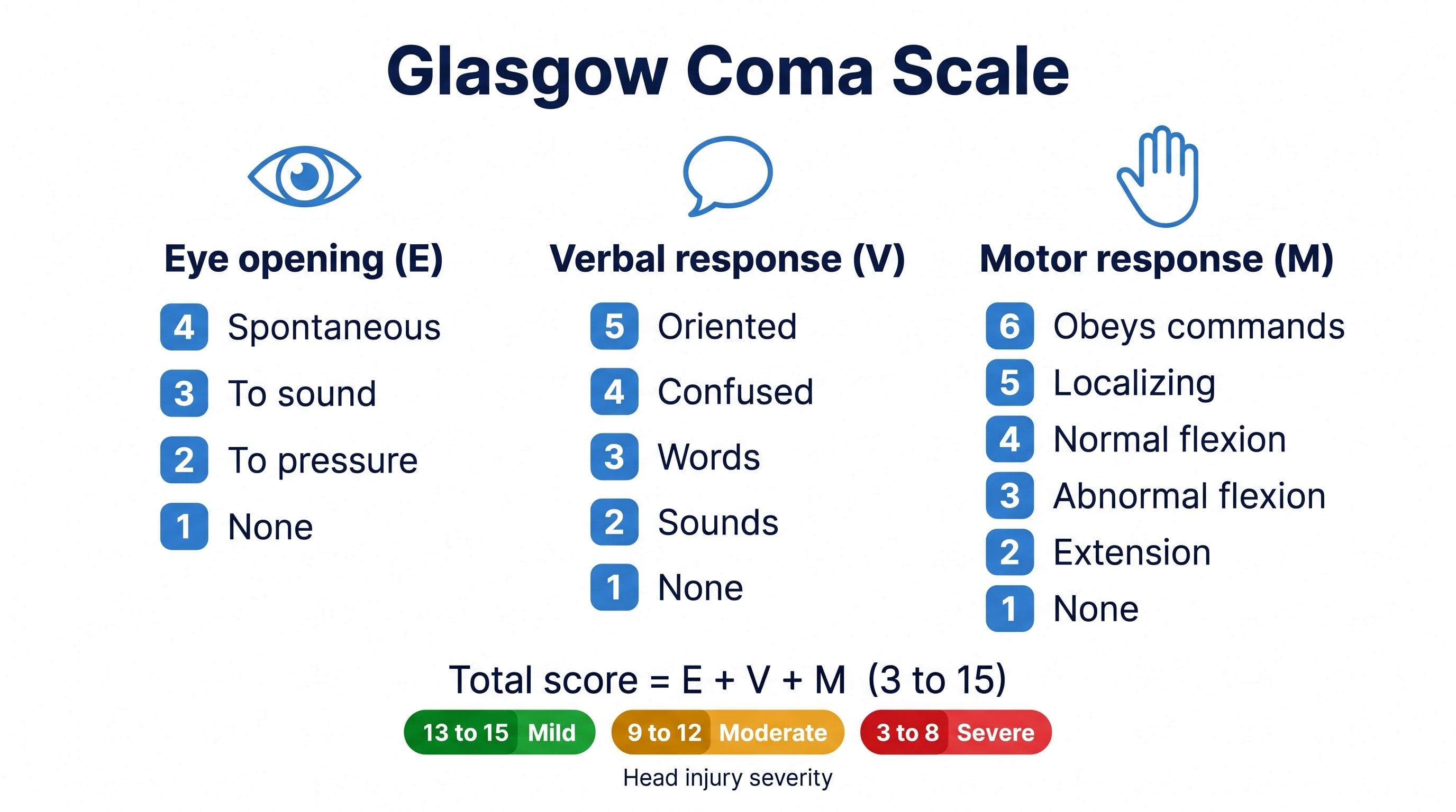
Glasgow Coma Scale Calculator
Score eye, verbal, and motor responses to get the GCS total, E V M code, severity range, and GCS-P.
Try it free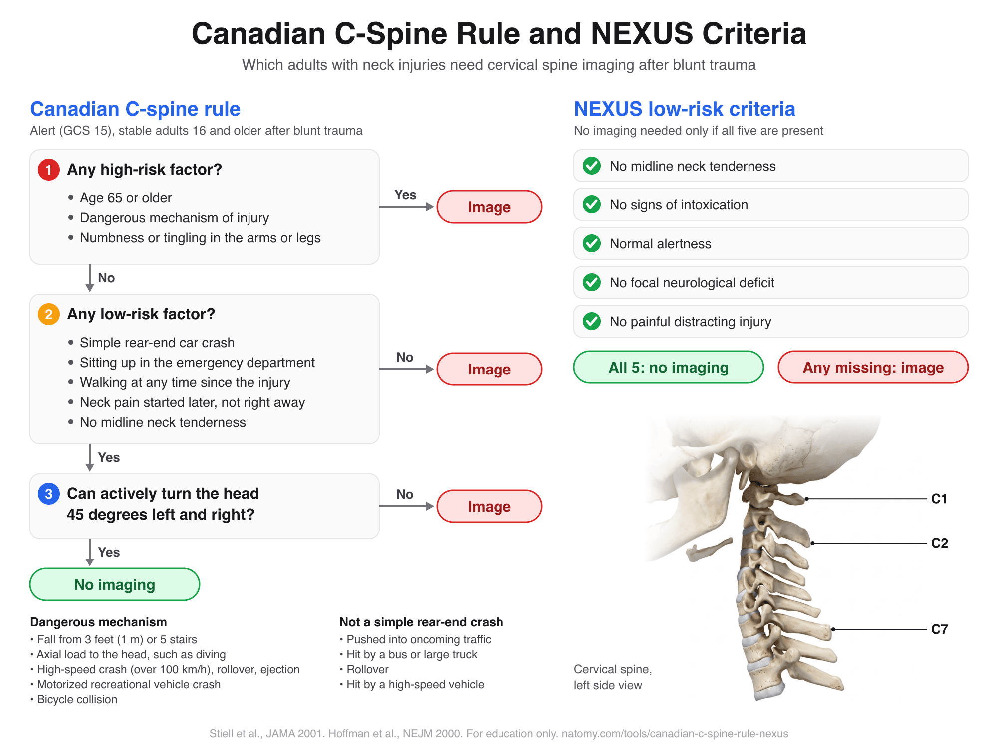
Canadian C-Spine Rule and NEXUS
Run the Canadian C-spine rule or the NEXUS criteria step by step to see which adults need neck imaging after trauma.
Try it free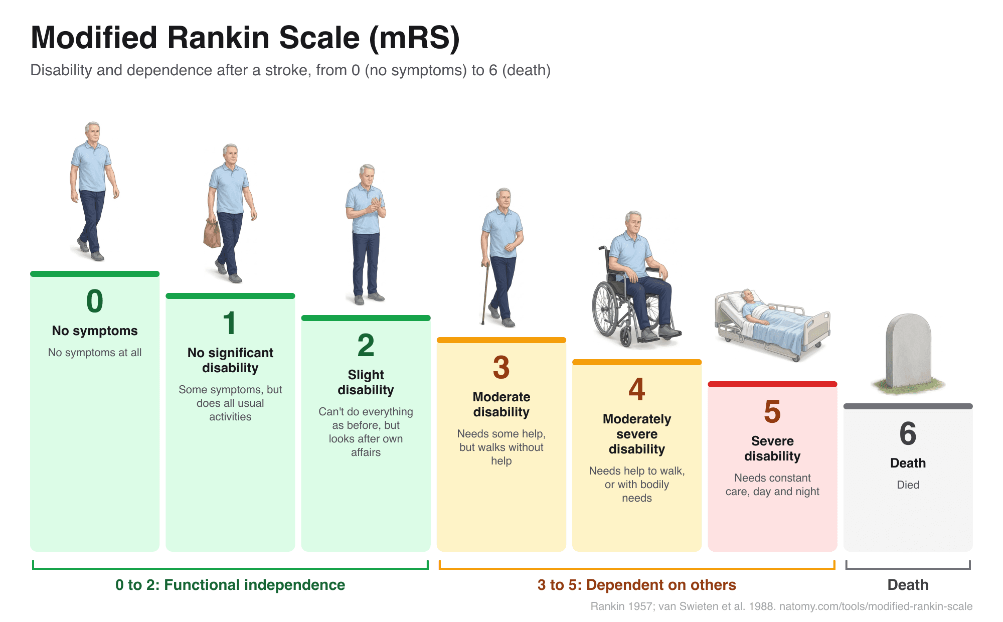
Modified Rankin Scale (mRS)
Illustrated mRS chart from 0 to 6 with a question-based score finder, what each grade means after a stroke, and a copyable note.
Try it free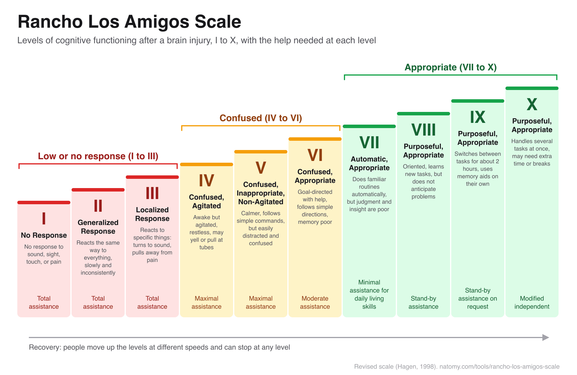
Rancho Los Amigos Scale
The 10 levels of cognitive functioning after a brain injury, with a level finder, what to expect, and how to help.
Try it free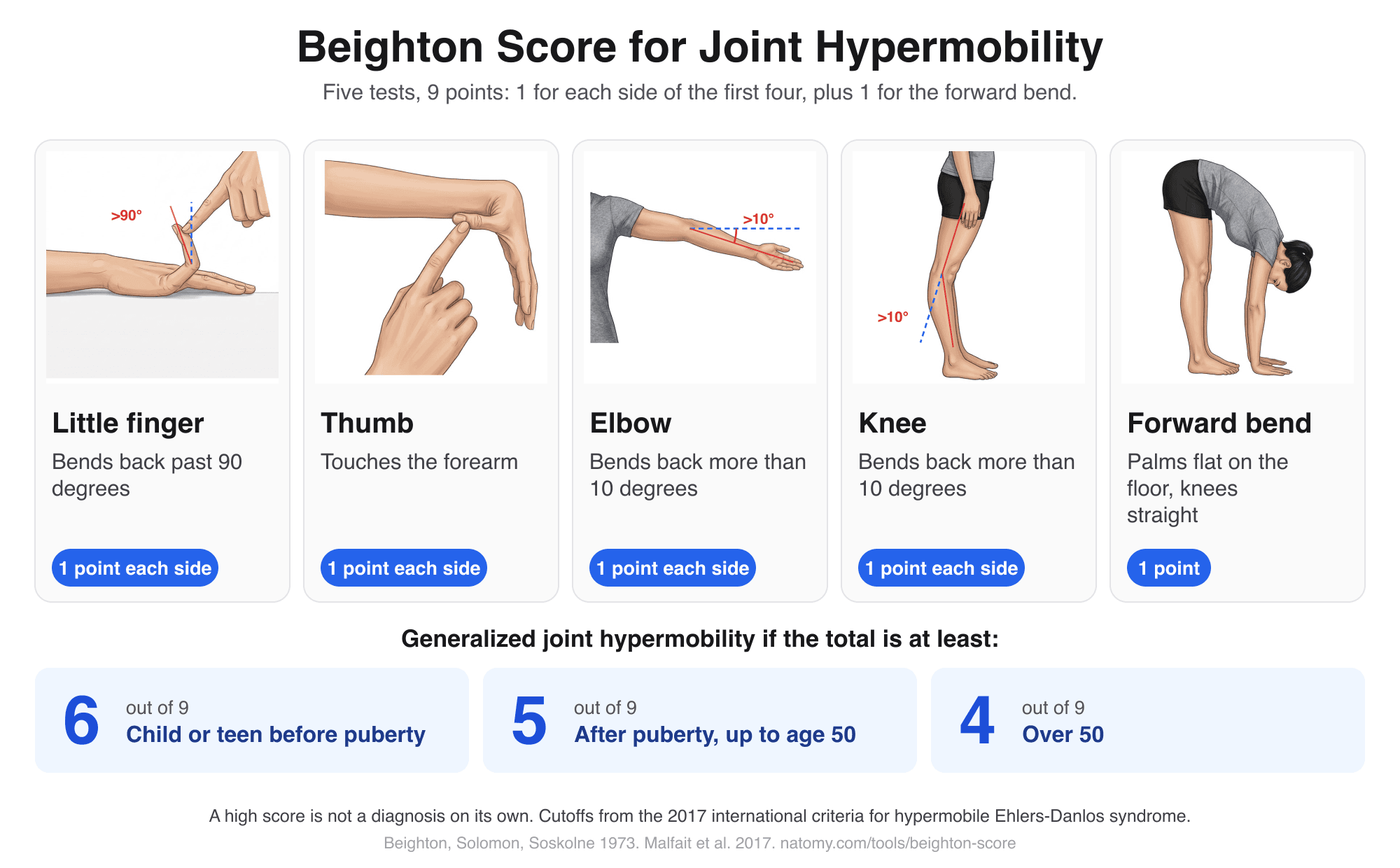
Beighton Score Calculator
Illustrated hypermobility test: score the 5 Beighton movements out of 9, compare with the age cutoff, and copy a note.
Try it free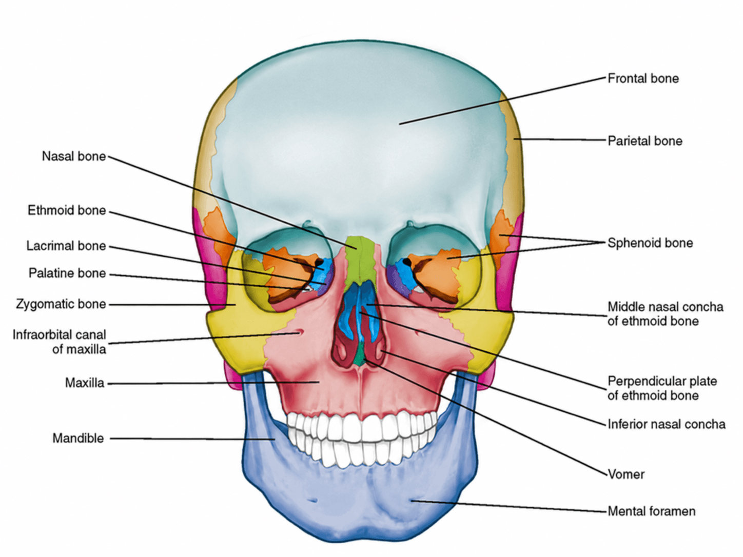
Anterior View of Skull Labeled
Free labeled and unlabeled anterior view of the skull, downloadable as PNG.
Try it free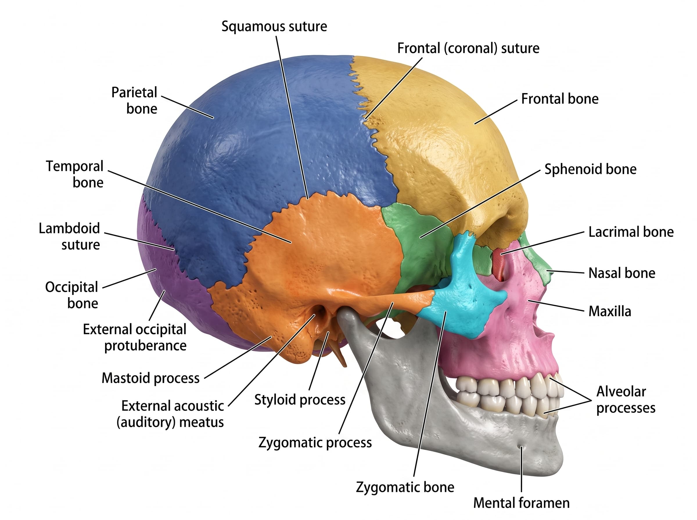
Lateral View of Skull Labeled
Free labeled and unlabeled lateral view of the skull, downloadable as PNG.
Try it free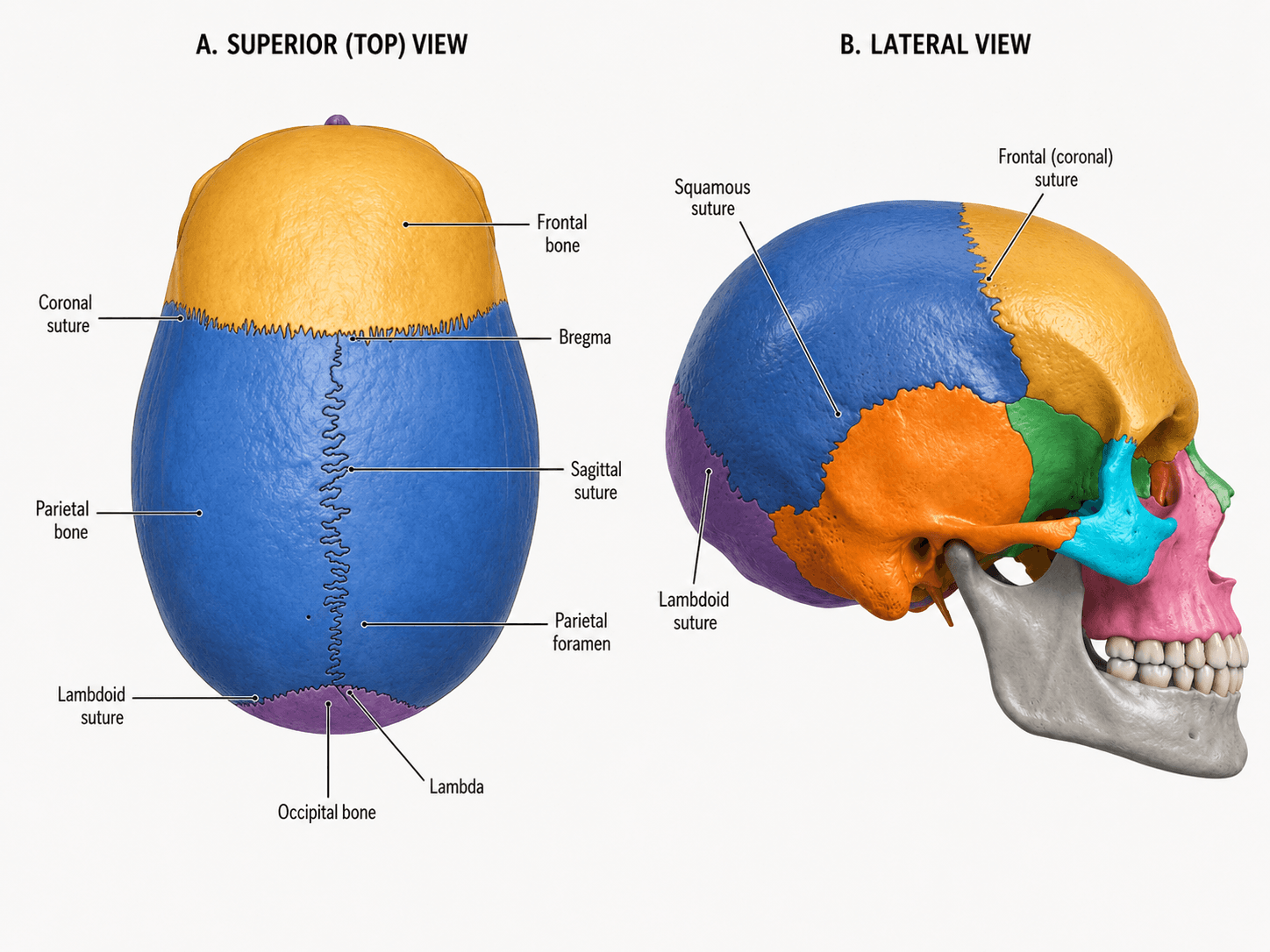
Skull Sutures Labeled
Free labeled and unlabeled skull sutures diagram, superior and lateral views, downloadable as PNG.
Try it free