Free Tools
Lateral View of Skull Labelled
A free, downloadable diagram of the lateral view of the skull, labelled with every major cranial bone, suture, and landmark. Switch to the unlabelled version of the same lateral skull view to quiz yourself before checking your answers.
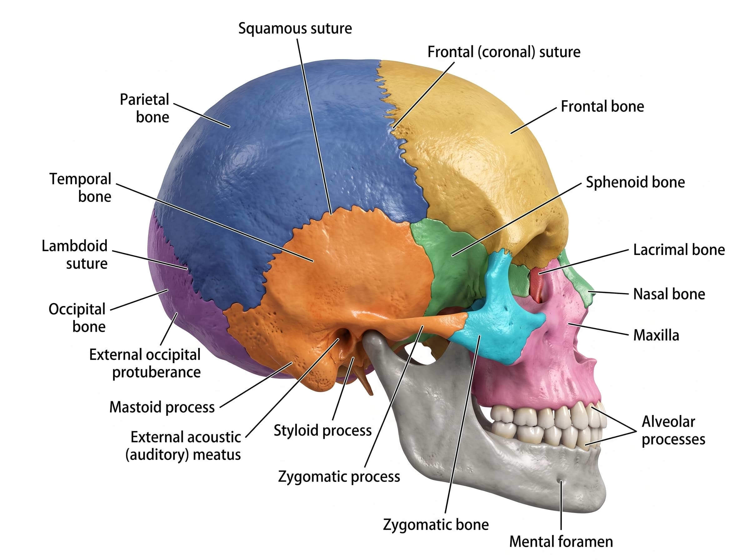
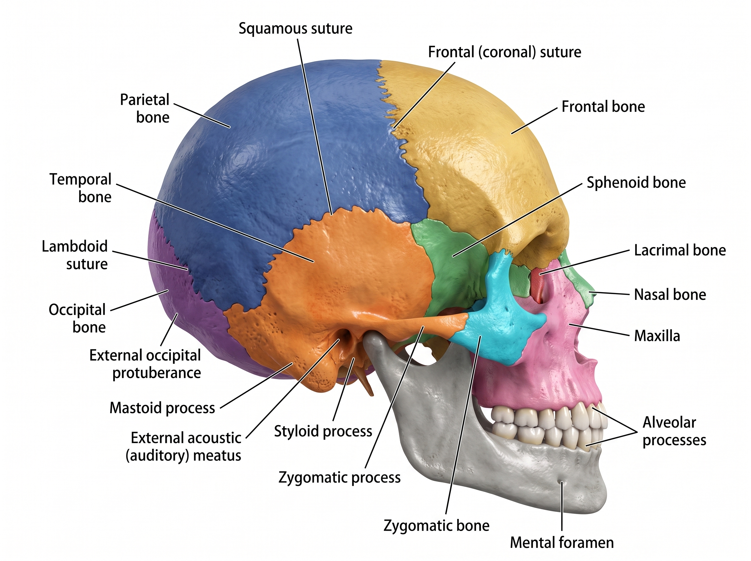
What does the lateral view of the skull show?
The lateral view looks at the skull from the side. It's the standard view for studying the temporal and occipital bones, the cranial sutures, and the landmarks clustered around the ear and jaw joint — structures that are largely hidden when looking at the skull head-on. Because the sutures and several small processes are only visible from this angle, the lateral view is frequently tested alongside the anterior view.
Cranial bones visible from the side
The lateral view shows five of the skull's eight cranial bones, including two — the temporal and occipital bones — that aren't visible from the front.
- Frontal bone
- Forms the forehead and the front of the cranial vault. In the lateral view it meets the parietal bone at the coronal suture.
- Parietal bone
- Forms the bulk of the upper side and roof of the skull. It borders the frontal bone at the coronal suture, the temporal bone at the squamous suture, and the occipital bone at the lambdoid suture.
- Temporal bone
- Forms the lower side wall of the skull around the ear. It carries several distinct landmarks, including the mastoid process, styloid process, zygomatic process, and the external acoustic meatus.
- Occipital bone
- Forms the back and part of the base of the skull. It meets the parietal bones at the lambdoid suture and carries the external occipital protuberance.
- Sphenoid bone
- A butterfly-shaped bone visible between the frontal, temporal, and parietal bones. It contributes to the lateral wall of the skull and the back of the eye socket.
Facial bones visible from the side
Fewer facial bones are visible in profile than from the front, but the ones that are visible show how the face attaches to the cranium.
- Nasal bone
- Forms the bridge of the nose, seen edge-on just below the frontal bone in the lateral view.
- Lacrimal bone
- A small bone forming part of the medial orbit wall, visible just behind the nasal bone.
- Zygomatic bone
- Forms the cheekbone. Together with the zygomatic process of the temporal bone, it forms the zygomatic arch that runs along the side of the face.
- Maxilla
- The upper jaw bone, forming the lower part of the face and part of the nasal cavity and orbit floor. It holds the upper teeth via its alveolar process.
Cranial sutures
Sutures are the fibrous joints between cranial bones. Three of the four major sutures are visible from the lateral view.
- Frontal (coronal) suture
- The joint running across the top of the skull where the frontal bone meets the two parietal bones.
- Squamous suture
- The curved joint where the temporal bone overlaps the parietal bone.
- Lambdoid suture
- The joint at the back of the skull where the parietal bones meet the occipital bone.
Landmarks and processes
These smaller bony features are frequently tested because they mark muscle attachment points and openings for nerves and vessels.
- External occipital protuberance
- A palpable bony bump on the occipital bone at the back of the head, anchoring neck ligaments and muscles.
- Mastoid process
- A cone-shaped projection of the temporal bone just behind the ear, providing attachment for several neck muscles.
- External acoustic (auditory) meatus
- The opening of the ear canal, part of the temporal bone, sitting between the mastoid and styloid processes.
- Styloid process
- A slender, pointed projection of the temporal bone that anchors muscles and ligaments of the tongue and neck.
- Zygomatic process
- A projection of the temporal bone that joins the zygomatic bone to complete the zygomatic arch.
- Alveolar processes
- The tooth-bearing ridges of the maxilla and mandible that hold the sockets for the upper and lower teeth.
- Mental foramen
- A small opening on the mandible, below the premolar teeth, through which the mental nerve and vessels pass to the chin and lower lip.
Lateral view vs. anterior view
The lateral view is best for the temporal and occipital bones, the cranial sutures, and landmarks like the mastoid and styloid processes. The anterior view is better suited for the orbits, nasal septum, and facial bones, since those structures face forward. Most anatomy courses expect you to identify structures from both views.
Tips for studying with this diagram
- Start with the labelled version to learn the position of each bone, suture, and landmark, then switch to the unlabelled version and try to name every structure from memory.
- Trace each suture with your eyes and name the two bones it joins — that's a common exam question format, and the skull sutures diagram breaks this down further.
- Download both PNGs so you can print them or drop them into flashcard software for spaced repetition.
- Pair this lateral view with the anterior view to build a full picture of the skull before an exam.
- For all 22 skull bones, the fontanelles and the foramina of the skull base, see the illustrated skull anatomy atlas page.
Need a custom illustration?
Natomy turns clinical photos and prompts into publication-ready medical illustrations in seconds — including custom anatomy diagrams beyond the lateral skull.
Try Natomy TodayFAQ
Is the lateral view of skull labelled diagram free to use?+
Yes. Both the labelled and unlabelled lateral view skull diagrams are free to download as PNG files for personal study, teaching, and educational use.
What view of the skull is this?+
This is the lateral (side) view of the skull, showing the temporal, parietal, and occipital bones along with the cranial sutures and landmarks around the ear and jaw.
Can I use the unlabelled version to quiz myself?+
Yes. The unlabelled lateral skull diagram keeps the same color-coded bones without text labels, so you can test your recall of each structure before checking the labelled version.
How is the lateral view different from the anterior view?+
The lateral view is best for studying the temporal and occipital bones, the cranial sutures, and landmarks around the ear such as the mastoid and styloid processes. The anterior (front) view is better suited for the orbits, nasal cavity, and facial bones.
Why do the sutures matter on a lateral skull diagram?+
The coronal, squamous, and lambdoid sutures are the fibrous joints between the cranial bones. They're commonly tested because their names and locations mark the borders between the frontal, parietal, temporal, and occipital bones.
Can I generate diagrams of other views or structures?+
Natomy can generate custom medical illustrations from clinical photos or prompts, including other skull views and anatomical structures beyond the lateral skull.
Related free tools

AI Storyboard Generator
Free AI tool to turn any science/medical topic into a multi-panel illustrated storyboard.
Try it free
Medical Image Finder
Free tool that searches the web and finds real medical images — scans, diagrams, and illustrations — from reputable sources. Rates images on how accurate they are.
Try it free
Medical Image Accuracy Checker
Free AI tool that reviews a medical or scientific image for anatomical accuracy and lists any specific issues found.
Try it free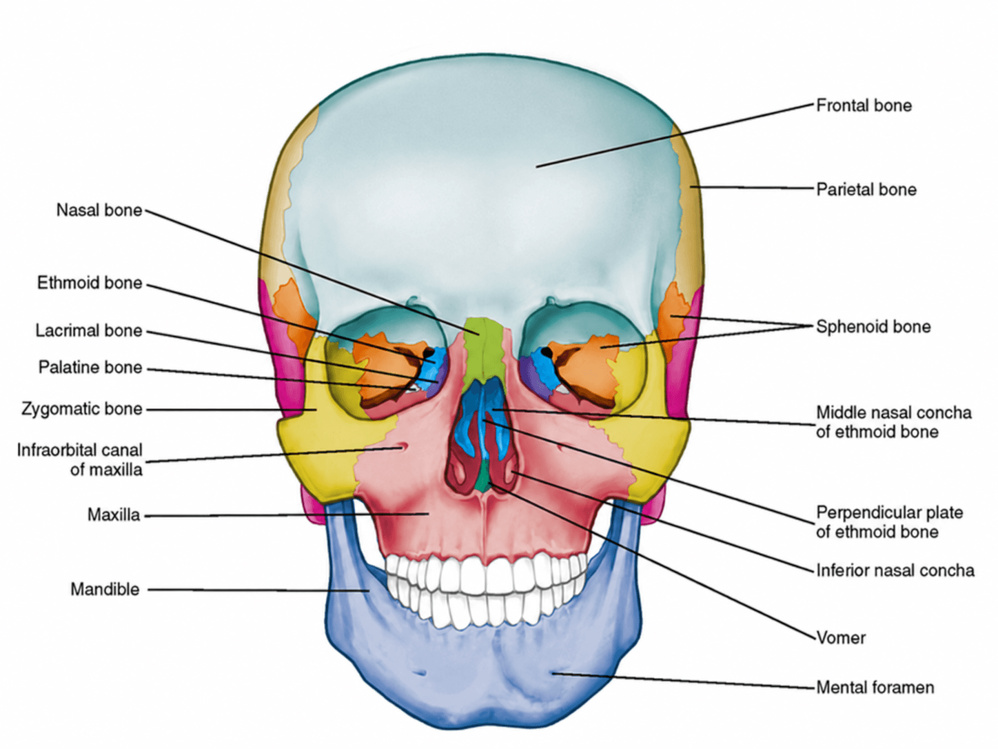
Anterior View of Skull Labelled
Free labelled and unlabelled anterior view of the skull, downloadable as PNG.
Try it free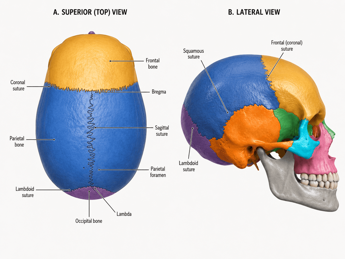
Skull Sutures Labelled
Free labelled and unlabelled skull sutures diagram, superior and lateral views, downloadable as PNG.
Try it free