Free Tools
Anterior View of Skull Labelled
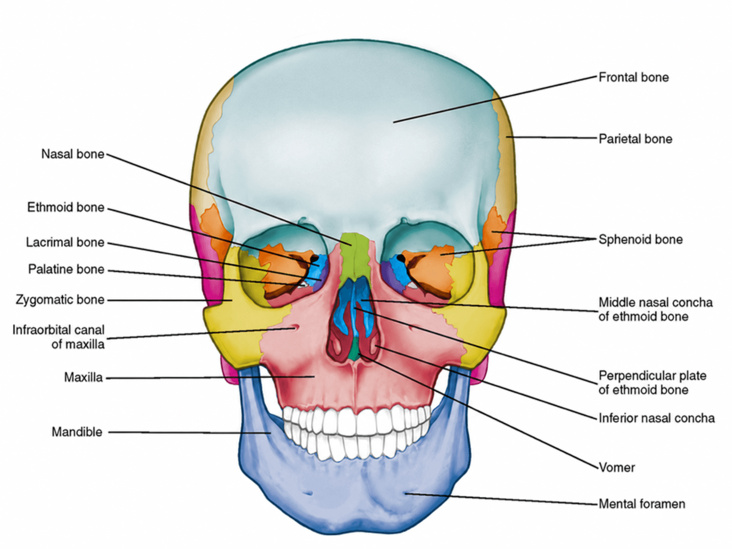
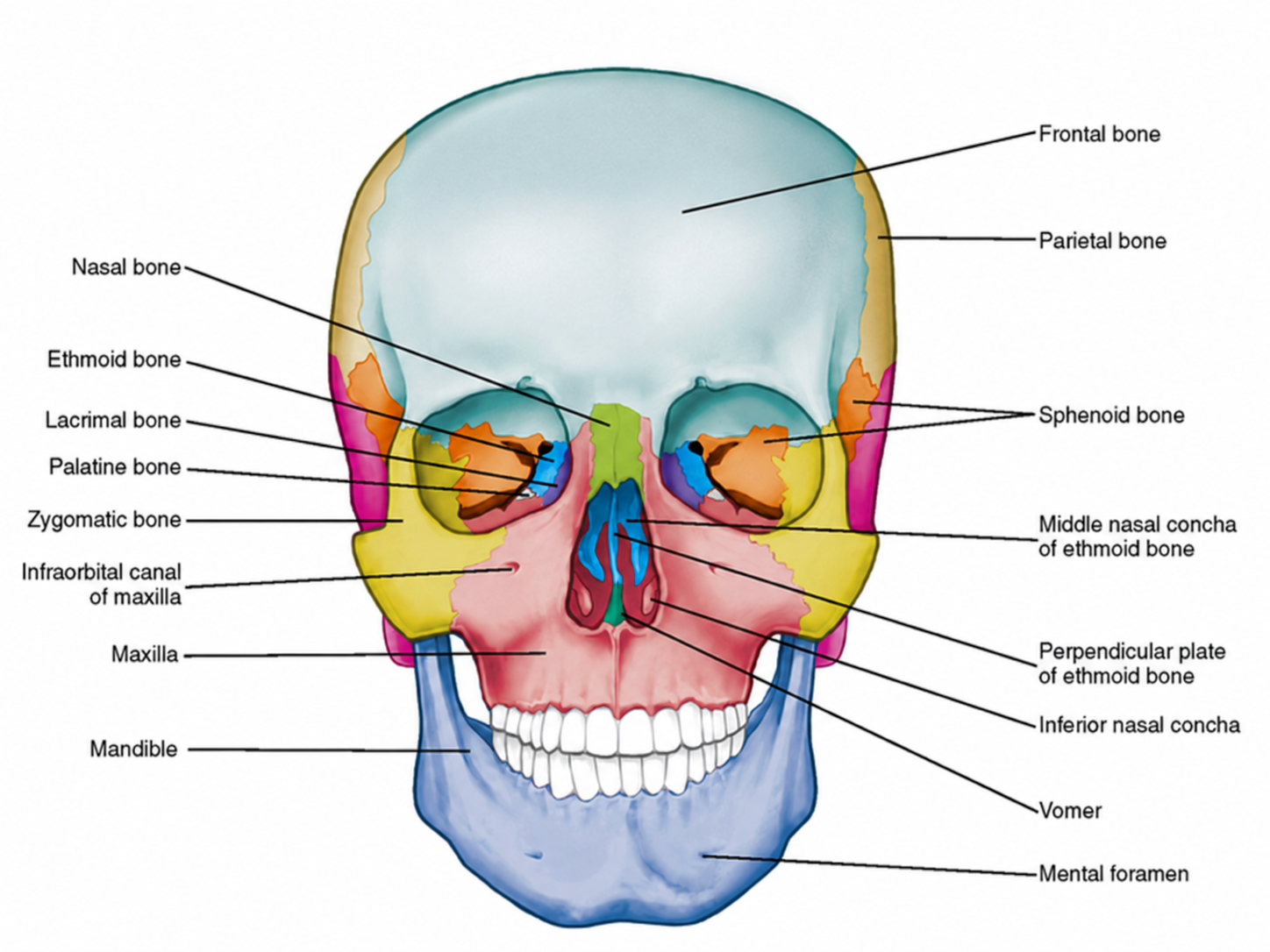
A free, downloadable diagram of the anterior view of the skull, labelled with every major cranial and facial bone. Switch to the unlabelled version of the same anterior skull view to quiz yourself before checking your answers.
What does the anterior view of the skull show?
The anterior view looks at the skull head-on, the way you'd see someone's face. It's the standard view for studying the orbits (eye sockets), the nasal cavity and its septum, and the bones that give the face its shape — the zygomatic bones, maxilla, and mandible. Because so many structures converge around the orbits and nose, the anterior view is one of the most commonly tested perspectives in anatomy courses.
Cranial bones visible from the front
The skull's eight cranial bones enclose and protect the brain. From the anterior view, only four are visible — the rest sit toward the side and back of the head.
- Frontal bone
- Forms the forehead and the upper wall of both eye sockets. It's the most prominent cranial bone visible from the front and fuses from two halves during early childhood.
- Parietal bone
- A pair of bones forming the sides and crown of the cranium. Only their front edges are visible in the anterior view, joining the frontal bone at the coronal suture.
- Sphenoid bone
- A butterfly-shaped bone at the base of the skull. A small portion is visible in the posterior wall of each orbit, between the frontal and zygomatic bones.
- Ethmoid bone
- A lightweight bone between the orbits that contributes to the nasal cavity, the medial orbit wall, and the nasal septum via its perpendicular plate.
Facial bones visible from the front
The facial bones give the lower skull its shape and house the eyes, nasal cavity, and teeth. Most of them are visible, at least in part, from the anterior view.
- Nasal bone
- A pair of small bones forming the bridge of the nose, just below the frontal bone and above the cartilage of the nasal tip.
- Lacrimal bone
- The smallest bone of the face. It sits in the medial wall of the orbit and houses part of the tear (lacrimal) drainage pathway.
- Zygomatic bone
- Commonly called the cheekbone. It forms the lateral wall and floor of the orbit and gives the cheek its prominence.
- Maxilla
- The upper jaw bone. It forms the floor of the orbit, the sides of the nasal cavity, and holds the upper teeth.
- Infraorbital canal of maxilla
- A canal running along the floor of the orbit through the maxilla, carrying the infraorbital nerve and blood vessels to the face.
- Palatine bone
- A small L-shaped bone that contributes to the back of the nasal cavity and the floor of the orbit, mostly hidden behind the maxilla.
- Vomer
- A thin, single midline bone forming the lower and back part of the nasal septum, visible through the nasal opening.
- Inferior nasal concha
- An independent, curved bone forming a shelf along the lower lateral wall of the nasal cavity that helps warm and humidify inhaled air.
- Middle nasal concha of ethmoid bone
- Part of the ethmoid bone, this projects into the nasal cavity above the inferior concha and performs a similar filtering function.
- Perpendicular plate of ethmoid bone
- The vertical portion of the ethmoid bone that forms the upper part of the nasal septum, continuous with the vomer below.
- Mandible
- The lower jaw and the only mobile bone of the skull. It holds the lower teeth and forms the chin and jawline.
- Mental foramen
- A small opening on the front of the mandible, below the premolar teeth, through which the mental nerve and vessels pass to the chin and lower lip.
Anterior view vs. lateral view
The anterior view is best for the orbits, nasal septum, and facial bones, since these structures face forward. The lateral (side) view is better suited for studying the temporal and occipital bones, the zygomatic arch, the temporomandibular joint, and the cranial sutures — none of which are fully visible from the front. Most anatomy courses expect you to be able to identify structures from both views.
Tips for studying with this diagram
- Start with the labelled version to learn the position of each bone, then switch to the unlabelled version and try to name every structure from memory.
- Group bones by category — cranial vs. facial — rather than memorizing them in a single long list.
- Download both PNGs so you can print them or drop them into flashcard software for spaced repetition.
- Pair this anterior view with a lateral view and a skull sutures diagram to build a full picture of the skull before an exam.
- For all 22 skull bones, the fontanelles and the foramina of the skull base, see the illustrated skull anatomy atlas page.
Need a custom illustration?
Natomy turns clinical photos and prompts into publication-ready medical illustrations in seconds — including custom anatomy diagrams beyond the anterior skull.
Try Natomy TodayFAQ
Is the anterior view of skull labelled diagram free to use?+
Yes. Both the labelled and unlabelled anterior view skull diagrams are free to download as PNG files for personal study, teaching, and educational use.
What view of the skull is this?+
This is the anterior (front-facing) view of the skull, showing the frontal, parietal, zygomatic, nasal, and facial bones along with the mandible, as if looking directly at someone's face.
Can I use the unlabelled version to quiz myself?+
Yes. The unlabelled anterior skull diagram keeps the same color-coded bones without text labels, so you can test your recall of each structure before checking the labelled version.
How is the anterior view different from the lateral view?+
The anterior view faces the skull head-on and is best for studying the orbits, nasal cavity, and facial bones. The lateral (side) view instead shows the temporal and occipital bones, the zygomatic arch, and the cranial sutures more clearly.
Why are some cranial bones, like the temporal and occipital bones, missing from this diagram?+
The temporal and occipital bones sit toward the side and back of the skull, so they're largely hidden behind the frontal and parietal bones when viewed from the front. They're visible on lateral or posterior skull views.
Can I generate diagrams of other views or structures?+
Natomy can generate custom medical illustrations from clinical photos or prompts, including other skull views and anatomical structures beyond the anterior skull.
Related free tools

AI Storyboard Generator
Free AI tool to turn any science/medical topic into a multi-panel illustrated storyboard.
Try it free
Medical Image Finder
Free tool that searches the web and finds real medical images — scans, diagrams, and illustrations — from reputable sources. Rates images on how accurate they are.
Try it free
Medical Image Accuracy Checker
Free AI tool that reviews a medical or scientific image for anatomical accuracy and lists any specific issues found.
Try it free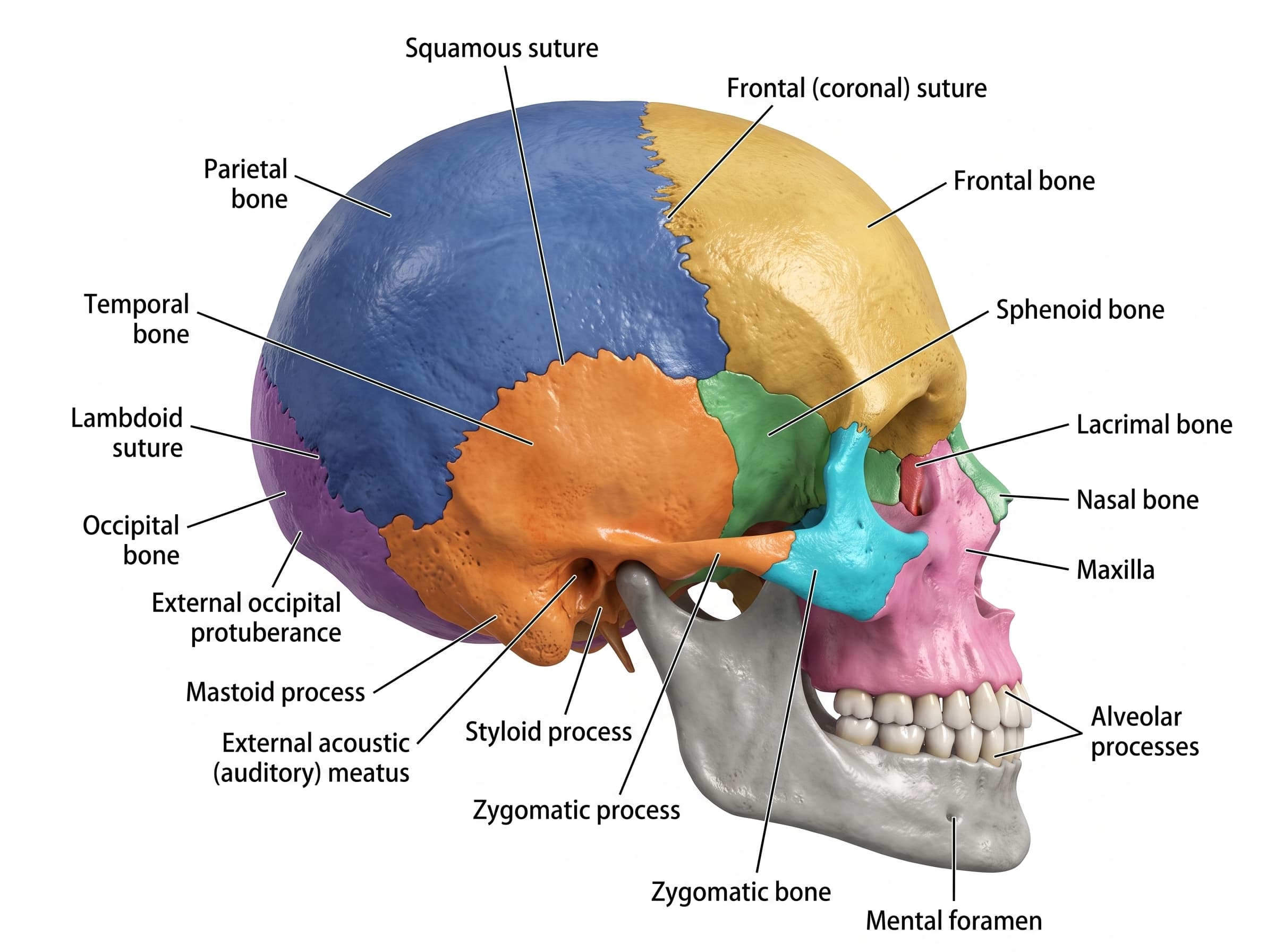
Lateral View of Skull Labelled
Free labelled and unlabelled lateral view of the skull, downloadable as PNG.
Try it free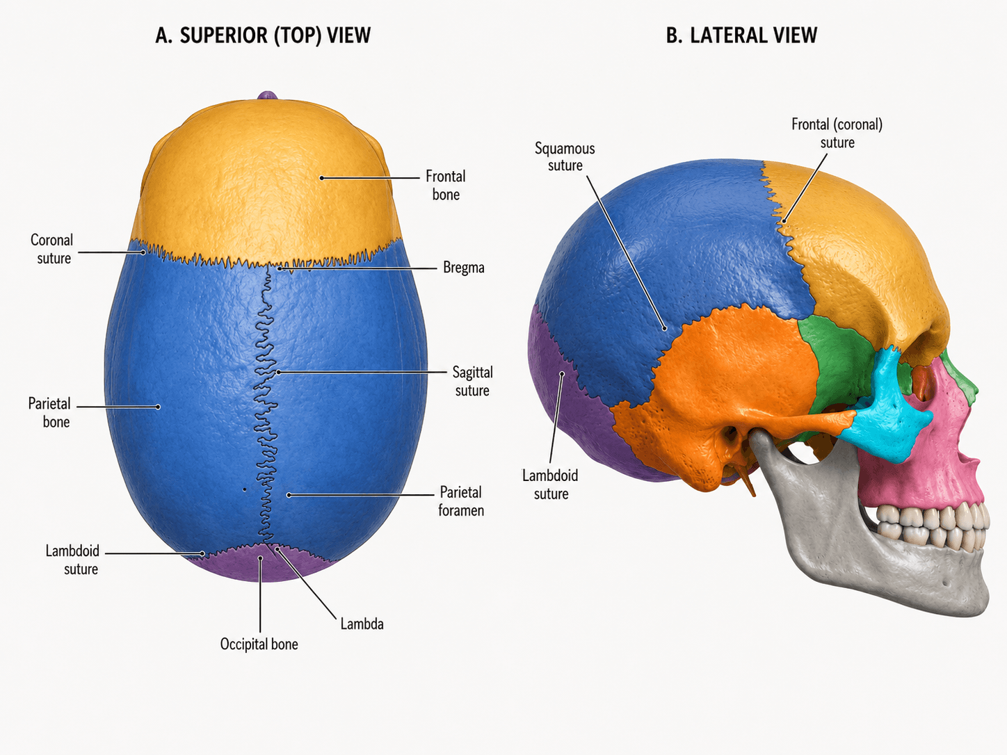
Skull Sutures Labelled
Free labelled and unlabelled skull sutures diagram, superior and lateral views, downloadable as PNG.
Try it free