Free Tools
View DICOM files online, in 3D
Open a CT or MRI from a hospital disc or a download and see it in 3D, right in your browser on Mac, Windows, or Linux. There is nothing to install, no account, and your scan never leaves your computer. The viewer opens with a sample aortic dissection CT; open your own scan to replace it.
How to open a DICOM CD or folder
- If your scan came on a CD or USB drive, copy the whole folder to your computer first. Imaging discs usually contain a folder named DICOM and a file named DICOMDIR.
- Click DICOM folder and choose that folder, or drag the folder onto the viewer. NIfTI (.nii, .nii.gz) and NRRD files work too.
- If the disc holds several series, pick one from the Series menu. The largest series, usually the main thin-slice scan, opens first.
- Drag to rotate and scroll to zoom. Switch to Slices to scroll through the scan, or click Label anatomy on a CT to color the organs, bones, and vessels.
What you can do
- 3D volume rendering
- Rotate the whole scan in 3D with presets for contrast-filled vessels, bone, and organs. A cut-away slider opens the body from the front, side, or top.
- Slice view
- Axial, coronal, and sagittal slices side by side, with sensible CT windows for each preset.
- AI anatomy labeling (CT)
- One click labels around 100 structures, including organs, vessels, vertebrae, and ribs, and turns them into smooth, color-coded 3D surfaces. The model runs on your own computer.
- Show, hide, and explode
- Toggle groups such as bones or vessels, or single structures like the liver or aorta. Click any structure to see its name, or pull organs apart with the exploded view.
- Save images
- Export the current view as a PNG for a patient handout, a presentation, or a case file.
Private by design
Most online DICOM viewers upload your scan to a server. This one does not. The DICOM files are converted with WebAssembly inside the browser tab, the 3D view is drawn by your graphics card, and the AI model runs locally. Natomy never receives the images or the patient details stored in them, which makes it a practical way to look at a scan when you cannot share it.
Who it's for
- Surgeons and physicians
- Show a patient their own anatomy in 3D during a consult, without opening the hospital PACS or installing a workstation.
- Personal injury and medical malpractice attorneys
- Review the CT and MRI discs that arrive with medical records and save clear 3D views of fractures, vessels, and organs for case preparation. Natomy can then turn them into demonstrative medical illustrations.
- Patients
- Open the disc from your hospital visit on a Mac or Windows laptop and see your scan in 3D. Your images are not uploaded anywhere.
- Educators and students
- Explore real cross-sectional anatomy in 3D, with every major structure labeled and clickable.
For viewing and education only. Not a medical device and not for diagnosis.
Turn a scan into a medical illustration
Natomy turns X-rays, CT and MRI views, clinical photos, and prompts into clear, labeled medical illustrations for patient education, publications, and medical-legal exhibits. See examples in the Natomy atlas.
Try Natomy TodayFAQ
Is this DICOM viewer free?+
Yes. The viewer is free to use and does not require an account or a download.
Are my scans uploaded to Natomy?+
No. Your DICOM files are read and converted inside your browser, and the 3D rendering and AI labeling also run on your own computer. Nothing from your scan is sent to Natomy or any other server.
Does it work on a Mac?+
Yes. It runs in a modern desktop browser such as Chrome, Edge, or Firefox on macOS and Windows, so you can view DICOM files on a Mac without installing Horos or OsiriX.
How do I view a CT or MRI CD from the hospital?+
Copy the folder from the disc to your computer, then click DICOM folder and select it, or drag it onto the viewer. The viewer finds the image series inside, including compressed JPEG and JPEG 2000 DICOM files.
Can it open MRI scans?+
Yes. MRI and CT both open in 3D and in the slice view. AI anatomy labeling currently works on CT scans only.
How does the AI anatomy labeling work?+
A segmentation model trained on a public dataset of labeled CT scans assigns each part of the scan to a structure, such as the liver, aorta, or a vertebra, and the viewer builds a smooth 3D surface for each one. It takes about one to three minutes depending on your computer. The result is an approximation, so compare it with the slice view.
Can I use it to diagnose a condition?+
No. The viewer is for viewing, education, and communication only. It is not a medical device and is not intended for diagnosis or treatment decisions.
Which file types are supported?+
DICOM folders and files, including common compressed DICOM, plus NIfTI (.nii, .nii.gz) and NRRD volumes.
Sample scan: type B aortic dissection CTA, case 11 of Mayer et al., "Aortic Dissection Dataset and Segmentations", figshare 2024 (doi:10.6084/m9.figshare.22269091), CC BY 4.0. AI anatomy model: MONAI whole-body CT segmentation, trained on the TotalSegmentator dataset.
Related free tools
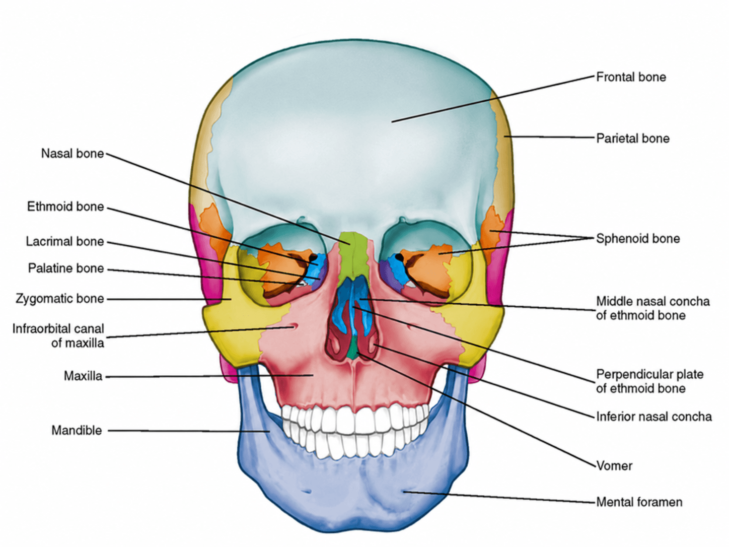
Anterior View of Skull Labeled
Free labeled and unlabeled anterior view of the skull, downloadable as PNG.
Try it free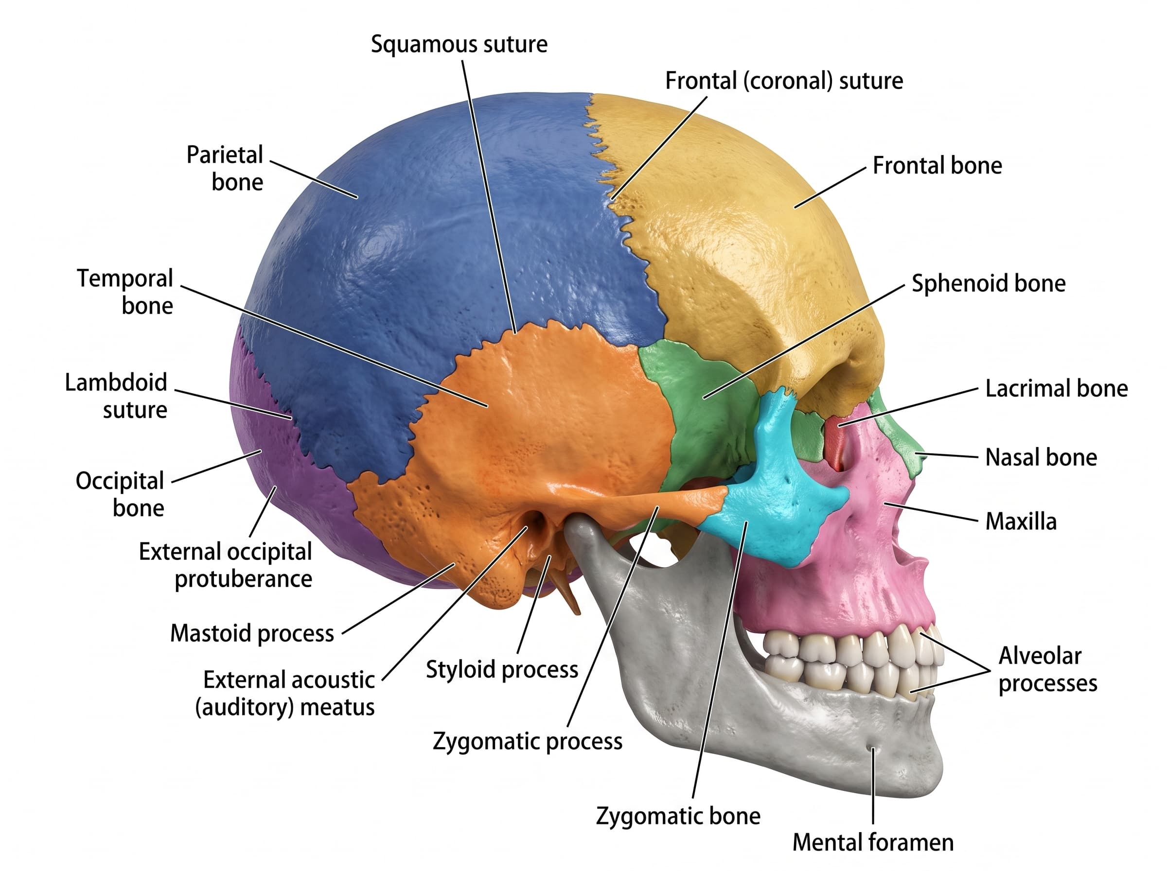
Lateral View of Skull Labeled
Free labeled and unlabeled lateral view of the skull, downloadable as PNG.
Try it free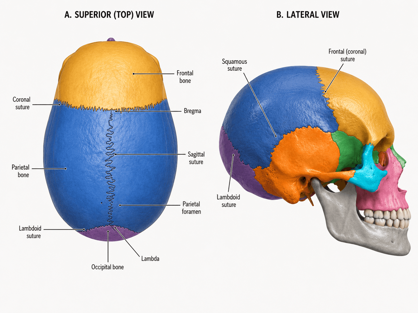
Skull Sutures Labeled
Free labeled and unlabeled skull sutures diagram, superior and lateral views, downloadable as PNG.
Try it free