Free Tools
Interactive Dermatome Map
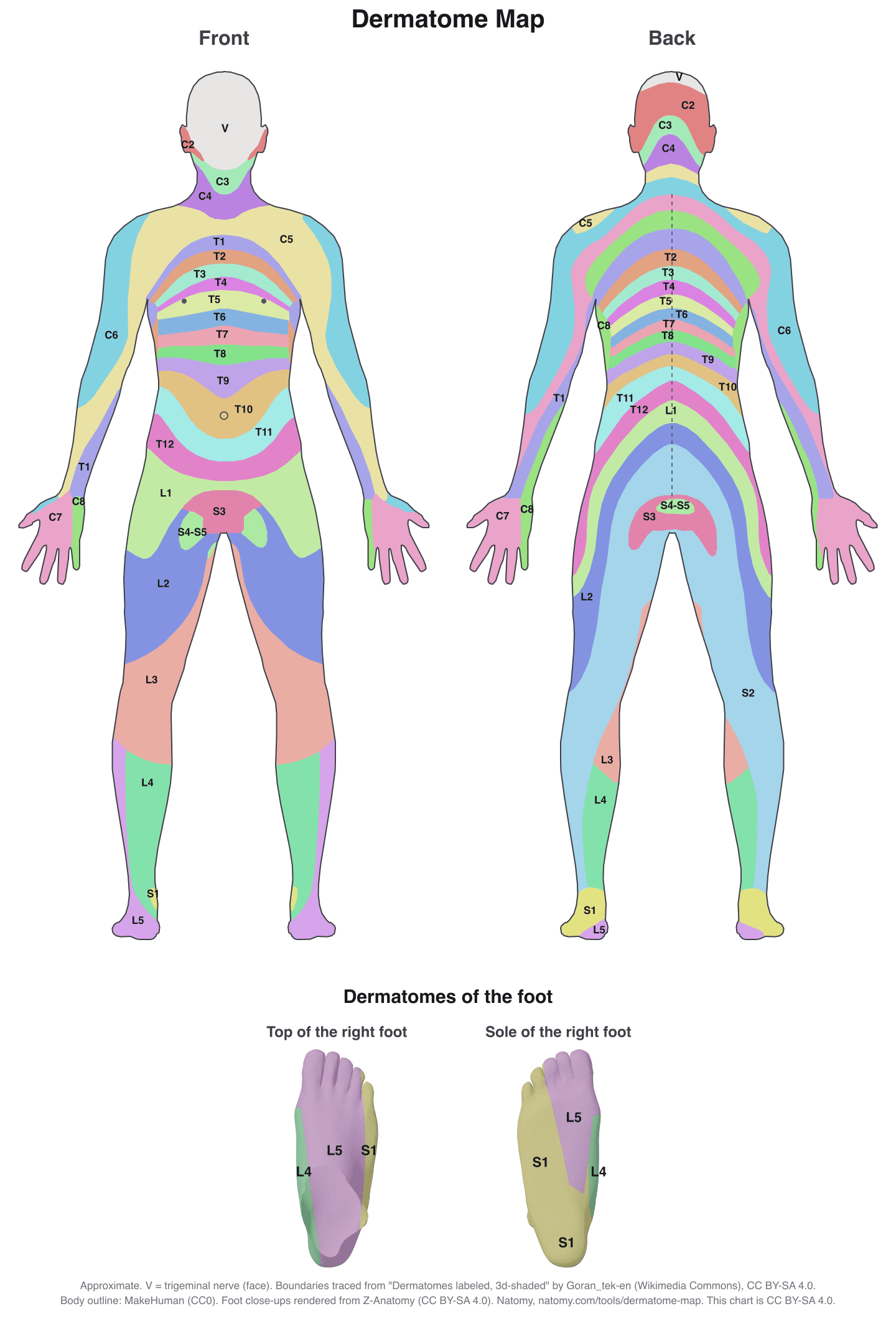
Hover or tap any part of the body to see which spinal nerve root supplies that skin, where the dermatome is tested, the muscles and reflex from the same root, and the disc level that usually affects it. Search by symptom location, or download the full dermatome chart.
Patient's right is on the left of the front view. Boundaries are approximate and overlap in real people.
Dermatomes of the foot
L4 runs along the inner edge, L5 covers the top of the foot and the big toe, and S1 covers the outer edge, the little toe, the heel, and most of the sole. Hover or tap an area.
Foot renders from Z-Anatomy (CC BY-SA 4.0). Boundaries approximate.
What the dermatome map shows
Each spinal nerve root carries sensation from one strip of skin, its dermatome. On the trunk the dermatomes run in bands that follow the ribs. On the limbs they stretch out into long strips, because the arms and legs grow out of a few segments of the embryo. When a disc herniation, bone spur, or injury presses on a root, the numbness, tingling, or burning pain follows that root's dermatome, which is why clinicians use the map to find the level of the problem.
Cervical dermatomes (C2 to C8)
The cervical roots supply the back of the head, the neck, the shoulders, and the arms. C1 usually has no skin area, so the map starts at C2. From C5 down, the roots run into the arm: C5 and C6 along the thumb side, C7 down the middle to the middle finger, and C8 along the little finger side. A pinched nerve in the neck (cervical radiculopathy) usually shows up as pain or numbness along one of these strips, most often C6 or C7.
| Root | Skin area | Muscle test | Reflex |
|---|---|---|---|
| C2 | Back of the head | Neck flexion | None |
| C3 | Neck | Neck side bending | None |
| C4 | Base of the neck and top of the shoulders | Shoulder shrug, diaphragm | None |
| C5 | Shoulder and upper outer arm | Elbow flexion (biceps), deltoid | Biceps |
| C6 | Outer forearm and thumb side of the hand | Wrist extension | Brachioradialis |
| C7 | Middle finger and back of the arm | Elbow extension (triceps) | Triceps |
| C8 | Little finger side of the hand and forearm | Finger flexion | None |
Thoracic dermatomes (T1 to T12)
The thoracic roots wrap around the chest and abdomen in bands that follow the ribs, sloping down a little toward the front. T1 and T2 reach into the inner arm and armpit. The easiest bands to place are T4 at the nipple line, T6 at the bottom of the breastbone, and T10 at the belly button; T12 ends just above the groin. Shingles (herpes zoster) is the classic thoracic dermatome rash: a painful band on one side of the trunk that stops at the midline.
Lumbar dermatomes (L1 to L5)
The lumbar roots supply the groin, the front and sides of the thigh, the knee, the shin, and the top of the foot. On the back they curve over the hips and buttocks. Lumbar disc herniations are the most common cause of radiating leg pain, and the dermatome that hurts or goes numb is the quickest pointer to the level: front of the thigh for L2 and L3, inner shin for L4, outer shin and top of the foot for L5.
| Root | Skin area | Muscle test | Reflex |
|---|---|---|---|
| L1 | Groin and the top of the thigh | Hip flexion (with L2) | Cremasteric |
| L2 | Front and outer thigh | Hip flexion | Cremasteric |
| L3 | Lower thigh and the knee | Knee extension | Knee jerk |
| L4 | Inner shin, inner ankle, inner edge of the foot | Ankle dorsiflexion | Knee jerk |
| L5 | Outer shin, top of the foot, big toe | Big toe extension | None |
The L5 dermatome
L5 is the dermatome people look up most, because the L4-L5 disc is one of the most common places for a disc to herniate. The L5 strip runs down the outer side of the lower leg, across the top of the foot, and into the big toe and the web space between the first and second toes. L5 radiculopathy typically causes pain from the buttock down the outer leg, numbness on the top of the foot, and weakness lifting the big toe or the foot (foot drop in severe cases). The knee and ankle reflexes are usually normal, which helps tell it apart from L4 (knee jerk) and S1 (ankle jerk) problems.
Upper limb dermatomes: arm and hand (C5 to T1)
| Root | Skin area | Muscle test | Reflex |
|---|---|---|---|
| C5 | Shoulder and upper outer arm | Elbow flexion (biceps) | Biceps |
| C6 | Outer forearm, thumb, index finger | Wrist extension | Brachioradialis |
| C7 | Middle finger | Elbow extension (triceps) | Triceps |
| C8 | Ring and little fingers, inner hand | Finger flexion | None |
| T1 | Inner arm and forearm | Finger abduction | None |
Leg and lower extremity dermatomes (L2 to S1)
In the leg the dermatomes spiral from the hip down to the foot: L2 and L3 on the thigh, L3 over the knee, L4 on the inner shin, L5 on the outer shin, and S1 on the outer foot and heel. S2 runs down the back of the thigh and calf.
| Root | Skin area | Muscle test | Reflex |
|---|---|---|---|
| L2 | Front and outer thigh | Hip flexion | None |
| L3 | Lower thigh and the knee | Knee extension | Knee jerk |
| L4 | Inner shin and inner ankle | Ankle dorsiflexion | Knee jerk |
| L5 | Outer shin, top of the foot, big toe | Big toe extension | None |
| S1 | Outer foot, little toe, heel, sole | Ankle plantar flexion | Ankle jerk |
Dermatomes of the foot
Three roots share the foot, which is why foot numbness is one of the most useful clues in sciatica. L4 covers the inner edge of the foot and the inner ankle. L5 covers the top of the foot and the big toe, including the web space between the first and second toes. S1 covers the outer edge of the foot, the little toe, the heel, and most of the sole. Numbness on top of the foot and the big toe points to L5, often from an L4-L5 disc herniation, while numbness of the outer foot and sole points to S1, often from an L5-S1 herniation. The map shows the top and the sole of the foot in close-up.
Landmarks to remember
- C4
- Top of the shoulder
- T4
- Nipple line
- T6
- Xiphoid (bottom of the breastbone)
- T10
- Belly button
- T12 / L1
- Groin and inguinal ligament
- S4-S5
- Around the anus (saddle area)
Printable dermatome chart
Front and back views with every dermatome labeled. Free to download and print for clinic, class, or a case file. Shared under CC BY-SA 4.0; please credit Natomy and Goran_tek-en when you reuse it.
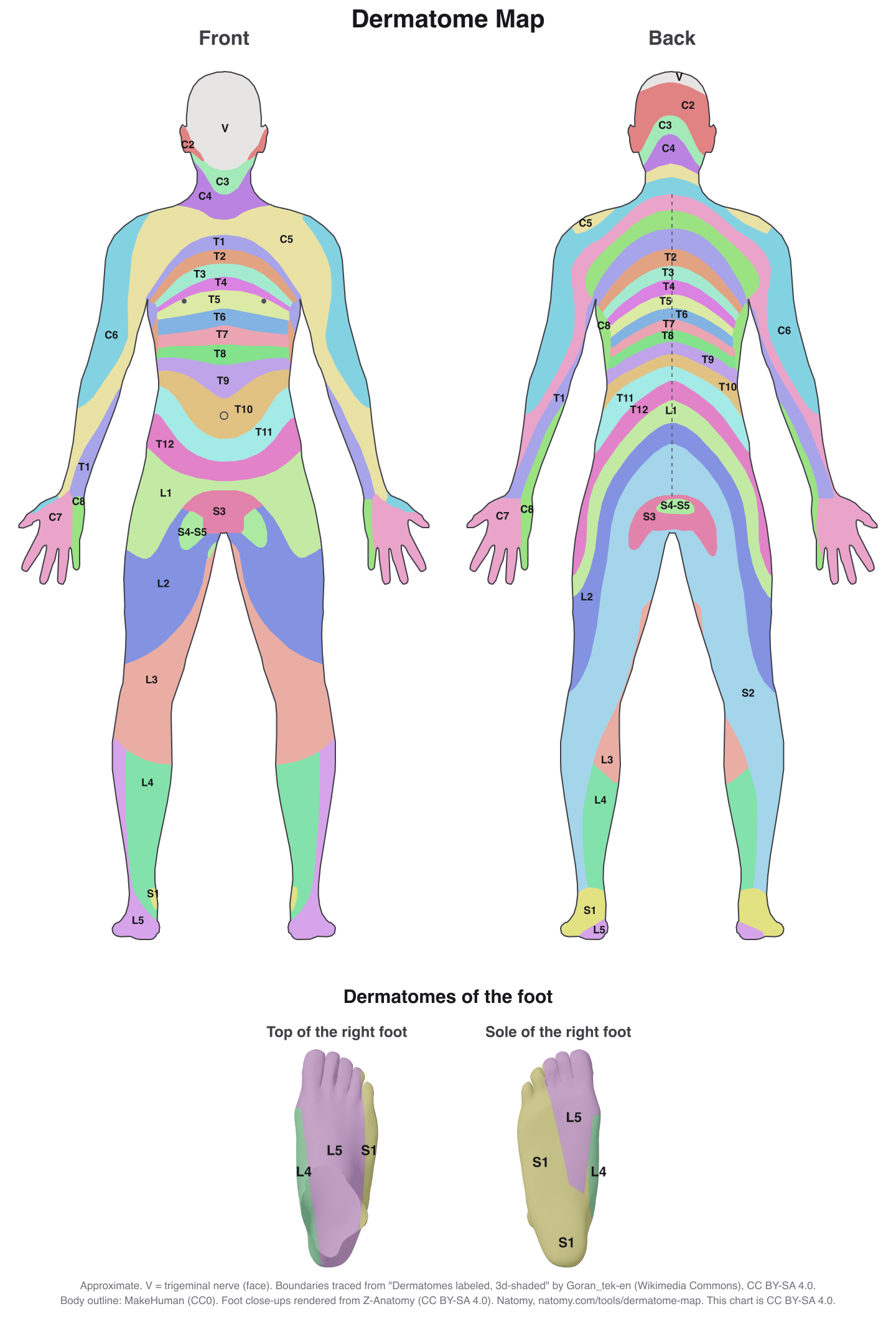
How dermatomes are tested
In the standard spinal cord injury exam (ISNCSCI), each dermatome has a key point that is tested with light touch and a pin prick and compared with the face, such as the back of the thumb for C6 or the outer heel for S1. The map shows the key point for every root. A full exam adds muscle strength (myotomes) and reflexes, because a single root problem often causes only patchy sensory changes. The myotome chart covers the key muscle for each root and the 0 to 5 strength grades.
Dermatomes in radiculopathy and injury cases
For surgeons and physicians, the map is a quick way to explain to a patient why a disc in the neck causes numbness in the hand, or why sciatica runs down to the foot. For personal injury and medical malpractice attorneys, it helps line up the symptoms in the records with the level shown on an MRI, such as L5 symptoms with an L4-L5 disc herniation.
Illustrate a nerve root injury
Natomy turns MRI views, reports, and prompts into clear, labeled medical illustrations of disc herniations, nerve root compression, and the dermatomes involved, for patient education and medical-legal exhibits. See examples in the Natomy atlas.
Try Natomy TodayFAQ
What is a dermatome?+
A dermatome is the area of skin whose sensation is carried by a single spinal nerve root. Each root leaves the spinal cord at one level, so the dermatomes form bands around the trunk and strips along the arms and legs. Numbness, tingling, or pain in one dermatome points to a problem at that root.
How many dermatomes are there?+
There are 31 pairs of spinal nerves (8 cervical, 12 thoracic, 5 lumbar, 5 sacral, and 1 coccygeal). C1 usually has no sensory fibers, so maps start at C2. That gives about 30 dermatomes on each side of the body. The face is supplied by the trigeminal nerve, a cranial nerve, so it is not part of the dermatome map.
What areas do C5, C6, C7, and C8 cover?+
C5 covers the outer upper arm, C6 the outer forearm, thumb, and index finger, C7 the middle finger, and C8 the ring and little fingers with the inner side of the hand. A C5-C6 disc herniation usually affects the C6 root, and a C6-C7 herniation the C7 root.
Which dermatomes are in the leg and foot?+
L2 and L3 cover the front and inner thigh down to the knee, L4 the inner shin and inner ankle, L5 the outer shin, the top of the foot, and the big toe, and S1 the outer foot, little toe, heel, and sole. S2 runs down the back of the thigh. An L4-L5 disc herniation usually affects L5, and an L5-S1 herniation affects S1.
What are the dermatomes of the foot?+
L4 supplies the inner edge of the foot and the inner ankle, L5 the top of the foot and the big toe, and S1 the outer edge, the little toe, the heel, and most of the sole. The S1 key test point is the outer heel, and the L5 key point is the top of the foot at the third toe joint.
What is the difference between a dermatome and a myotome?+
A dermatome is the skin a nerve root supplies; a myotome is the group of muscles it supplies. Clinicians test both, plus reflexes, to work out which root is involved. The map shows all three for each root.
Which dermatome is at the nipple and the belly button?+
The nipple line is T4 and the belly button (umbilicus) is T10. These two landmarks are the quickest way to place a sensory level on the trunk.
Is a dermatome map exact?+
No. Neighboring dermatomes overlap, and the borders vary from person to person, so a single root problem rarely causes numbness over the whole area. The boundaries on this map are traced from a widely used illustrated dermatome chart on Wikimedia Commons, and the test points follow the ISNCSCI spinal cord injury exam. It is meant for education and communication, not diagnosis.
How is a dermatome different from a peripheral nerve area?+
A nerve root and a peripheral nerve can cover overlapping skin but in different patterns. For example, numbness in the thumb and index finger can come from the C6 root in the neck or from the median nerve at the wrist (carpal tunnel syndrome). Muscle strength, reflexes, and nerve studies help tell them apart.
Sources: dermatome boundaries traced and adapted from "Dermatomes labeled, male front/back 3d-shaded" by Goran_tek-en, Wikimedia Commons, licensed CC BY-SA 4.0 (warped to a different body, recoloured, and combined zones split to single roots). Test points from the American Spinal Injury Association's International Standards for Neurological Classification of Spinal Cord Injury (ISNCSCI). Body outline from the MakeHuman base mesh (CC0). Foot close-ups rendered from the Z-Anatomy model (CC BY-SA 4.0). Boundaries are approximate. For education only; not a substitute for clinical assessment. The dermatome map and chart are shared under CC BY-SA 4.0.
Related free tools
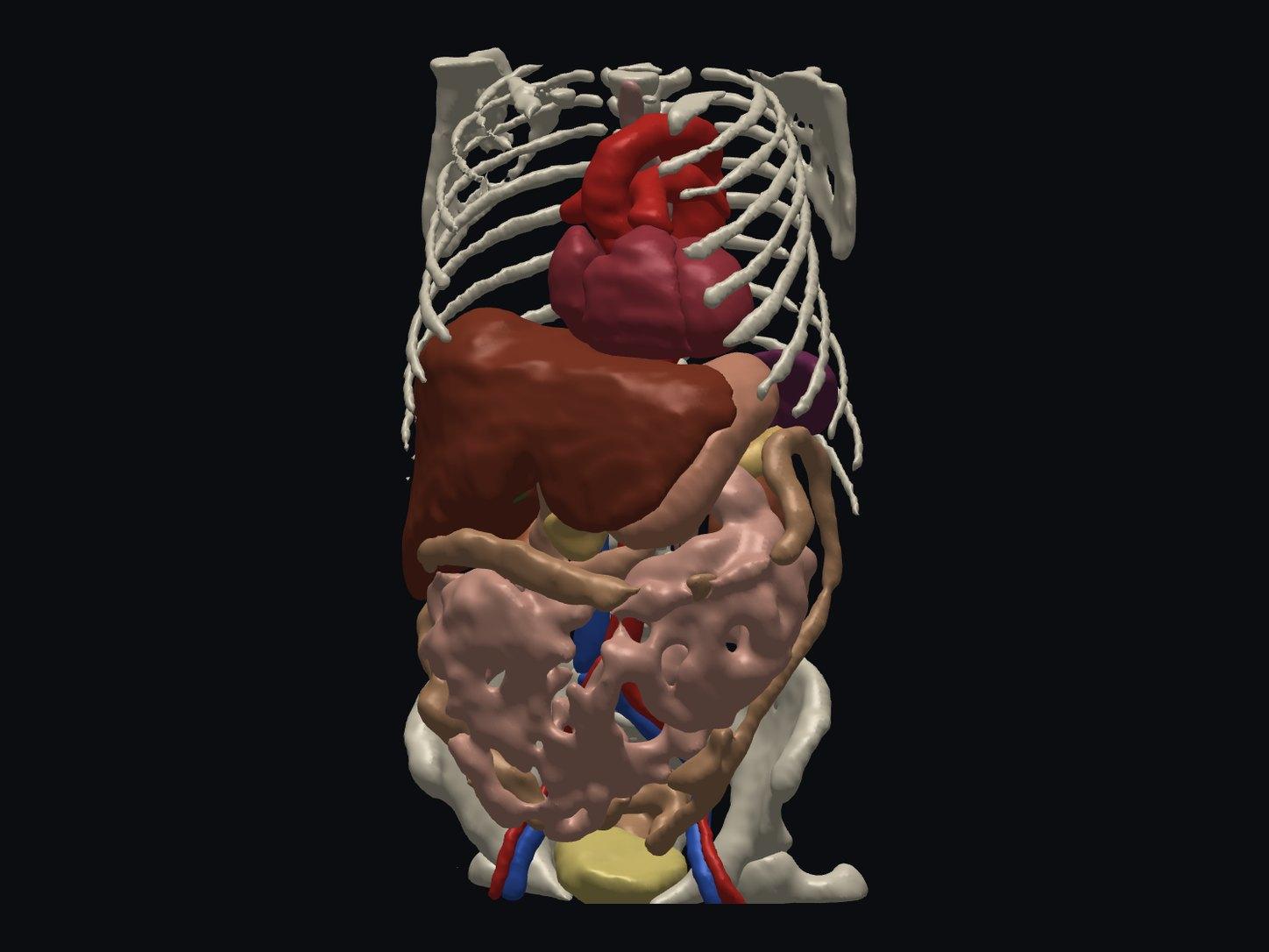
Free Online DICOM Viewer (3D)
Open a CT or MRI DICOM folder in your browser and view it in 3D, with AI anatomy labeling. Nothing is uploaded.
Try it free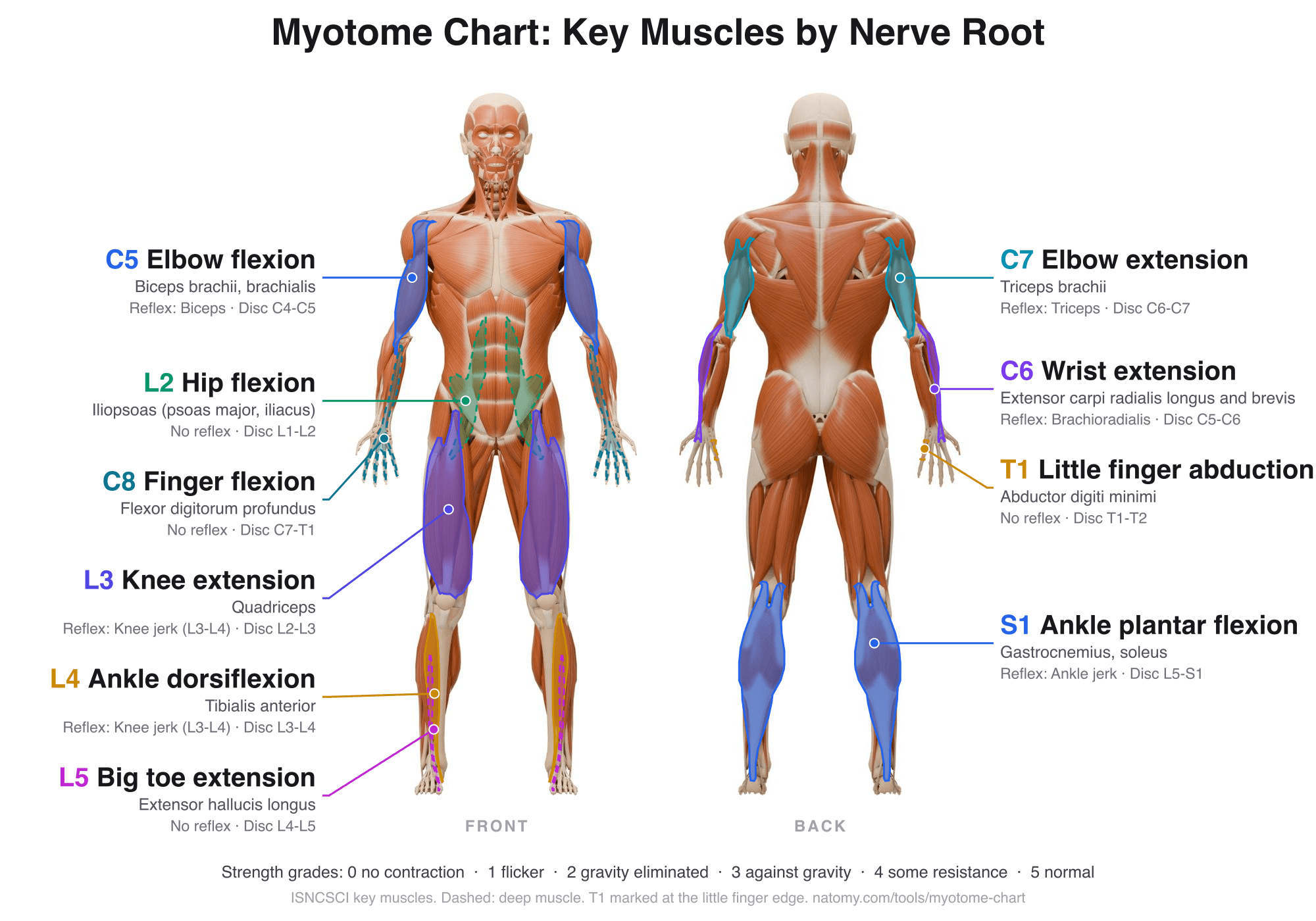
Myotome Chart and Motor Score
Tap a nerve root to see its key muscle, test position, and reflex. Grade strength 0 to 5 for the ISNCSCI motor score.
Try it free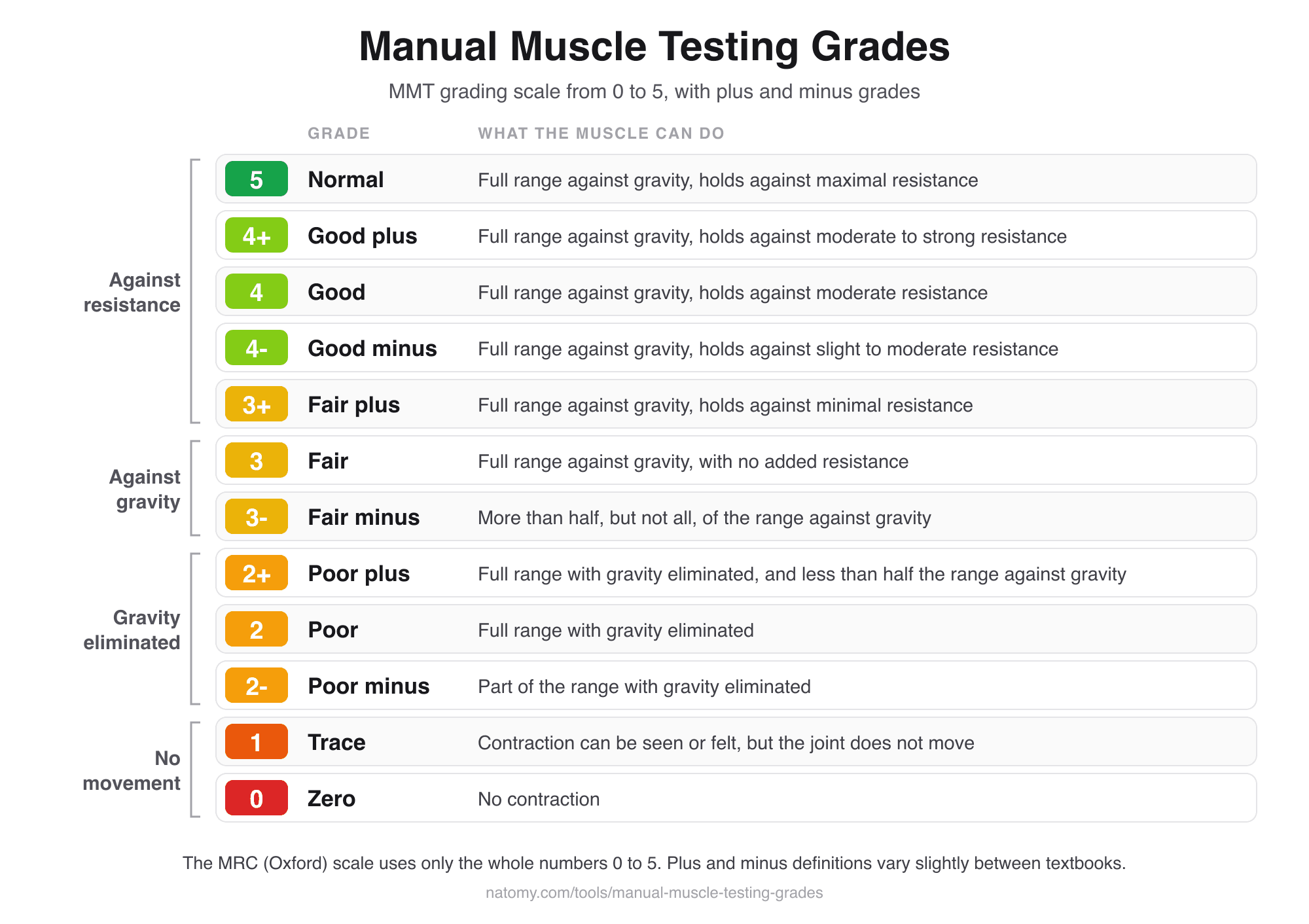
Manual Muscle Testing Grades
MMT grading scale chart from 0 to 5 with plus and minus grades, and a grade finder that writes the note for you.
Try it free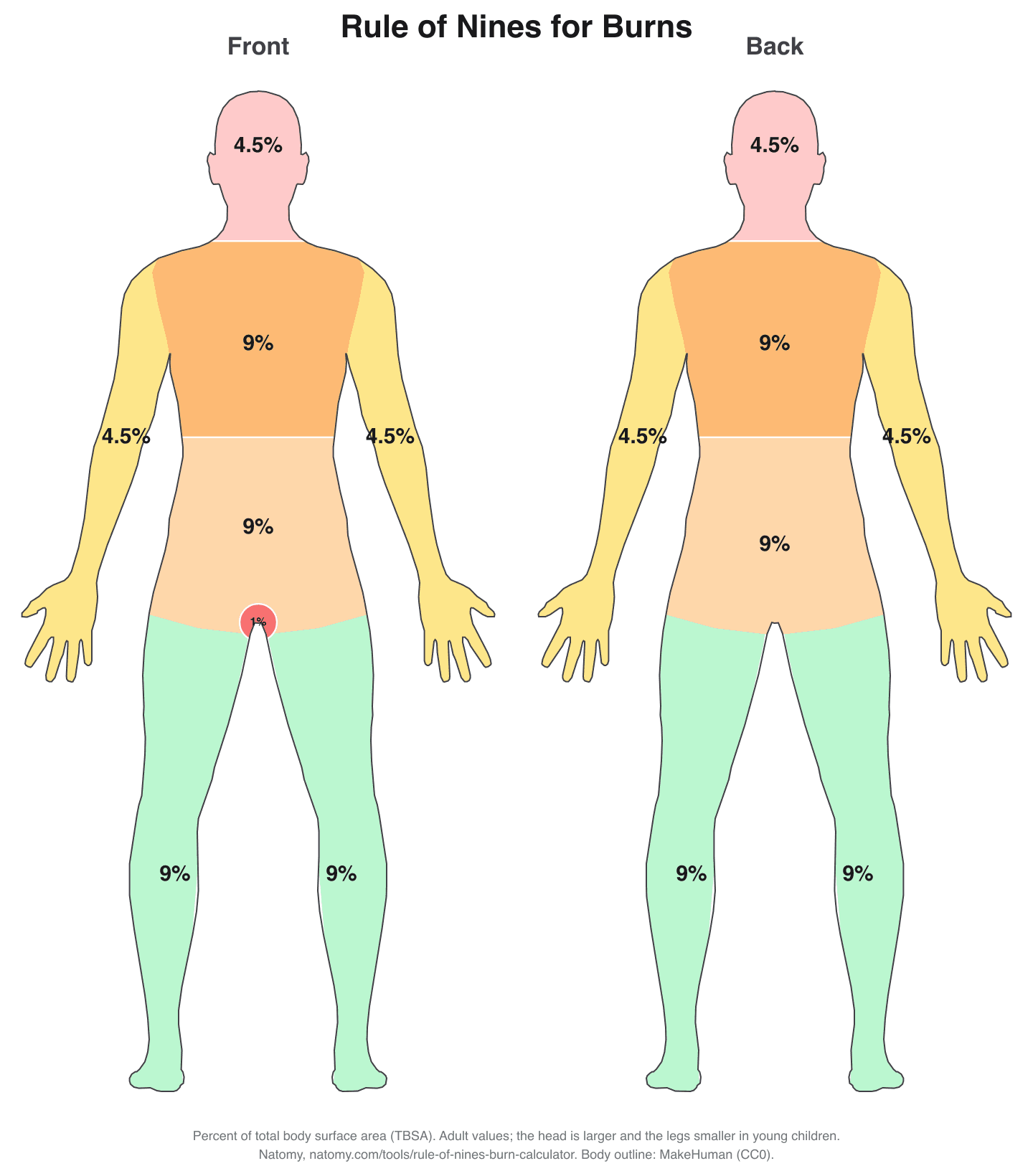
Rule of Nines Burn Calculator
Click the burned areas to get the total body surface area (TBSA), with the Parkland formula and a printable burn chart.
Try it free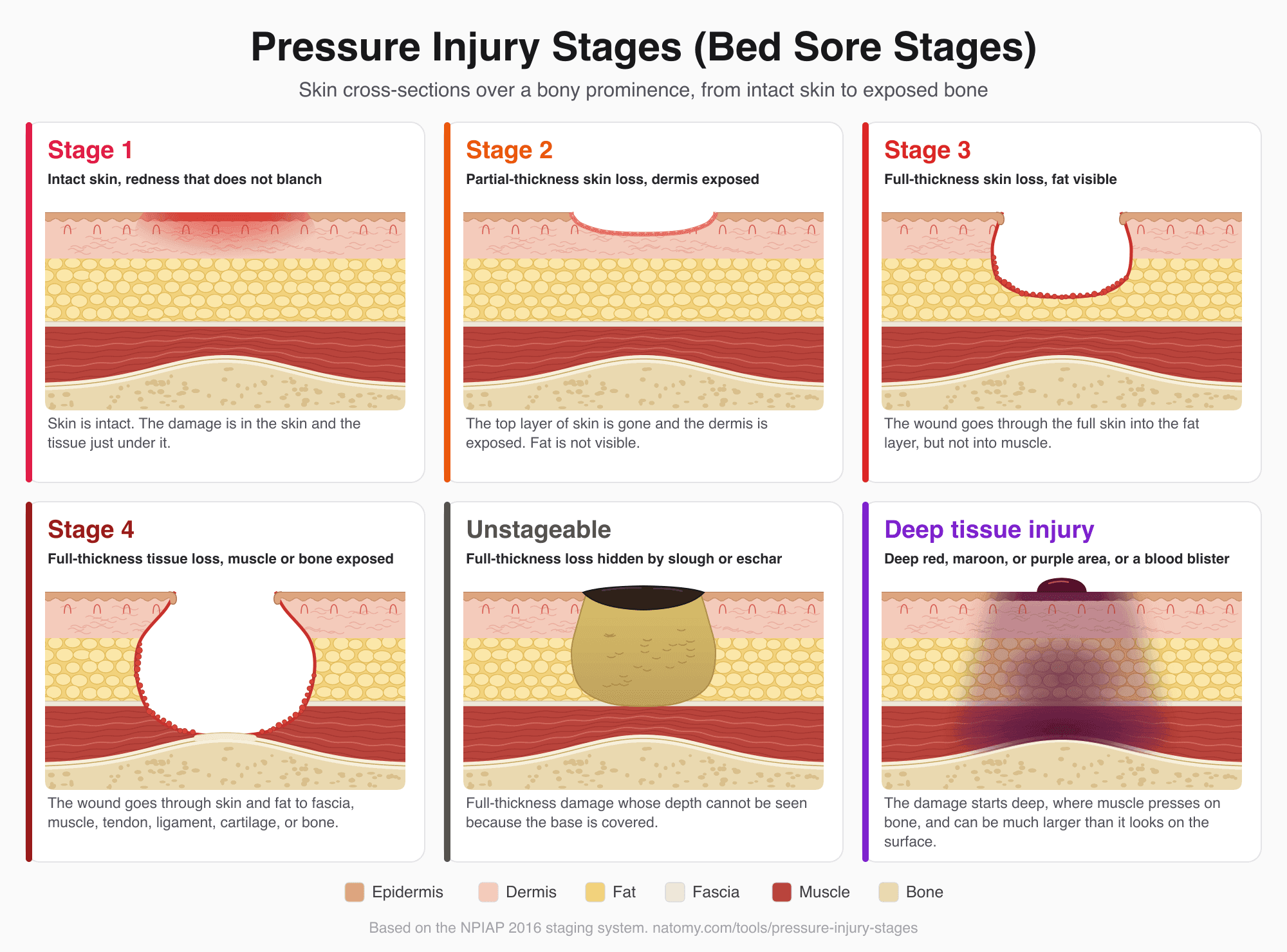
Pressure Injury (Bed Sore) Stages
Illustrated pressure ulcer stages chart, Stage 1 to 4, unstageable, and deep tissue injury, with a stage finder and wound note.
Try it free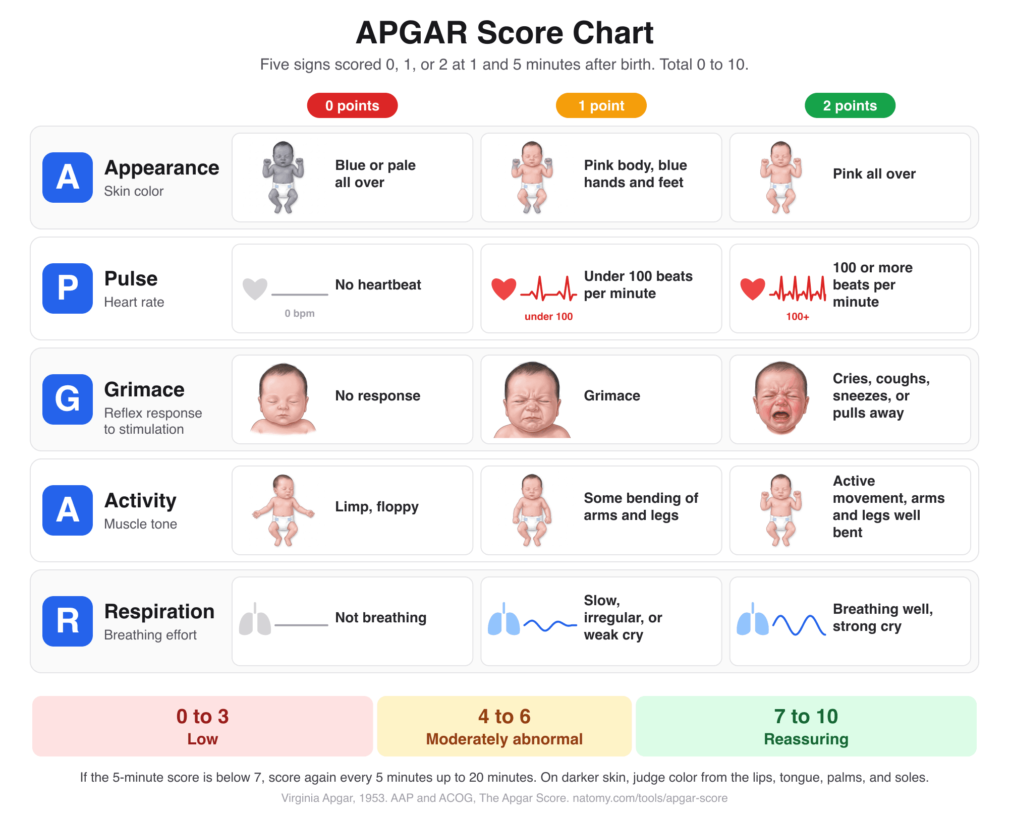
APGAR Score Chart and Calculator
Score the 5 newborn signs at 1, 5, and 10 minutes with an illustrated chart, what normal and low scores mean, and a note for the record.
Try it free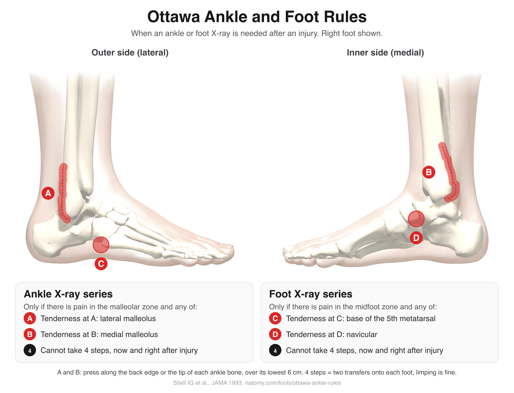
Ottawa Ankle Rules
Tap the tender spots on an illustrated ankle and foot to see if the Ottawa rules call for an X-ray. Includes the Ottawa knee rule.
Try it free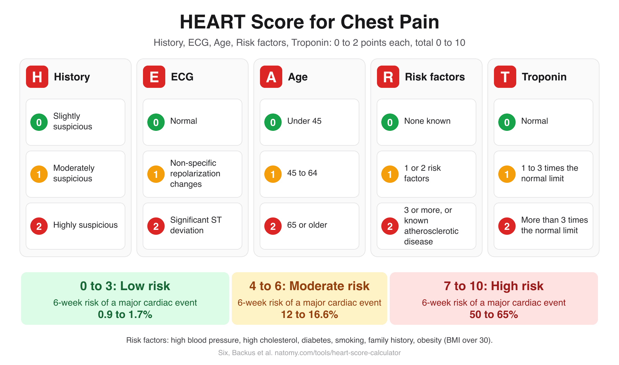
HEART Score Calculator
Score history, ECG, age, risk factors, and troponin for chest pain to get the HEART score, risk group, and 6-week MACE risk.
Try it free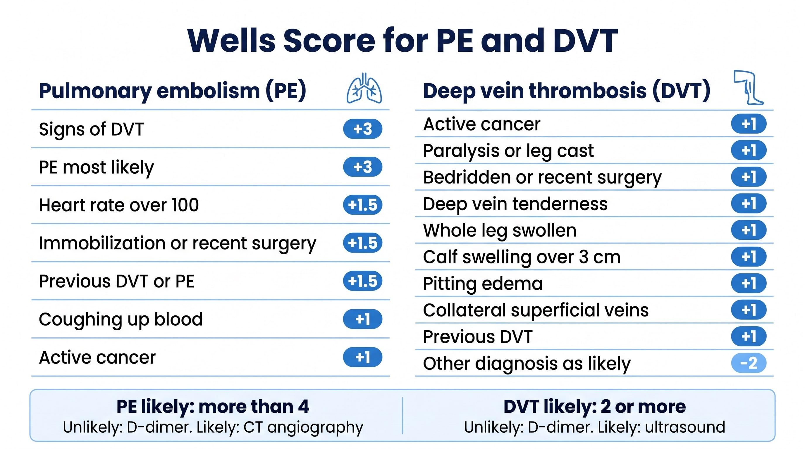
Wells Score Calculator (PE and DVT)
Check the Wells criteria for pulmonary embolism or deep vein thrombosis to get the score, the risk group, and the next test.
Try it free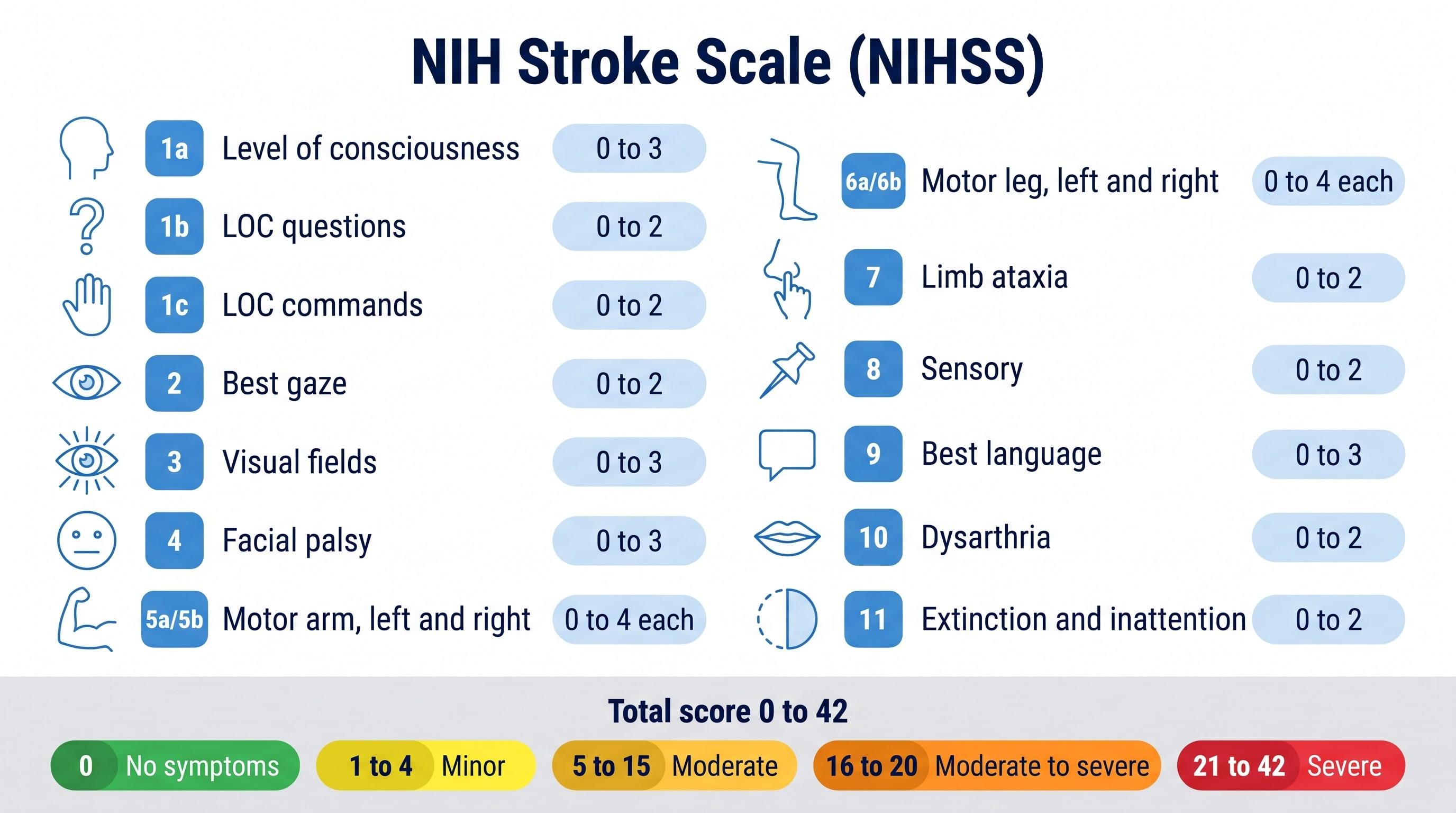
NIHSS Calculator (NIH Stroke Scale)
Score all 15 NIH Stroke Scale items to get the NIHSS total from 0 to 42 and the stroke severity range.
Try it free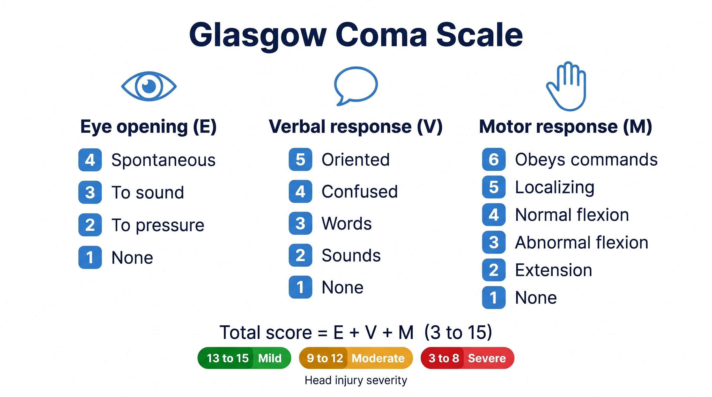
Glasgow Coma Scale Calculator
Score eye, verbal, and motor responses to get the GCS total, E V M code, severity range, and GCS-P.
Try it free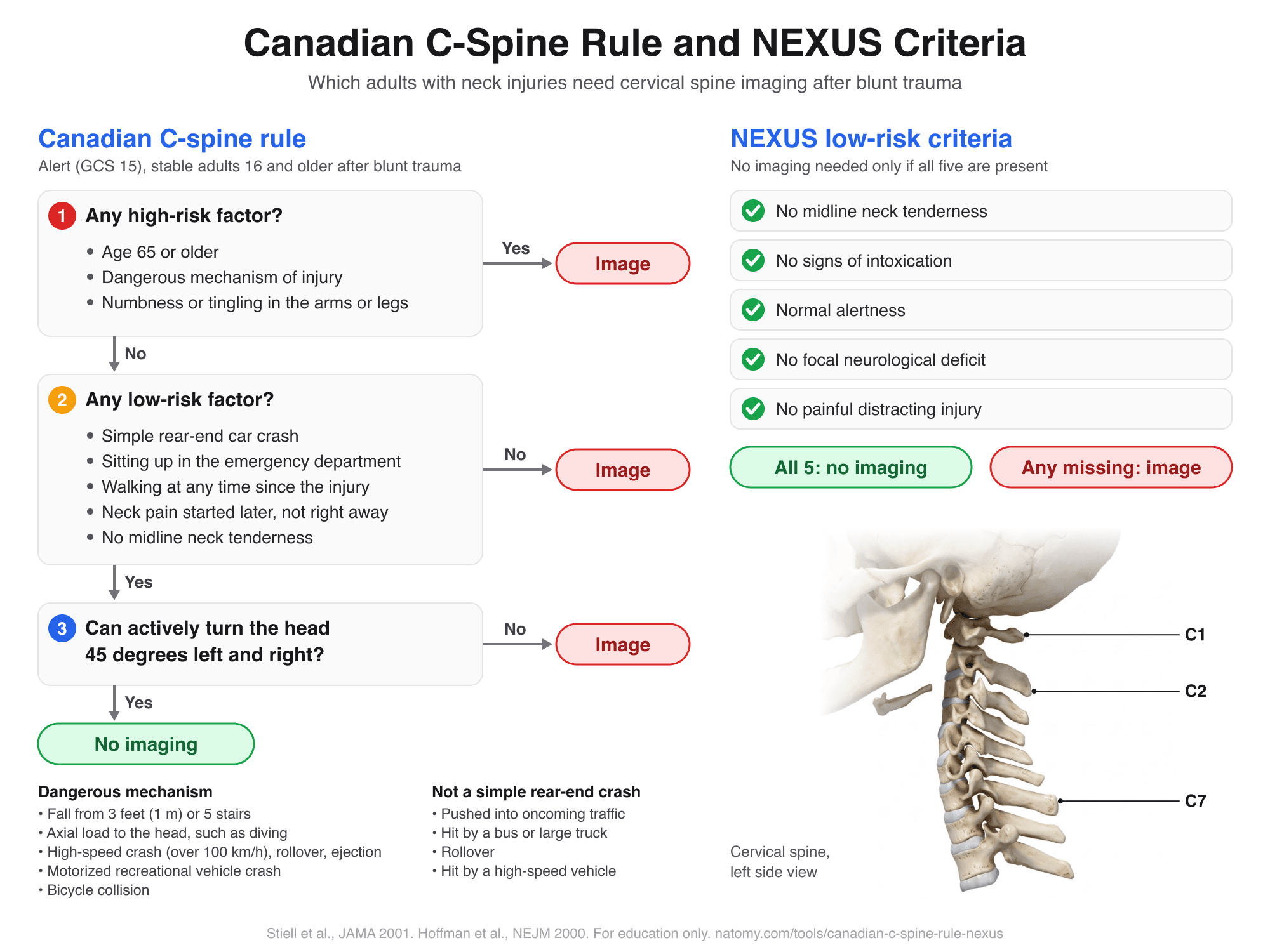
Canadian C-Spine Rule and NEXUS
Run the Canadian C-spine rule or the NEXUS criteria step by step to see which adults need neck imaging after trauma.
Try it free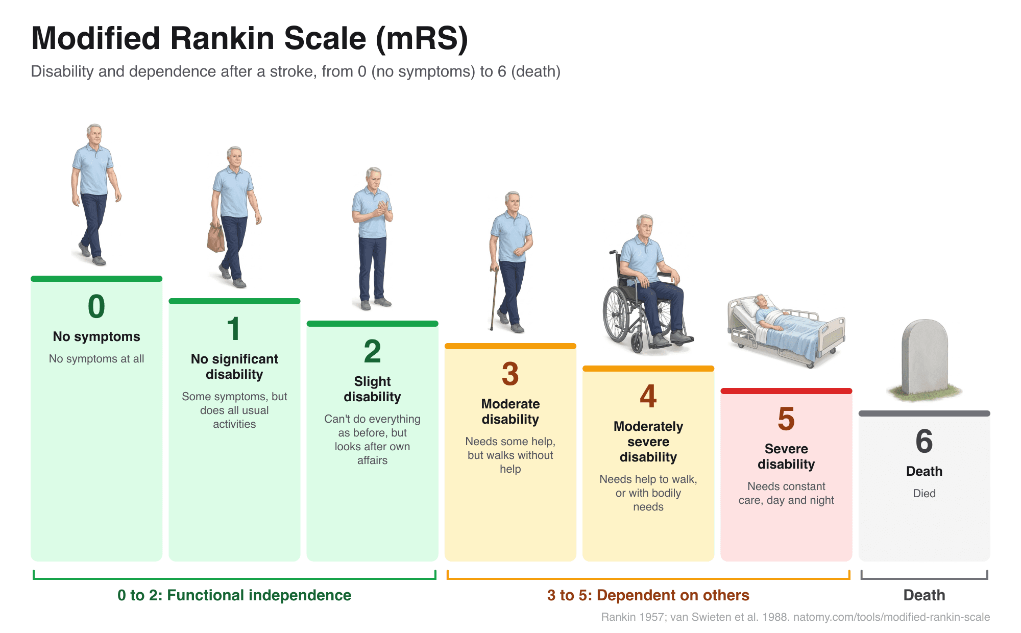
Modified Rankin Scale (mRS)
Illustrated mRS chart from 0 to 6 with a question-based score finder, what each grade means after a stroke, and a copyable note.
Try it free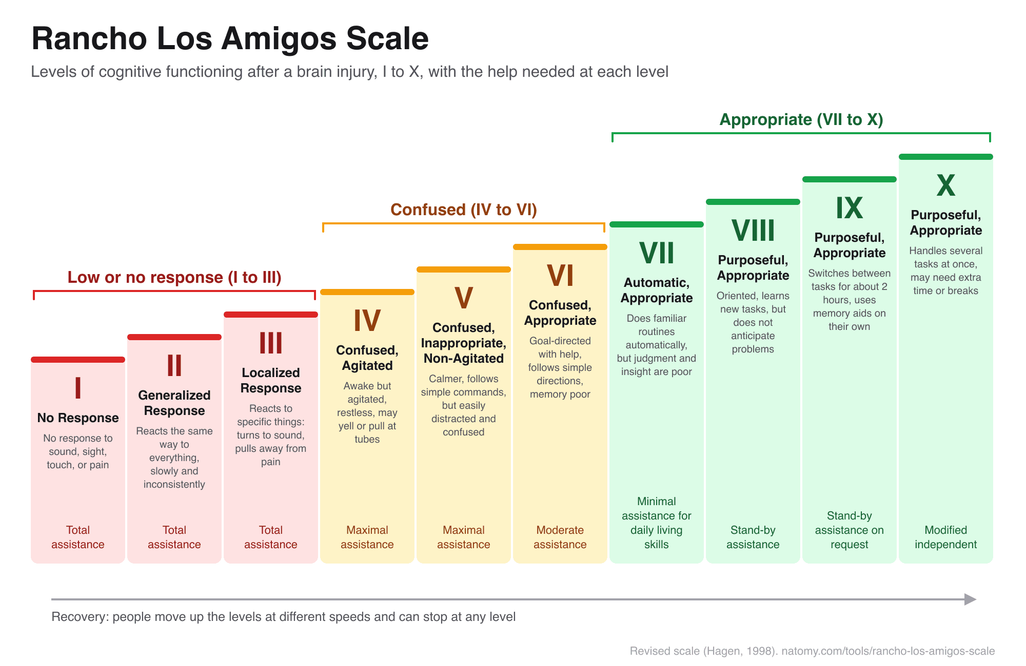
Rancho Los Amigos Scale
The 10 levels of cognitive functioning after a brain injury, with a level finder, what to expect, and how to help.
Try it free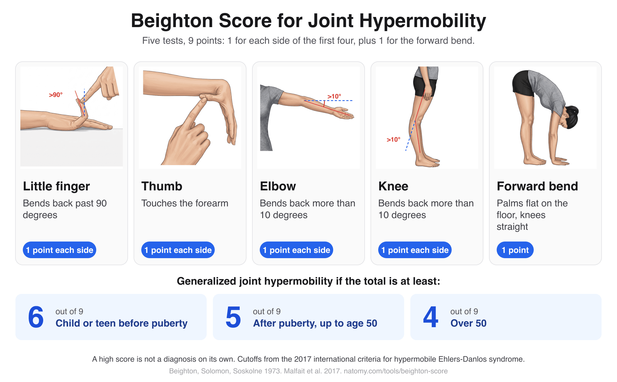
Beighton Score Calculator
Illustrated hypermobility test: score the 5 Beighton movements out of 9, compare with the age cutoff, and copy a note.
Try it free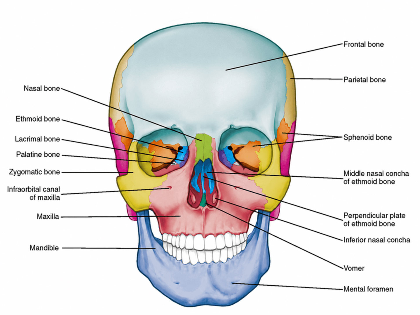
Anterior View of Skull Labeled
Free labeled and unlabeled anterior view of the skull, downloadable as PNG.
Try it free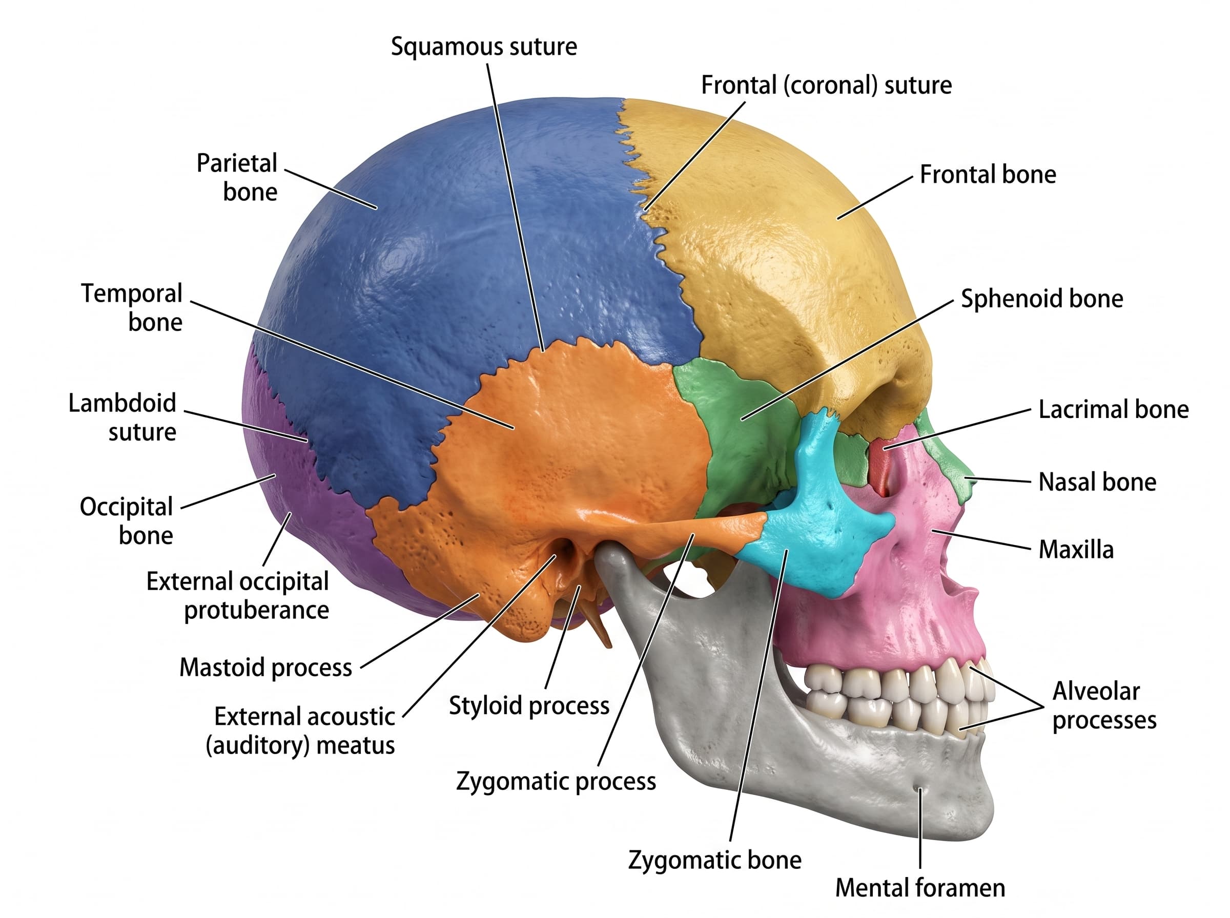
Lateral View of Skull Labeled
Free labeled and unlabeled lateral view of the skull, downloadable as PNG.
Try it free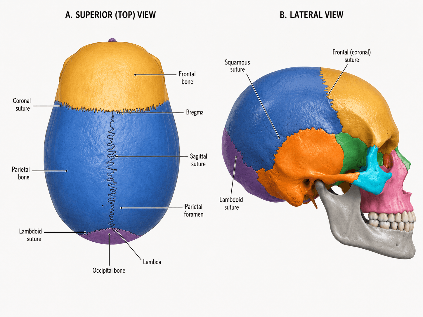
Skull Sutures Labeled
Free labeled and unlabeled skull sutures diagram, superior and lateral views, downloadable as PNG.
Try it free