Free Tools
Myotome Chart: Upper and Lower Extremity Myotomes
Tap a nerve root or a muscle to see the key muscle for that myotome, how it is tested, its nerve, reflex, and disc level. Grade each muscle 0 to 5 on both sides to get the ISNCSCI motor score and motor level, then copy the result into your notes.
 RL
 LR
Tap a muscle or a root. Dashed outlines are deep muscles, covered by others.
Motor score
0 of 20 graded
| Root | Movement | Right | Left |
|---|---|---|---|
| Elbow flexion | |||
| Wrist extension | |||
| Elbow extension | |||
| Finger flexion | |||
| Little finger abduction | |||
| Hip flexion | |||
| Knee extension | |||
| Ankle dorsiflexion | |||
| Big toe extension | |||
| Ankle plantar flexion |
Upper limb (UEMS)
0 / 50
R 0 / L 0
Lower limb (LEMS)
0 / 50
R 0 / L 0
Total motor score
0 / 100
Upper + lower
Motor level, right
Grade all 10 muscles
Motor level, left
Grade all 10 muscles
Myotomes and the motor score in 70 seconds
Myotome chart (printable)
The 10 key muscles from the International Standards for Neurological Classification of Spinal Cord Injury (ISNCSCI), with the movement, reflex, and disc level for each root. Free to download and use in notes, teaching, and patient education.
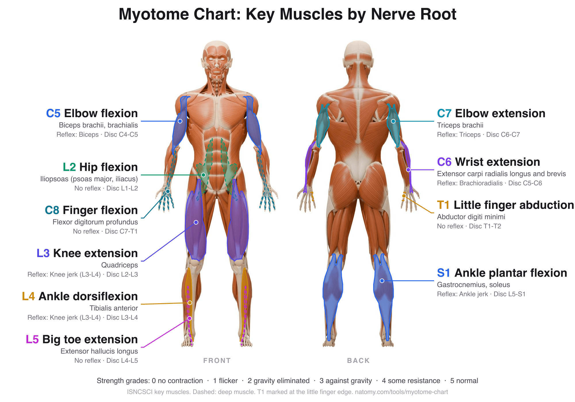
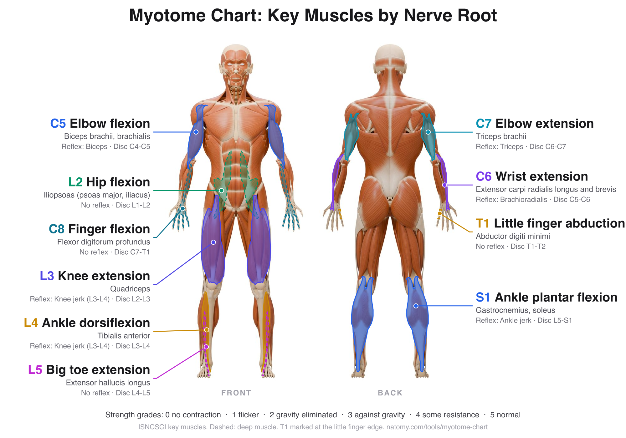
Upper extremity myotomes
The arm is supplied by the C5 to T1 roots through the brachial plexus. Each key muscle is tested in a set position so results can be compared between examiners and over time.
| Root | Movement | Key muscles | Reflex |
|---|---|---|---|
| C5 | Elbow flexion | Biceps brachii, brachialis | Biceps |
| C6 | Wrist extension | Extensor carpi radialis longus and brevis | Brachioradialis |
| C7 | Elbow extension | Triceps brachii | Triceps |
| C8 | Finger flexion | Flexor digitorum profundus (to the middle finger) | None |
| T1 | Little finger abduction | Abductor digiti minimi | None |
Lower extremity myotomes
The leg is supplied by the L2 to S1 roots through the lumbar and sacral plexuses. In lumbar disc herniations, the root below the disc is usually the one affected, so an L4-L5 disc weakens L5.
| Root | Movement | Key muscles | Reflex |
|---|---|---|---|
| L2 | Hip flexion | Iliopsoas (psoas major, iliacus) | None |
| L3 | Knee extension | Quadriceps | Knee jerk (L3-L4) |
| L4 | Ankle dorsiflexion | Tibialis anterior | Knee jerk (L3-L4) |
| L5 | Big toe extension | Extensor hallucis longus | None |
| S1 | Ankle plantar flexion | Gastrocnemius, soleus | Ankle jerk |
Full list of myotomes
Outside the 10 ISNCSCI key muscles, screening exams often test the movements below. Most muscles get fibers from two or three roots, so lists differ a little between textbooks; C5, for example, is tested with elbow flexion in ISNCSCI and with shoulder abduction in many physical therapy screens.
| Root | Movement tested |
|---|---|
| C1-C2 | Neck flexion |
| C3 | Neck side bending |
| C4 | Shoulder shrug (and the diaphragm, C3-C5) |
| C5 | Shoulder abduction (deltoid) and elbow flexion |
| C6 | Elbow flexion and wrist extension |
| C7 | Elbow extension and wrist flexion |
| C8 | Finger flexion, thumb extension |
| T1 | Finger spreading (hand intrinsic muscles) |
| T2-T12 | Intercostal and abdominal muscles |
| L1-L2 | Hip flexion |
| L3 | Knee extension |
| L4 | Ankle dorsiflexion |
| L5 | Big toe extension |
| S1 | Ankle plantar flexion, foot eversion, hip extension |
| S2 | Knee flexion |
| S3-S5 | Anal sphincter, bladder, and bowel (no limb movement) |
Muscle strength grading (0 to 5)
Strength is graded on the Medical Research Council (MRC) scale, also called the Oxford scale, which ISNCSCI uses for the key muscles. Grades 4 and 5 are tested against resistance in the muscle's test position. For plus and minus grades and test positions, see the manual muscle testing grades chart.
| Grade | Meaning |
|---|---|
| 0 | Total paralysis |
| 1 | Contraction can be felt or seen, but no movement |
| 2 | Full range of motion with gravity eliminated |
| 3 | Full range of motion against gravity, no added resistance |
| 4 | Full range against gravity and moderate resistance |
| 5 | Full range against full resistance |
| NT | Not testable, for example because of a cast, pain, or contracture |
Motor score and motor level
Adding the grades of the five arm muscles on both sides gives the upper extremity motor score (UEMS, out of 50), and the five leg muscles give the lower extremity motor score (LEMS, out of 50). The motor level on each side is the lowest key muscle graded at least 3 with all the key muscles above it graded 5. With the sensory exam from the dermatome map, these give the neurological level of injury and the ASIA Impairment Scale (AIS) grade.
Myotomes in radiculopathy and injury cases
For surgeons and physicians, a quick myotome screen shows which root a disc herniation or stenosis is compressing, and serial grades track recovery after surgery. For personal injury and medical malpractice attorneys, the strength grades in the records show whether weakness matches the level on the MRI, such as weak big toe extension with an L4-L5 herniation, and whether it was documented before and after an event.
Illustrate a nerve root injury
Natomy turns MRI views, reports, and prompts into clear, labeled medical illustrations of disc herniations, nerve root compression, and the muscles affected, for patient education and medical-legal exhibits. See examples in the Natomy atlas.
Try Natomy TodayFAQ
What is a myotome?+
A myotome is the group of muscles supplied by a single spinal nerve root. Weakness of the movement a root controls, such as ankle dorsiflexion for L4, points to a problem at that root or its level of the spinal cord. Myotomes are the motor counterpart of dermatomes, the skin areas each root supplies.
What is the difference between a myotome and a dermatome?+
A dermatome is the area of skin a nerve root carries sensation from, and a myotome is the group of muscles the same root moves. A full neurological exam tests both, plus reflexes. Sensory changes from one root are often patchy, so muscle weakness and reflexes help confirm the level.
What are the upper extremity myotomes?+
In the standard spinal cord injury exam (ISNCSCI), the upper extremity myotomes are C5 elbow flexion, C6 wrist extension, C7 elbow extension, C8 flexion of the middle fingertip, and T1 little finger abduction. Many physical therapy screens test C5 with shoulder abduction instead.
What are the lower extremity myotomes?+
The lower extremity myotomes are L2 hip flexion, L3 knee extension, L4 ankle dorsiflexion, L5 big toe extension, and S1 ankle plantar flexion. S2 is often tested with knee flexion. An L4-L5 disc herniation usually weakens big toe extension (L5), and an L5-S1 herniation weakens plantar flexion (S1).
What does 4/5 muscle strength mean?+
On the 0 to 5 scale, 4 out of 5 means the muscle moves the joint through its full range against gravity and against moderate resistance, but not against full resistance. 5/5 is normal strength, 3/5 is movement against gravity only, and 0/5 is no contraction at all. Some examiners add plus and minus grades, such as 4+, to show finer differences.
How is the ISNCSCI motor score calculated?+
Each of the 10 key muscles is graded 0 to 5 on both sides. The five arm muscles add up to the upper extremity motor score (UEMS, 50 maximum for both arms) and the five leg muscles to the lower extremity motor score (LEMS, 50 maximum). The total motor score is out of 100.
What is the motor level?+
The motor level is the lowest key muscle graded 3 or more, provided every key muscle above it is graded 5. It is set for each side. Where there are no key muscles to test, from C1 to C4 and from T2 to L1, the motor level is taken from the sensory level.
Why do lawyers look at muscle strength grades?+
In spine and nerve injury claims, documented strength grades show whether weakness was present, how severe it was, and whether it matches the level of injury on the MRI. A change from 5/5 to 4/5 at L5 after an accident, for example, supports a nerve root injury at L4-L5. Serial grades also show recovery or decline over time.
Sources: key muscles, test positions, and scoring from the American Spinal Injury Association's International Standards for Neurological Classification of Spinal Cord Injury (ISNCSCI, 2019 revision). Strength grades from the Medical Research Council scale (Aids to the Examination of the Peripheral Nervous System). Figures rendered from Alex Lashko's 3D écorché model; the model has no small hand muscles, so T1 (abductor digiti minimi) is marked at the little finger edge of the hand. Muscles get fibers from more than one root and myotomes vary between people. For education and documentation only; not a substitute for clinical assessment.
Related free tools
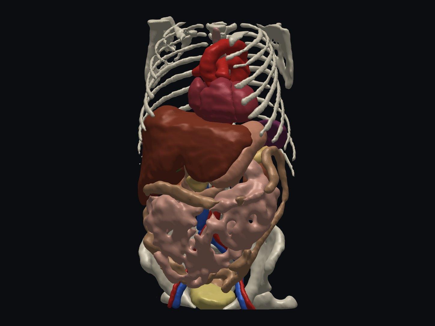
Free Online DICOM Viewer (3D)
Open a CT or MRI DICOM folder in your browser and view it in 3D, with AI anatomy labeling. Nothing is uploaded.
Try it free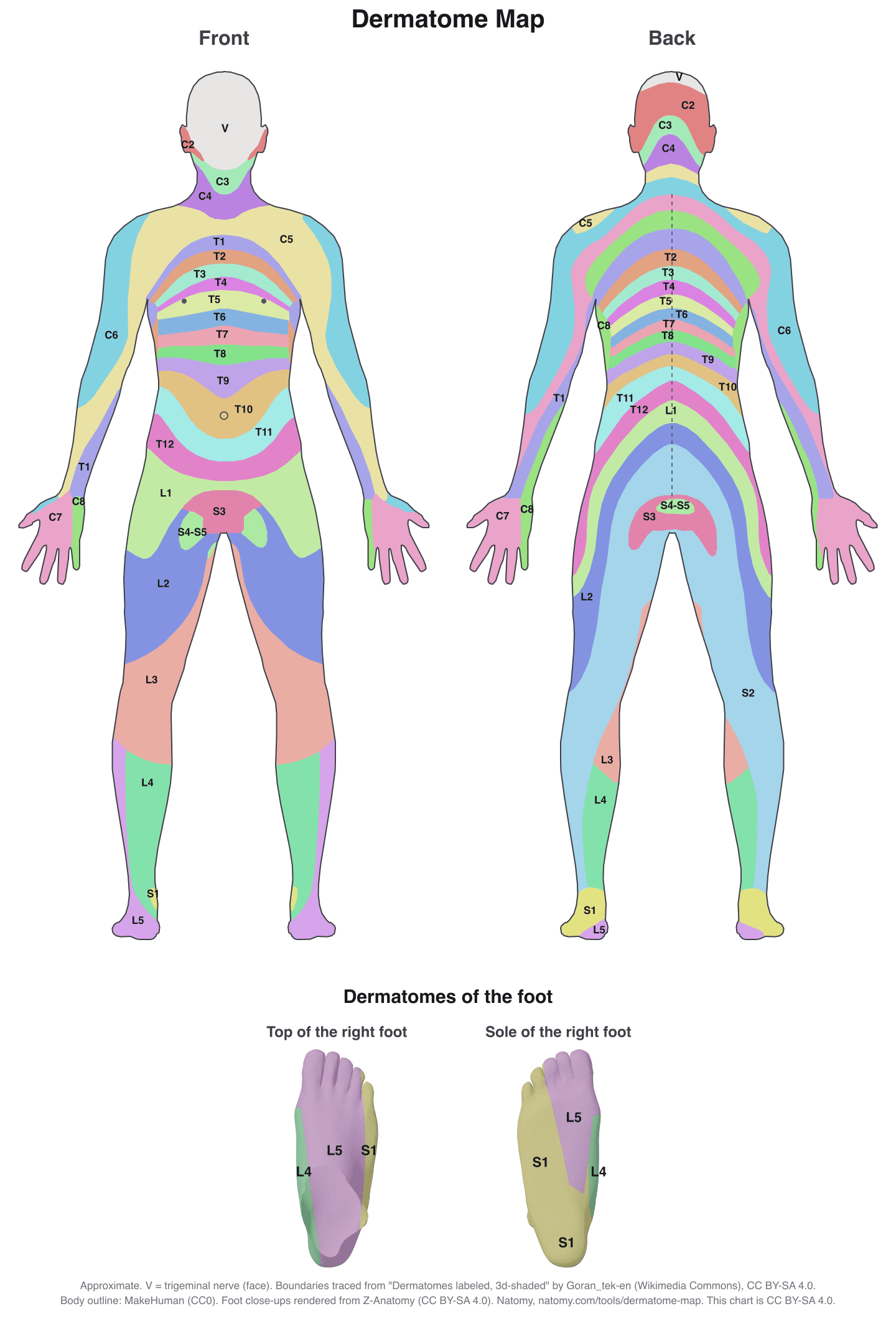
Interactive Dermatome Map
Tap the body to see each nerve root's skin area, test point, myotome, reflex, and disc level. Free printable chart.
Try it free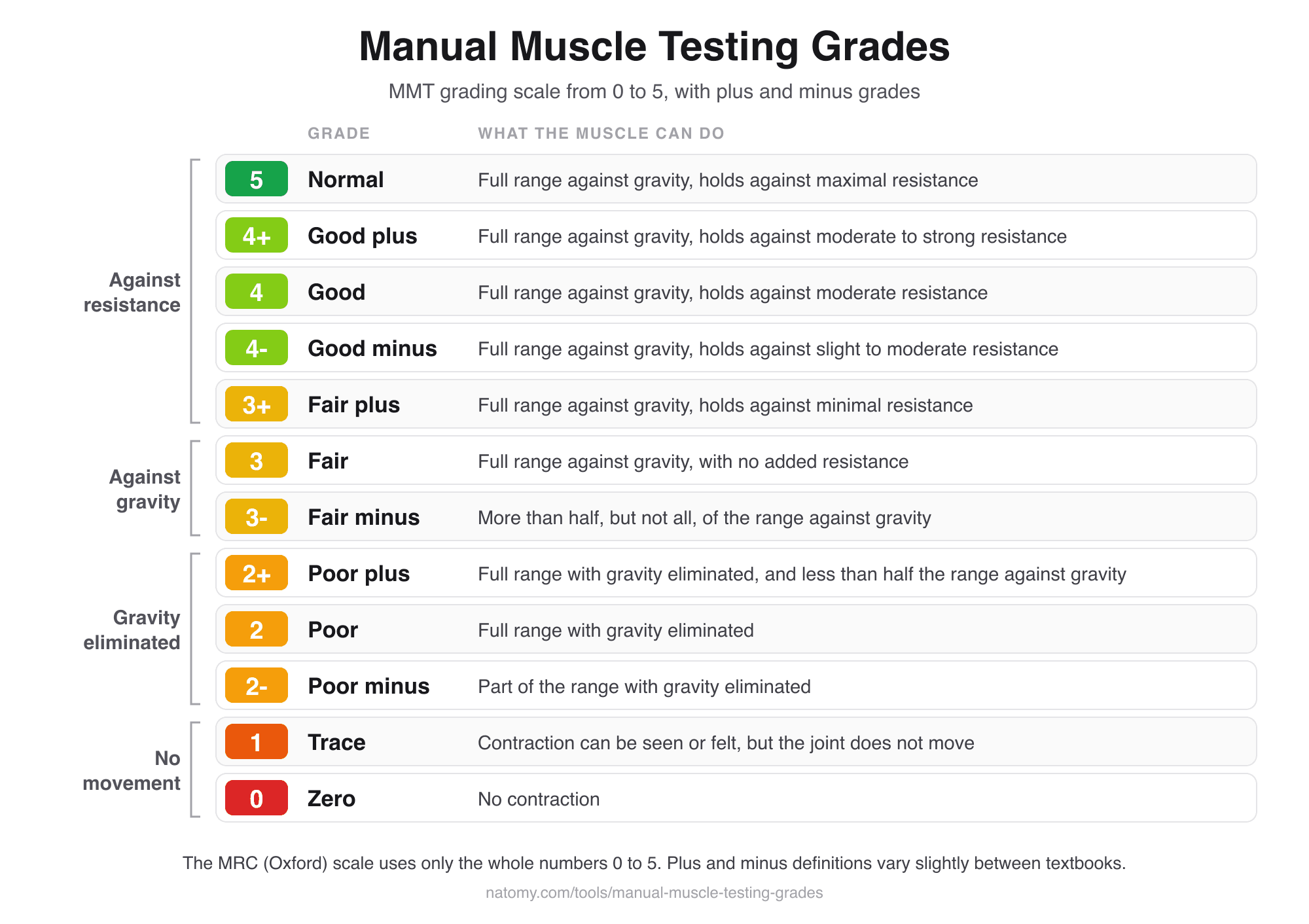
Manual Muscle Testing Grades
MMT grading scale chart from 0 to 5 with plus and minus grades, and a grade finder that writes the note for you.
Try it free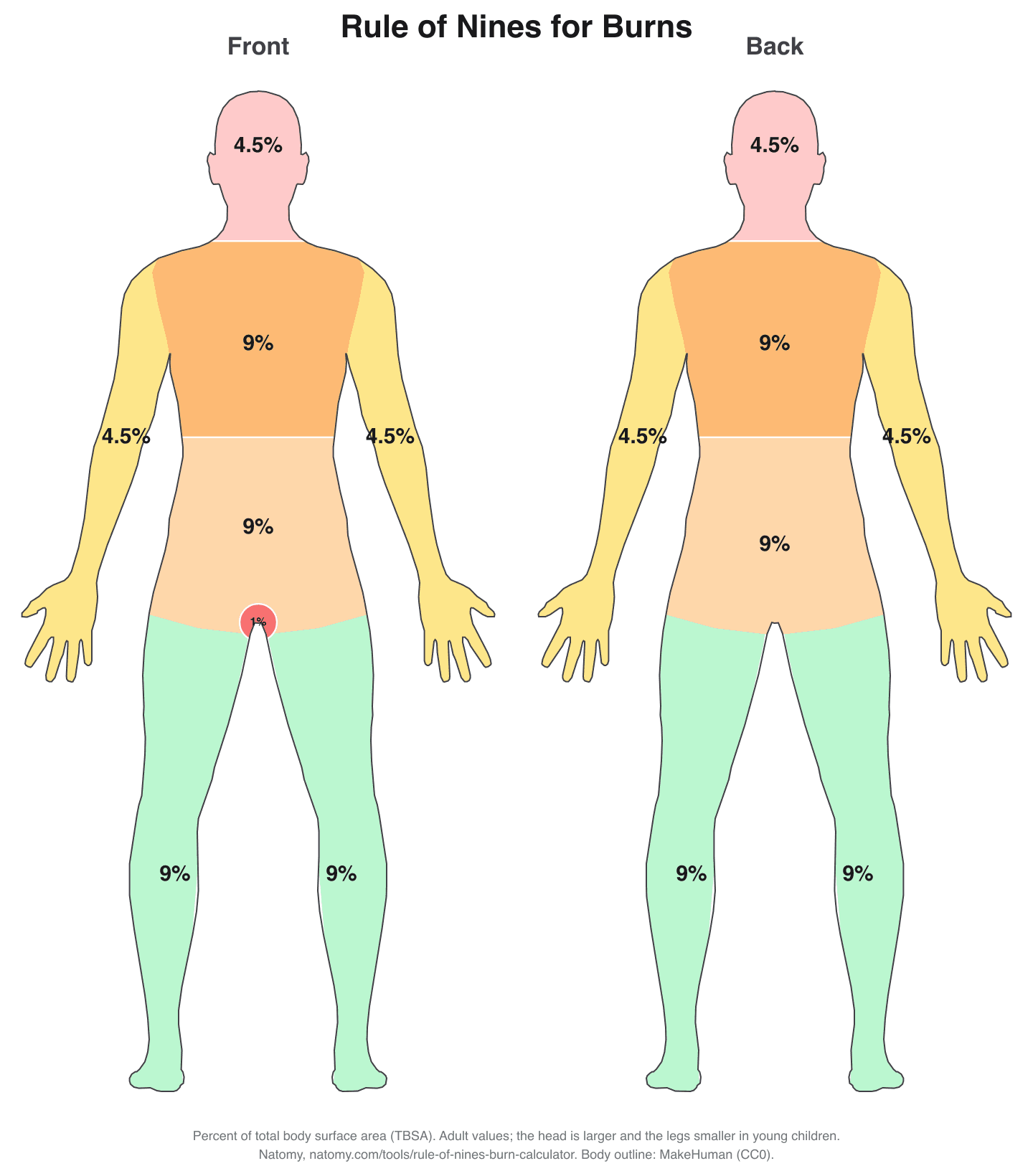
Rule of Nines Burn Calculator
Click the burned areas to get the total body surface area (TBSA), with the Parkland formula and a printable burn chart.
Try it free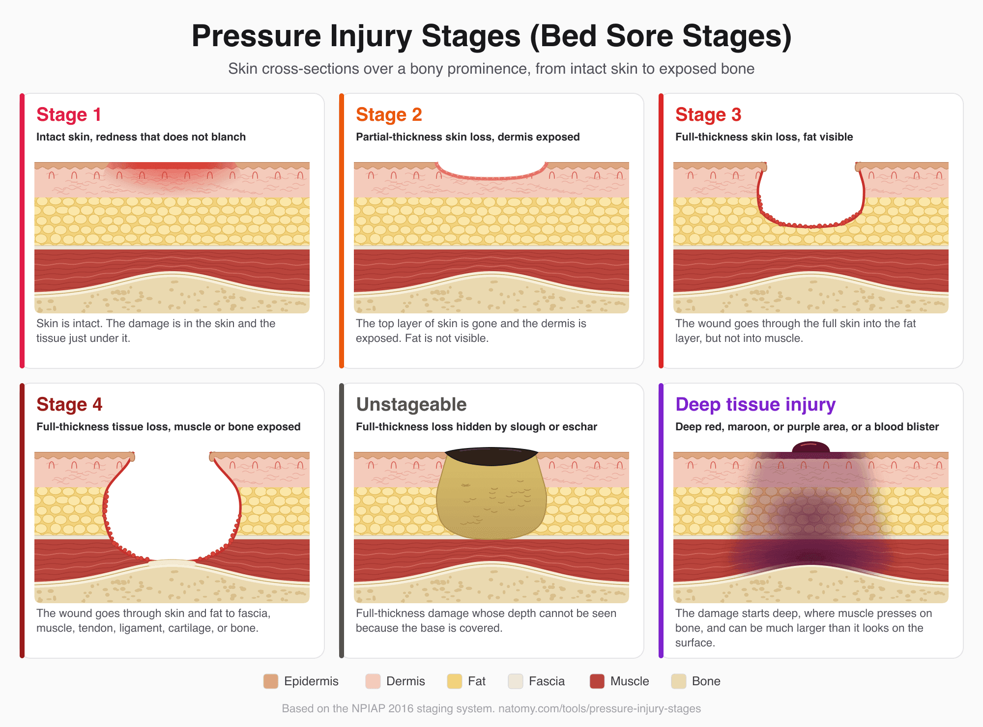
Pressure Injury (Bed Sore) Stages
Illustrated pressure ulcer stages chart, Stage 1 to 4, unstageable, and deep tissue injury, with a stage finder and wound note.
Try it free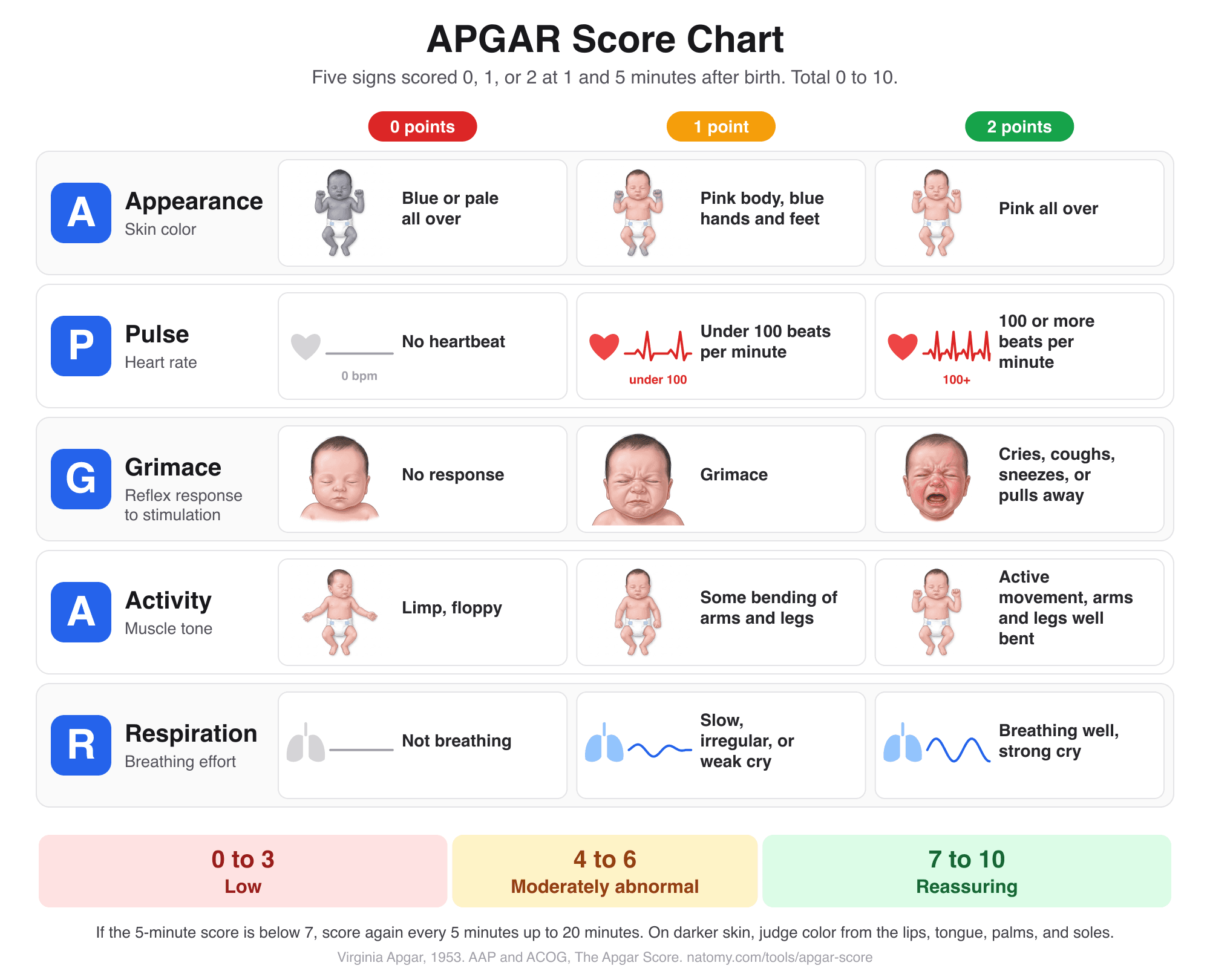
APGAR Score Chart and Calculator
Score the 5 newborn signs at 1, 5, and 10 minutes with an illustrated chart, what normal and low scores mean, and a note for the record.
Try it free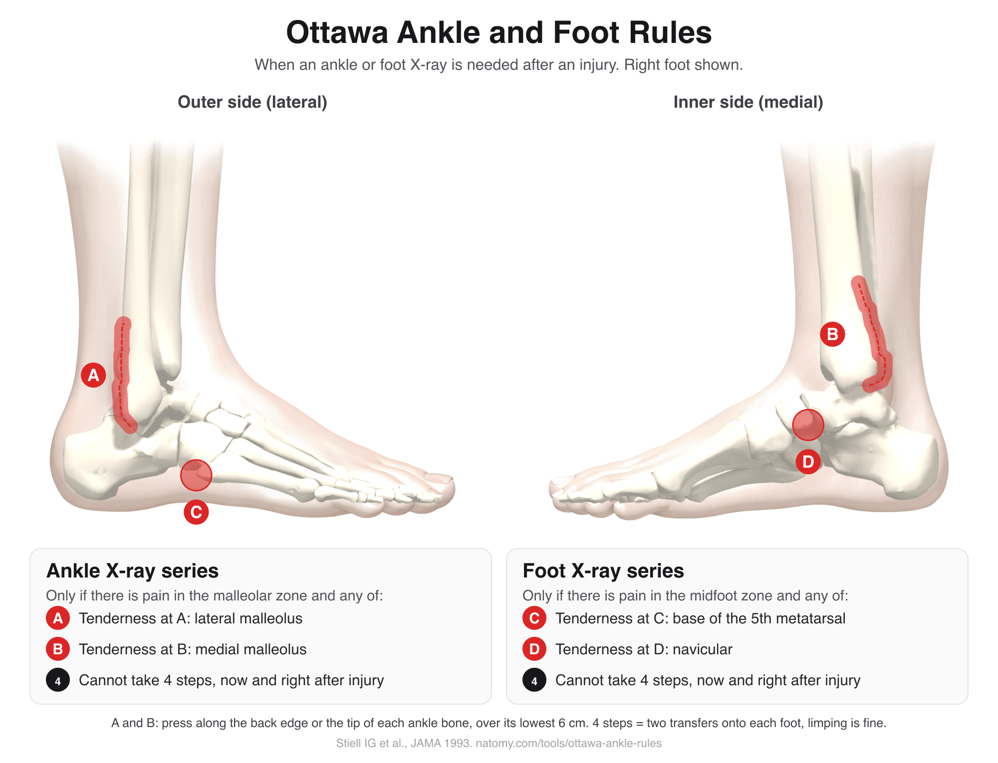
Ottawa Ankle Rules
Tap the tender spots on an illustrated ankle and foot to see if the Ottawa rules call for an X-ray. Includes the Ottawa knee rule.
Try it free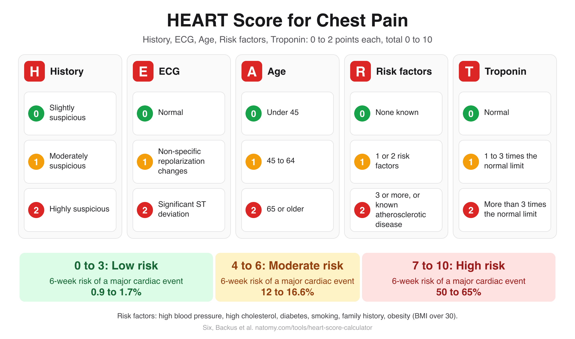
HEART Score Calculator
Score history, ECG, age, risk factors, and troponin for chest pain to get the HEART score, risk group, and 6-week MACE risk.
Try it free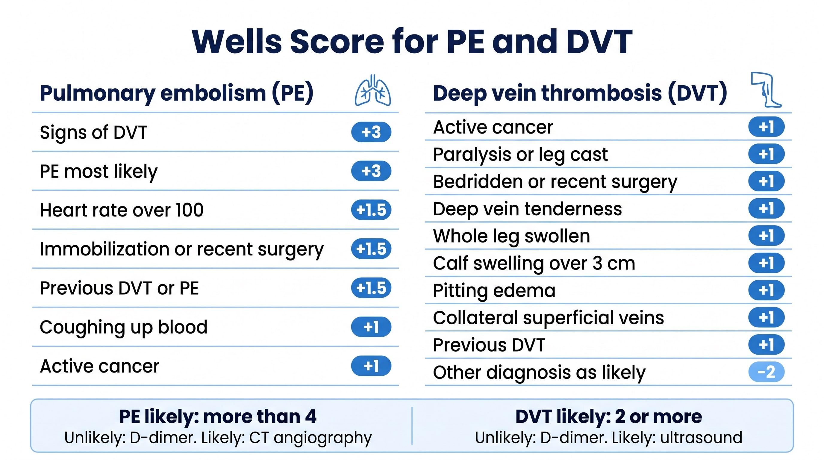
Wells Score Calculator (PE and DVT)
Check the Wells criteria for pulmonary embolism or deep vein thrombosis to get the score, the risk group, and the next test.
Try it free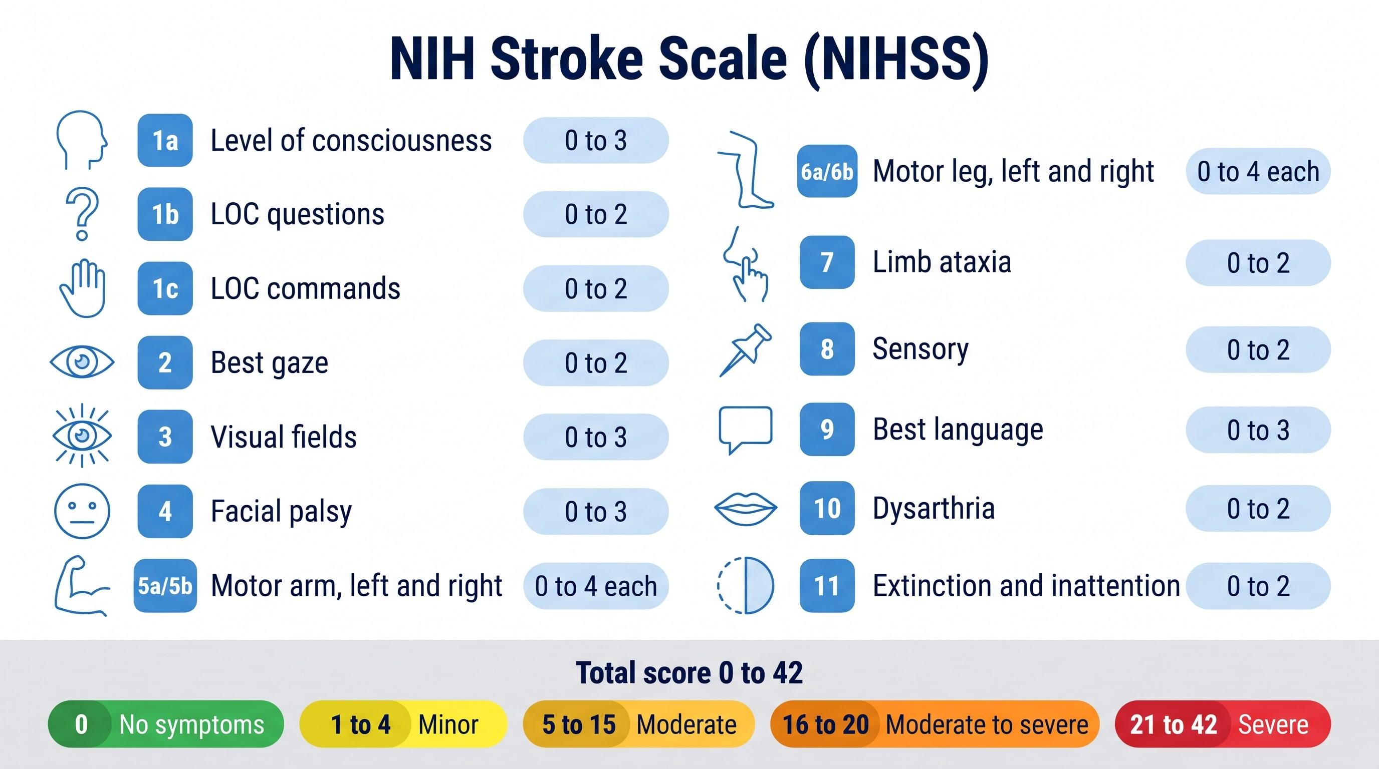
NIHSS Calculator (NIH Stroke Scale)
Score all 15 NIH Stroke Scale items to get the NIHSS total from 0 to 42 and the stroke severity range.
Try it free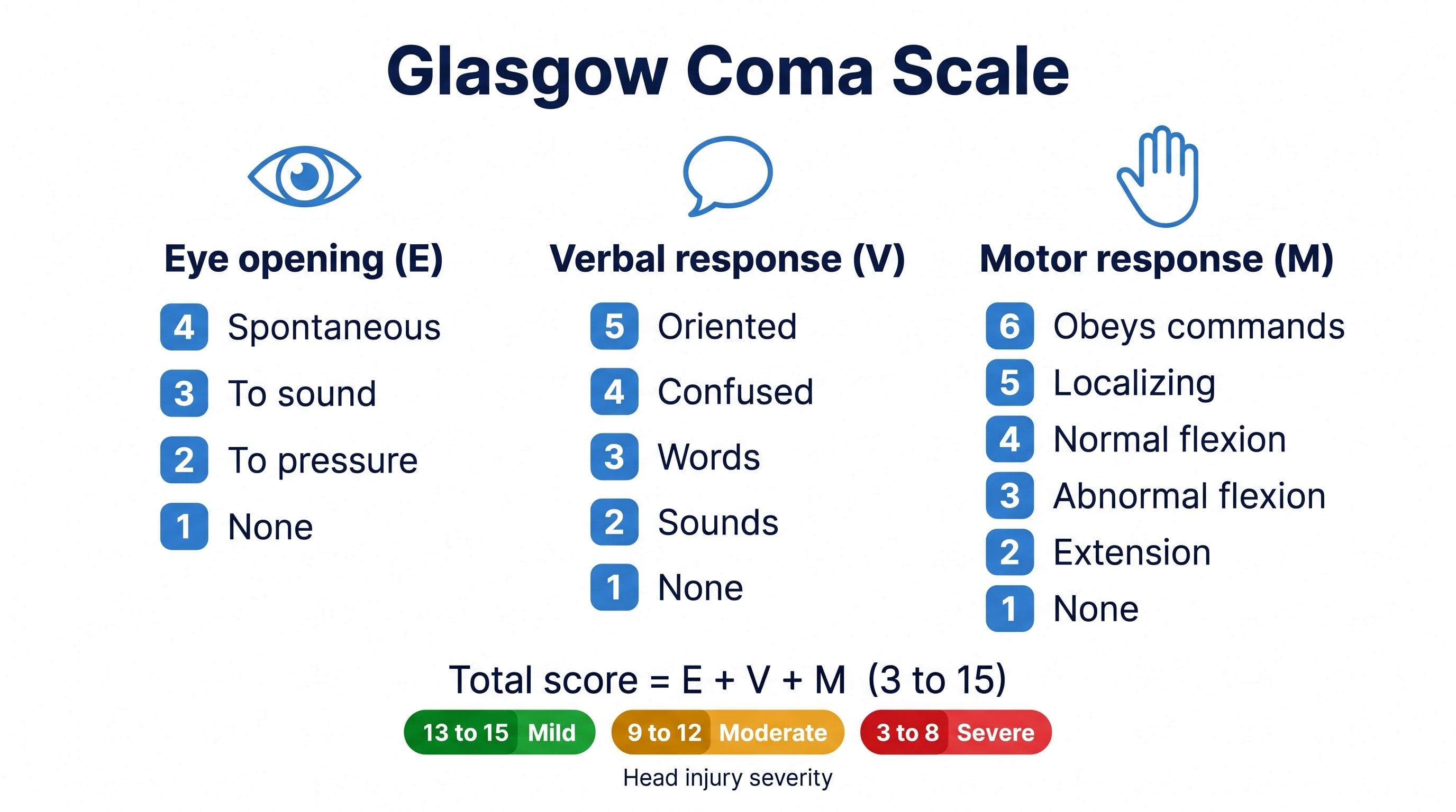
Glasgow Coma Scale Calculator
Score eye, verbal, and motor responses to get the GCS total, E V M code, severity range, and GCS-P.
Try it free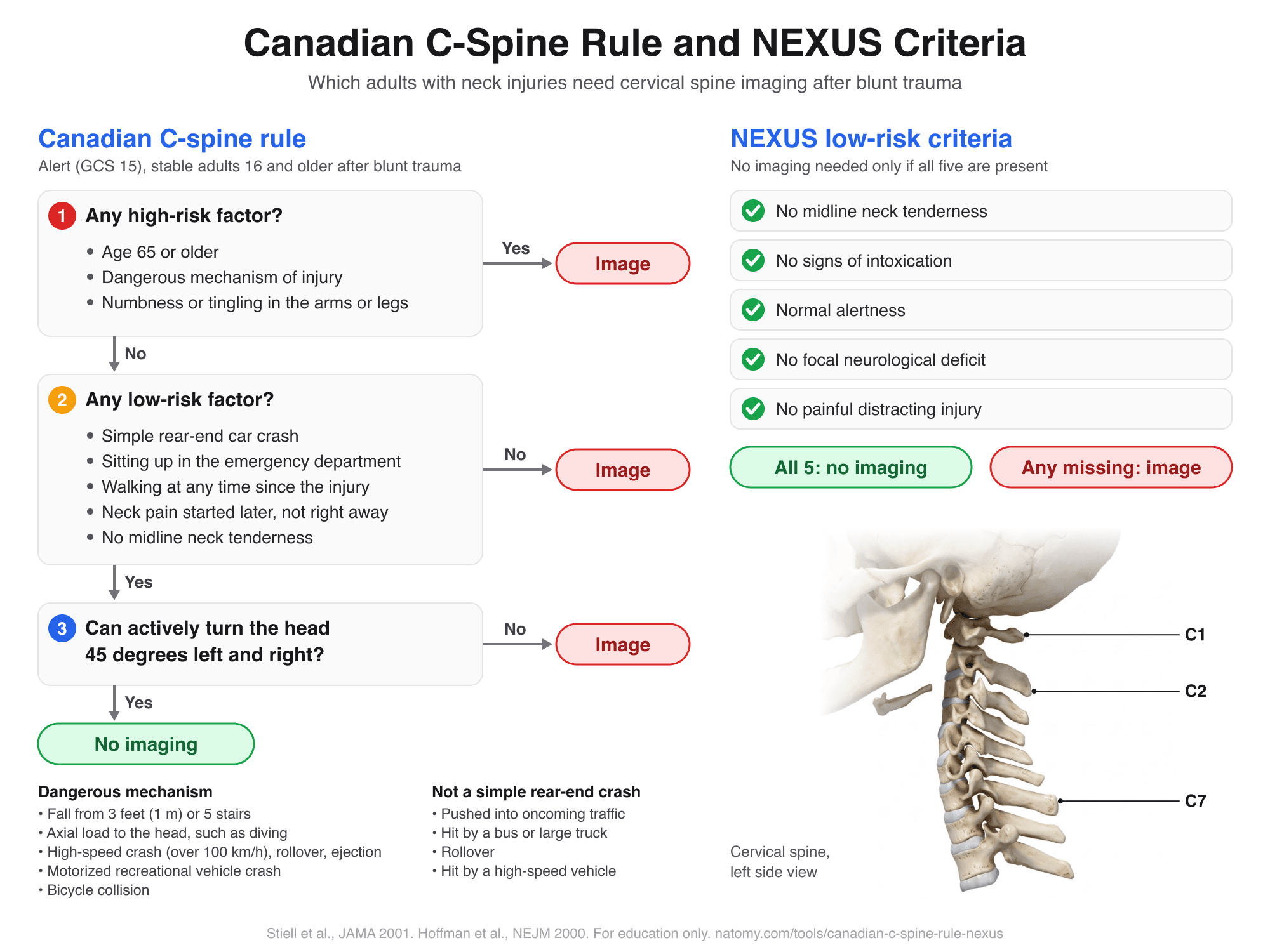
Canadian C-Spine Rule and NEXUS
Run the Canadian C-spine rule or the NEXUS criteria step by step to see which adults need neck imaging after trauma.
Try it free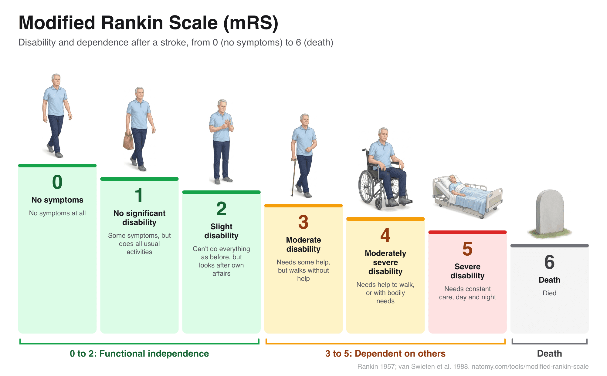
Modified Rankin Scale (mRS)
Illustrated mRS chart from 0 to 6 with a question-based score finder, what each grade means after a stroke, and a copyable note.
Try it free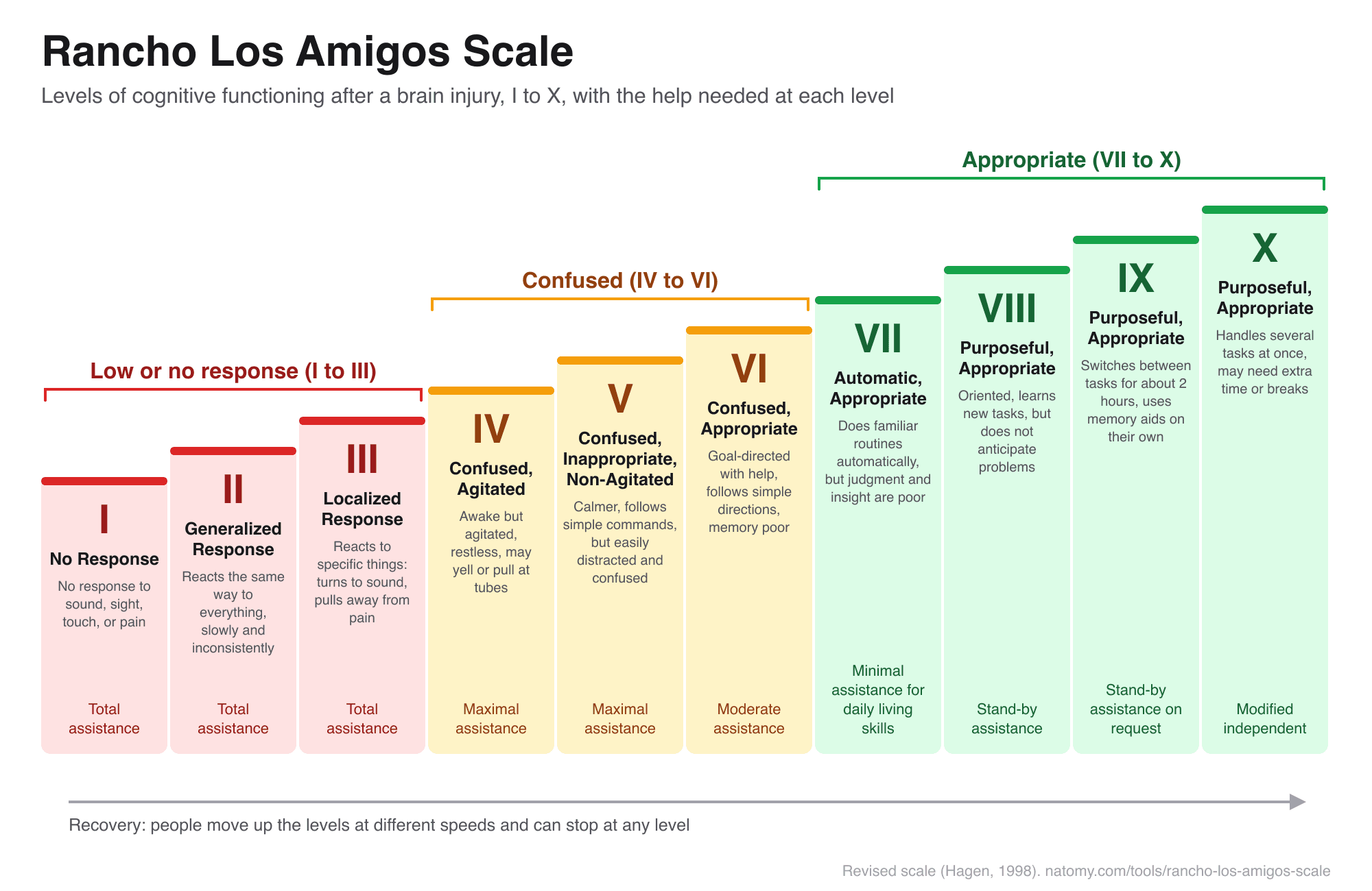
Rancho Los Amigos Scale
The 10 levels of cognitive functioning after a brain injury, with a level finder, what to expect, and how to help.
Try it free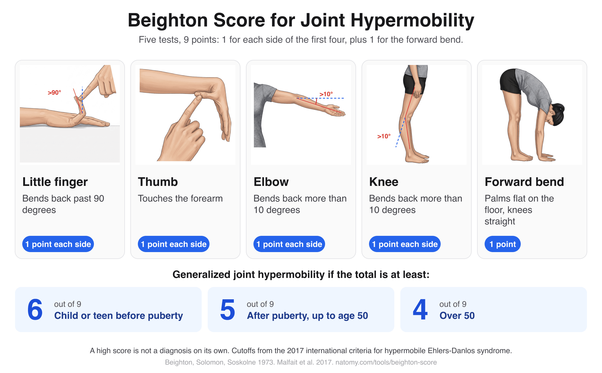
Beighton Score Calculator
Illustrated hypermobility test: score the 5 Beighton movements out of 9, compare with the age cutoff, and copy a note.
Try it free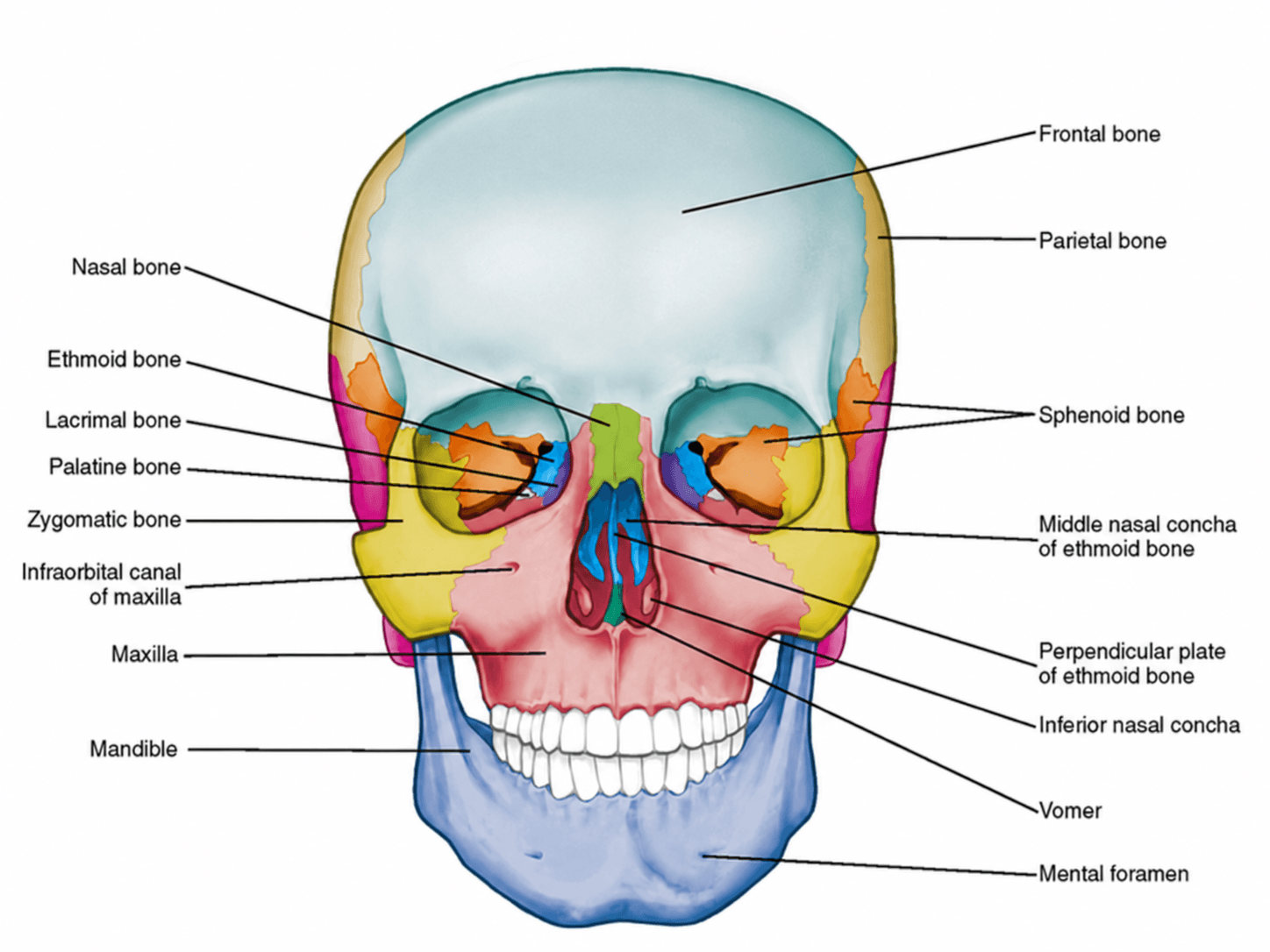
Anterior View of Skull Labeled
Free labeled and unlabeled anterior view of the skull, downloadable as PNG.
Try it free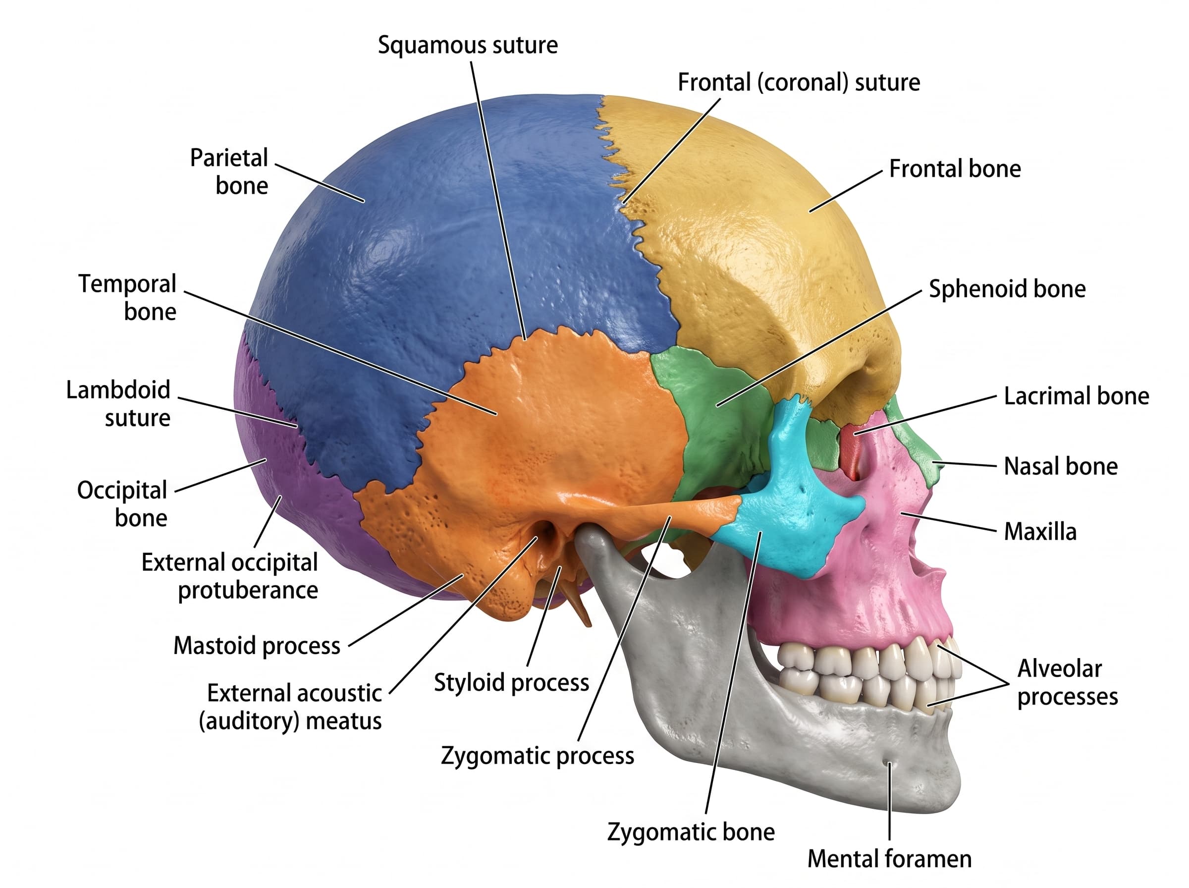
Lateral View of Skull Labeled
Free labeled and unlabeled lateral view of the skull, downloadable as PNG.
Try it free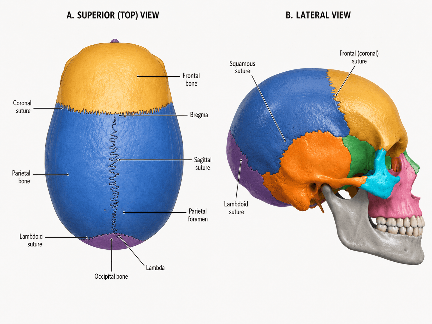
Skull Sutures Labeled
Free labeled and unlabeled skull sutures diagram, superior and lateral views, downloadable as PNG.
Try it free