Fractured Patella: X-ray, Surgery & Recovery
A patella fracture is a break of the kneecap, often straight across from a fall or dashboard injury.
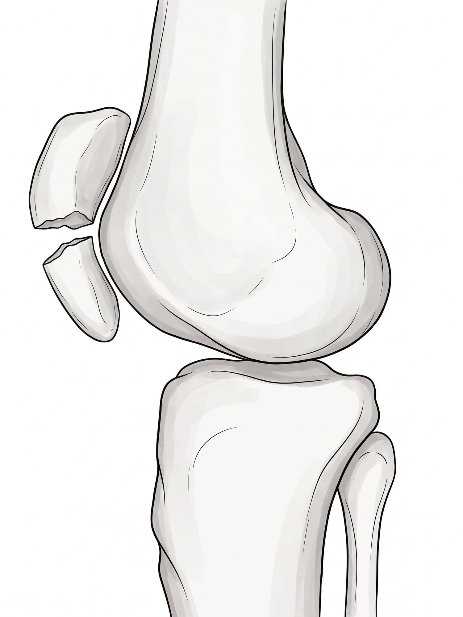
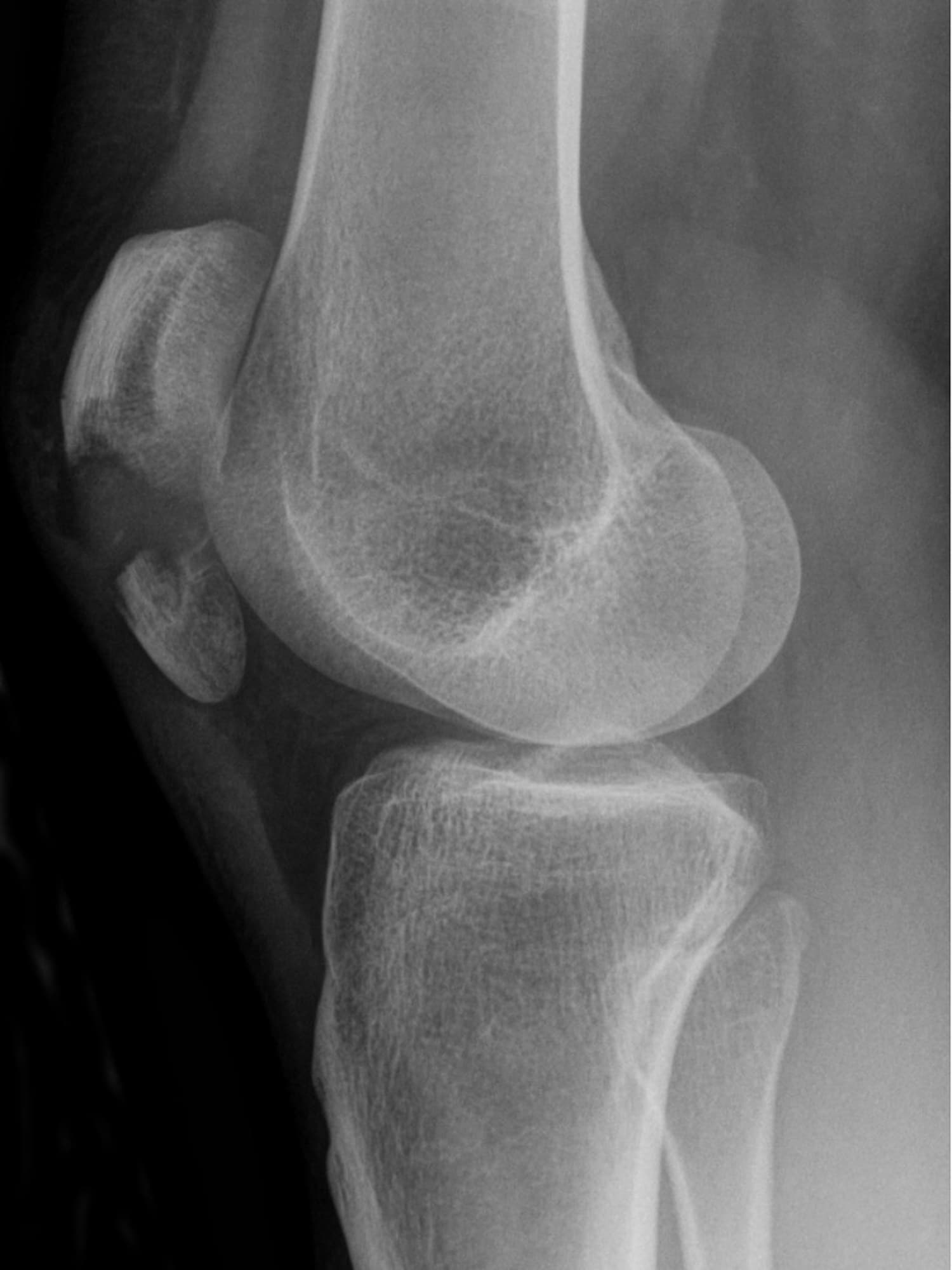
A patella fracture is a break of the kneecap, the flat triangular bone at the front of the knee. Because the kneecap is part of the mechanism that straightens the leg, a displaced patella fracture usually means you cannot straighten your knee. The most common pattern breaks it straight across into an upper and a lower piece. The figure above shows a real side view X-ray of a transverse patella fracture with a medical illustration drawn from it: drag the slider to compare them.
The kneecap and the extensor mechanism
The patella sits inside the tendon of the quadriceps, the large muscle at the front of the thigh. Above it, the quadriceps tendon attaches to its upper edge; below it, the patellar tendon runs from its lower tip to the tibia. Together they form the extensor mechanism, the chain that straightens the knee. The patella acts as a lever that increases the quadriceps' pull. When it breaks and the pieces separate, the chain is interrupted. For the full anatomy of the knee, see knee anatomy.
Types of patella fracture
- Transverse: straight across, into upper and lower pieces, like the one above. The most common type.
- Comminuted (stellate): broken into several pieces, usually from a direct blow. See comminuted fracture.
- Vertical: a split running up and down, often stable.
- Pole (sleeve) fractures: a piece of the upper or lower tip pulls off with the tendon.
- Osteochondral: a flake of bone and cartilage, often from a kneecap dislocation.
Each can be non-displaced (the pieces stay together) or displaced (the pieces separate, usually pulled apart by the quadriceps).
Patella fracture on X-ray
The side (lateral) view is the most useful. In the X-ray above, the kneecap is split into an upper fragment and a lower fragment with a clear gap between them, while the femur and tibia are intact. A front (AP) view and sometimes a skyline view (looking down the length of the thigh with the knee bent) are added.
Two measurements guide treatment:
| Measurement | Usually treated without surgery | Usually treated with surgery |
|---|---|---|
| Gap between the fragments | Less than about 3 mm | More than about 3 mm |
| Step in the joint surface | Less than about 2 mm | More than about 2 mm |
The most important test is clinical: can the patient lift the straightened leg? If not, the extensor mechanism is broken.
A bipartite patella, a normal variant in which the kneecap develops in two pieces, can look like a fracture on X-ray. It has smooth, rounded edges and is usually in the upper outer corner.
How a patella fracture happens
- Direct blow: a fall onto the knee, or the knee hitting the dashboard in a car crash. These often cause comminuted fractures.
- Indirect force: a sudden, forceful contraction of the quadriceps while the knee is bent, as when stumbling and trying to catch yourself. This tends to cause transverse fractures that pull apart.
Symptoms
- Pain and rapid swelling at the front of the knee
- Bruising over the kneecap
- Inability to straighten the knee or perform a straight leg raise
- A palpable gap in the kneecap when the fragments separate
- Difficulty walking
Treatment
- Brace locked straight: for non-displaced fractures with an intact extensor mechanism. Weight bearing is often allowed with the knee straight, and bending is increased gradually over about 6 weeks.
- Tension band wiring: the classic surgery for transverse fractures. Two pins or screws hold the pieces, and a figure-of-eight wire converts the quadriceps pull into compression across the fracture when the knee bends.
- Screws or plates: newer low-profile plates are used for comminuted fractures.
- Partial patellectomy: removing small, unrepairable fragments and reattaching the tendon to the remaining bone.
Recovery time
The bone heals in about 6 to 8 weeks. The brace is usually unlocked in stages to allow more bending, with physical therapy focused on quadriceps strength and range of motion. Most people return to everyday activity in 3 to 4 months, but kneeling, squatting and stairs can remain uncomfortable for 6 to 12 months.
Complications
- Knee stiffness and loss of bending
- Painful hardware: tension band wires sit just under the skin and are commonly removed once the bone heals
- Post-traumatic arthritis behind the kneecap (patellofemoral arthritis)
- Quadriceps weakness that can persist for months
- Loss of fixation or nonunion, more common in older patients
How a patella fracture is shown in a personal injury case
The dashboard knee injury is a classic of motor vehicle cases, and patella fractures also follow falls on stairs and slippery floors. A side view X-ray shows the break, but jurors do not see why a broken kneecap stops someone from straightening their leg. An illustration drawn from the plaintiff's X-ray, like the one above, shows the separated fragments and the tendons attached to each, which explains the loss of function, the tension band surgery and the months of therapy. For how these images are used at trial, see demonstrative evidence and medical legal illustration.
Related pages
FAQ
What does a fractured patella feel like?+
Sudden, sharp pain at the front of the knee, with swelling that comes on quickly as blood fills the joint. Bending the knee hurts, and with a displaced fracture you usually cannot straighten the leg or lift it straight off the bed. Sometimes a gap can be felt between the two pieces of the kneecap.
Can you walk with a fractured patella?+
With a non-displaced fracture and the knee held straight in a brace, many people can walk. With a displaced fracture the quadriceps can no longer straighten the knee, so walking normally is not possible until the kneecap is repaired.
How long does it take a patella fracture to heal?+
The bone usually heals in about 6 to 8 weeks. Getting back full knee bending and quadriceps strength takes longer: most people return to normal daily activities in 3 to 4 months, and full recovery, including stairs, kneeling and sport, can take 6 to 12 months.
Does a broken kneecap need surgery?+
Not always. A non-displaced fracture with an intact extensor mechanism, meaning the patient can still lift the straight leg, is usually treated in a knee brace locked straight. Surgery is recommended when the pieces are separated by more than about 3 mm, the joint surface has a step of more than about 2 mm, or the patient cannot straighten the knee.
When can I climb stairs after patella fracture surgery?+
Most people climb stairs one step at a time, leading with the good leg going up and the operated leg going down, from the first days after surgery with a brace and crutches. Climbing normally, one foot per step, usually returns once the knee bends past about 90 degrees and the quadriceps are strong, often around 3 months.
Do I need a brace for a fractured patella?+
Usually yes. A hinged knee brace locked straight protects the repair or the fracture for the first weeks, then is gradually unlocked to allow more bending as the bone heals, typically over 6 weeks.
What is the difference between a patella fracture and a dislocated kneecap?+
In a patella fracture the bone itself breaks. In a dislocation the intact kneecap slips out of its groove on the femur, usually to the outside. A dislocation can chip a small piece of bone off the patella, so both can show on the same X-ray.
Need an illustration of a patient's own X-ray?
Upload an X-ray, CT or MRI and turn it into a clear, labeled medical illustration for a consult, a chart or a case exhibit.
Try Natomy →