Tibial Plateau Fracture: X-ray, Surgery & Recovery
A tibial plateau fracture is a break into the top of the tibia at the knee joint. See one on X-ray beside a labeled illustration.
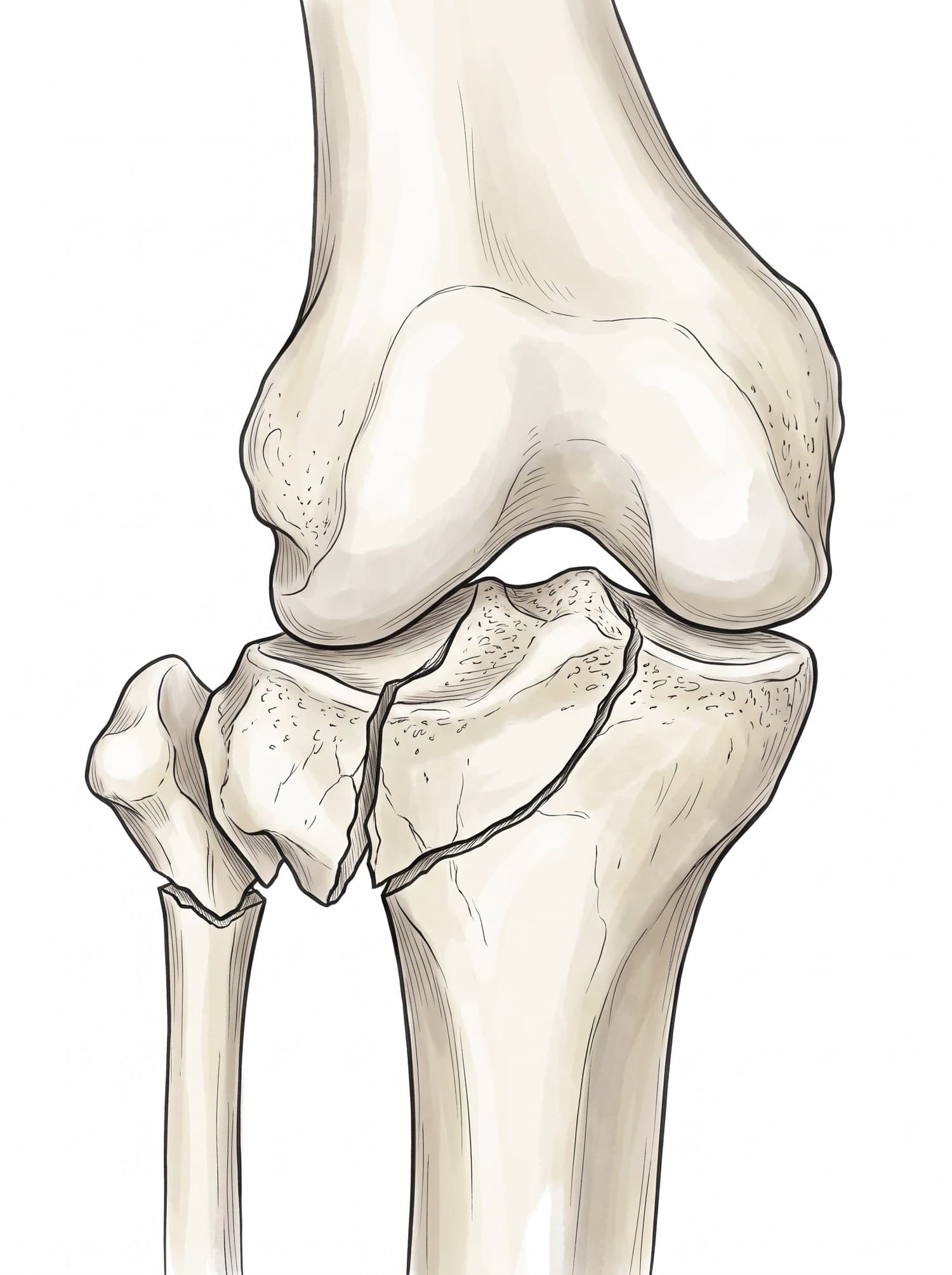
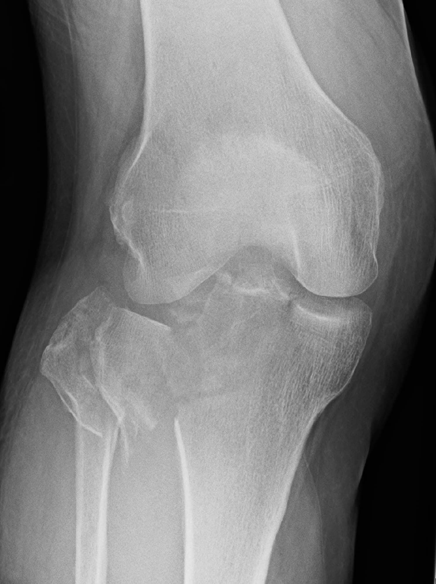
A tibial plateau fracture is a break into the top of the tibia, the flat surface that forms the lower half of the knee joint. Because the fracture enters the joint, treatment focuses on restoring a smooth cartilage surface to protect the knee from arthritis. The figure above shows a real X-ray of a severe tibial plateau fracture with a medical illustration drawn from it: drag the slider to compare them.
The tibial plateau
The top of the tibia widens into two shallow, cartilage-covered surfaces, the lateral plateau (outer side) and the medial plateau (inner side), separated by a bony ridge where the cruciate ligaments attach. The rounded ends of the femur rest on these surfaces, cushioned by the menisci. The fibula sits just below and outside the lateral plateau. For the full anatomy of the knee, see knee anatomy.
Types of tibial plateau fracture
Surgeons use the Schatzker classification:
| Type | Pattern | Typical cause |
|---|---|---|
| I | Split of the lateral plateau | Younger adults, strong bone |
| II | Split with depression of the lateral plateau | The most common type |
| III | Pure depression of the lateral plateau | Older adults with osteoporosis |
| IV | Medial plateau fracture | Higher energy; often with ligament injury |
| V | Both plateaus (bicondylar) | High energy |
| VI | Plateau separated from the shaft | High energy, often with soft tissue damage |
Lateral plateau fractures are far more common, because most blows come from the outside of the knee.
Tibial plateau fracture on X-ray
Front (AP) and side views of the knee are taken, and CT is almost always added to measure how far the joint surface has sunk and to plan surgery. On the X-ray above, the lateral plateau is split and pushed down in several pieces, the fracture runs into the knee joint, and the fibular neck just below the fibular head is broken too. This is a comminuted split-depression fracture.
| Finding | Why it matters |
|---|---|
| Depression of the joint surface | More than a few millimeters usually means surgery |
| Widening of the plateau | Leaves the femur unsupported and the knee unstable |
| One or both plateaus | Bicondylar fractures are more serious |
| Fibular neck fracture | Signals a higher-energy injury; the peroneal nerve runs here |
| Fat-fluid level in the joint (on a side view) | Marrow fat in the joint, a sign of a fracture into it |
How a tibial plateau fracture happens
The classic mechanism is a blow to the outside of the knee that drives the lateral femoral condyle into the lateral plateau, as when a car bumper strikes a pedestrian's knee. Falls from a height, motorcycle crashes and skiing injuries cause the same fracture. In older adults with osteoporosis, a fall from standing can depress the plateau.
Symptoms
- Pain and rapid swelling of the knee
- Inability to bear weight
- Bruising around the knee and down the leg
- A knee that feels unstable or gives way
- Numbness or weakness lifting the foot, if the peroneal nerve is injured
Treatment
- Hinged brace with protected weight bearing: for non-displaced, stable fractures.
- ORIF: elevating the depressed joint surface, supporting it with bone graft or substitute, and fixing it with plates and screws.
- External fixation first: for high-energy fractures with severe swelling, before definitive surgery.
- Arthroscopy-assisted fixation: in selected fractures, to check the joint surface and menisci.
- Rehabilitation: early knee motion, then gradual weight bearing over about 6 to 12 weeks.
Recovery time
Bone healing takes about 3 months. Most people walk without crutches by around 3 months and return to everyday activities in 4 to 6 months, but improvement in strength and motion can continue for 1 to 2 years. Higher-energy fractures and those with joint surface damage take longer.
Complications
- Post-traumatic arthritis, the main long-term risk when the joint surface heals unevenly
- Knee stiffness
- Meniscus and ligament injuries, which often accompany the fracture
- Compartment syndrome after high-energy fractures, a surgical emergency
- Wound problems and infection after surgery
- Peroneal nerve injury, causing foot drop
How a tibial plateau fracture is shown in a personal injury case
Tibial plateau fractures are among the most expensive lower-limb injuries in personal injury cases: pedestrian bumper strikes, motorcycle crashes and falls from scaffolding and ladders, often followed by surgery, months of limited walking and a risk of arthritis and later knee replacement. An illustration drawn from the patient's own imaging, like the one above, shows the depressed joint surface and why it had to be lifted and fixed, which supports claims for future medical care. For how these images are used at trial, see demonstrative evidence and medical legal illustration.
Related pages
FAQ
How long is recovery from a tibial plateau fracture?+
Most people are kept off the leg or on very limited weight for about 6 to 12 weeks while the joint surface heals, then gradually return to walking with physical therapy. Getting back to normal daily activities usually takes 4 to 6 months, and recovery of strength, motion and comfort often continues for a year or more.
What is life like 2 years after a tibial plateau fracture?+
Many people return to most of their activities, but stiffness, aching with weather or long walks, and some loss of high-impact activity are common. Fractures that damaged the joint surface, or that healed with a step in the cartilage, can lead to post-traumatic arthritis, which may eventually need a knee replacement.
Does a tibial plateau fracture need surgery?+
Not always. A fracture that is not displaced and leaves the joint surface smooth and stable can be treated in a hinged brace with protected weight bearing. Surgery (ORIF with plates and screws, sometimes with bone graft) is recommended when the joint surface is depressed or stepped, the knee is unstable, or both plateaus are broken.
What is ORIF for a tibial plateau fracture?+
Open reduction and internal fixation: the surgeon lifts the depressed joint surface back into place, often packs bone graft or bone substitute beneath it, and holds the pieces with a plate and screws. Severe fractures with badly swollen skin are sometimes held first with an external frame until the swelling settles.
Can you walk with a tibial plateau fracture?+
Usually not normally. Most people cannot bear weight comfortably, and walking on a displaced fracture can push the joint surface down further. After treatment, weight bearing is restricted for weeks and increased step by step under the surgeon's guidance.
What is a bicondylar tibial plateau fracture?+
A fracture that breaks both the inner (medial) and outer (lateral) plateaus, usually from a high-energy injury. In the Schatzker classification these are types V and VI. They are more often unstable and almost always need surgery.
What causes a tibial plateau fracture?+
A force that drives the femur down into the top of the tibia, often with the knee bent sideways: a car bumper striking the outside of the knee, a fall from a height, or a sports or skiing injury. In older adults with osteoporosis, a simple fall can be enough.
Need an illustration of a patient's own X-ray?
Upload an X-ray, CT or MRI and turn it into a clear, labeled medical illustration for a consult, a chart or a case exhibit.
Try Natomy →