Ankle Fracture: Types, X-ray & Recovery Time
An ankle fracture is a break of the fibula, tibia or both at the ankle joint. See a lateral malleolus fracture on X-ray beside a labeled illustration.
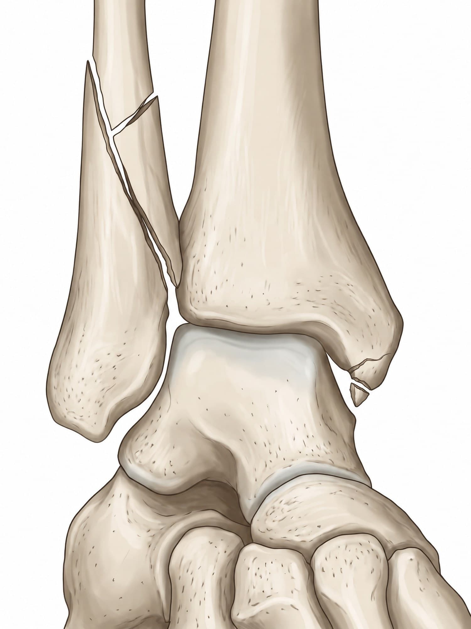
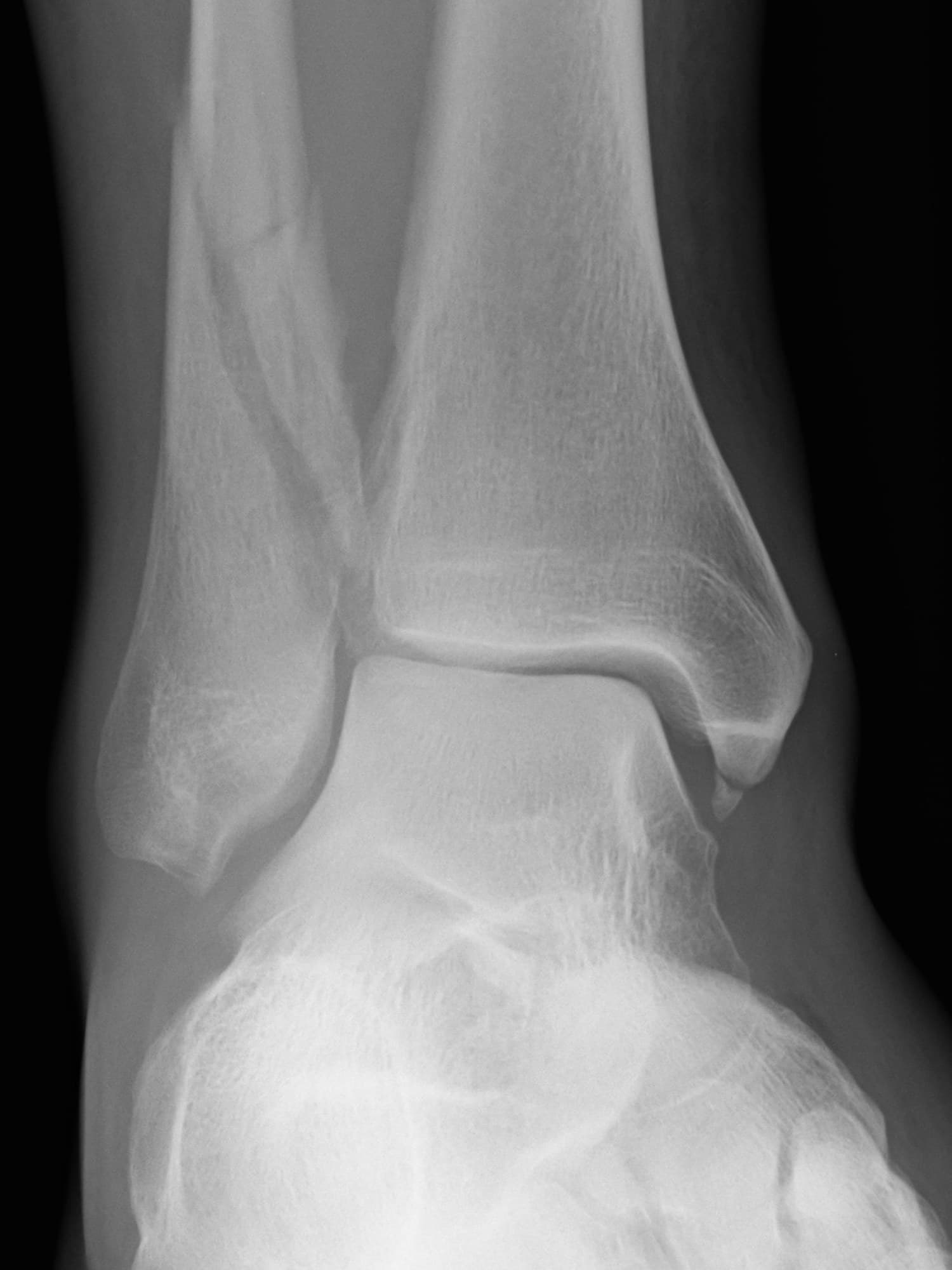
An ankle fracture is a break of one or more of the bones that form the ankle joint: the lower end of the fibula (the lateral malleolus), the lower end of the tibia (the medial malleolus and the back edge, the posterior malleolus), or a combination. It is one of the most common fractures in adults, usually from a twisting fall. The figure above shows a real X-ray of a right ankle fracture with a medical illustration drawn from it: drag the slider to compare them.
The bones of the ankle joint
The ankle joint is a hinge between three bones. The tibia and fibula form a bracket, called the mortise, that holds the top of the talus. The bony bumps on each side of the ankle are the ends of those two leg bones:
- Lateral malleolus: the outer bump, the lower end of the fibula.
- Medial malleolus: the inner bump, part of the tibia.
- Posterior malleolus: the back edge of the tibia's joint surface.
The tibia and fibula are tied together just above the joint by the syndesmosis, a set of strong ligaments. For the full anatomy, including the ligaments and tendons, see ankle anatomy.
Types of ankle fracture
Ankle fractures are named by how many malleoli are broken:
- Lateral malleolus fracture: the fibula alone, the most common type. If the joint is stable, it is often treated in a boot.
- Medial malleolus fracture: the tibia alone, less common on its own.
- Bimalleolar fracture: both the lateral and medial malleoli. The ring of bone and ligament around the talus is broken in two places, so the joint is unstable.
- Trimalleolar fracture: both malleoli plus the posterior malleolus. Usually from a severe twisting injury, and almost always treated with surgery.
Fibula fractures are also sorted by the Weber classification, by where the break sits compared with the syndesmosis: Weber A below it (usually stable), Weber B at its level (sometimes unstable), and Weber C above it (the syndesmosis is usually torn and the ankle is unstable).
Ankle fracture on X-ray
Three standard views are taken: front (AP), mortise (rotated slightly inward so the joint space shows evenly) and side (lateral). The X-ray above is an AP view of a right ankle. A long oblique (spiral) fracture runs down the fibula from a few centimeters above the joint to about the level of the syndesmosis, a Weber B to C pattern, with a small butterfly fragment broken off near its upper end. A small flake of bone has also pulled off the tip of the medial malleolus, where the deltoid ligament attaches.
What a radiologist checks on the X-ray:
| Finding | What it means |
|---|---|
| Height of the fibula fracture | Weber A, B or C, and how likely the syndesmosis is injured |
| Medial clear space (gap between the talus and medial malleolus) | Widening means the deltoid ligament is torn and the ankle is unstable |
| Tibiofibular overlap | Reduced overlap suggests a syndesmosis injury |
| Posterior malleolus fragment | Size of the piece of the tibia's back edge, best seen on the lateral view |
| Talar shift | Any sideways shift of the talus in the mortise |
When the injury is complex, especially with a posterior malleolus fragment, a CT scan shows each fragment before surgery.
How an ankle fracture happens
Most ankle fractures come from the foot rolling or twisting while the body keeps moving: a misstep off a curb or stair, a fall, or a sports injury. The direction of the twist decides the pattern, which is the basis of the Lauge-Hansen classification surgeons use. Higher energy injuries, such as car crashes and falls from a height, cause the more complex bimalleolar and trimalleolar fractures and fractures of the tibial joint surface itself (pilon fractures).
Symptoms
- Pain and swelling around the ankle, often immediately after the injury
- Bruising that spreads down into the foot over the next days
- Tenderness directly over the bone of one or both malleoli
- Difficulty or inability to put weight on the foot
- A visible deformity when the ankle is dislocated
The Ottawa ankle rules guide when an X-ray is needed: pain near the malleoli plus either bone tenderness at the back edge or tip of a malleolus, or being unable to take four steps.
Treatment
The goal is a talus that sits centered in the mortise, because even a small shift raises the pressure on the joint cartilage.
- Boot or cast: for stable fractures, usually an isolated lateral malleolus fracture with a normal medial clear space. Weight bearing often starts early in a boot.
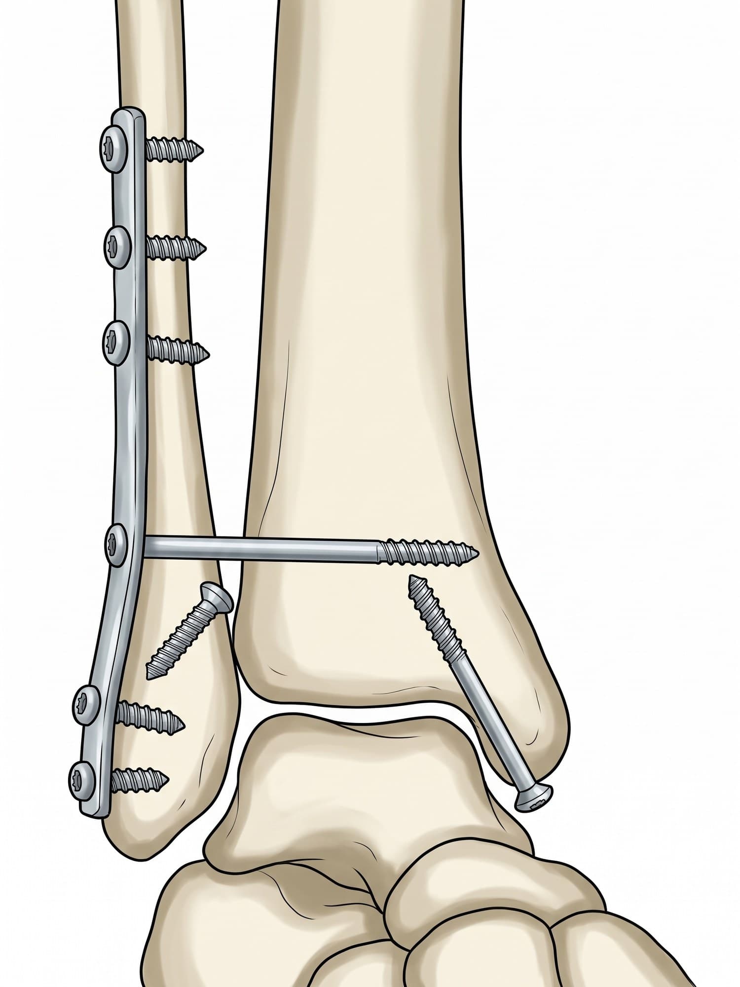
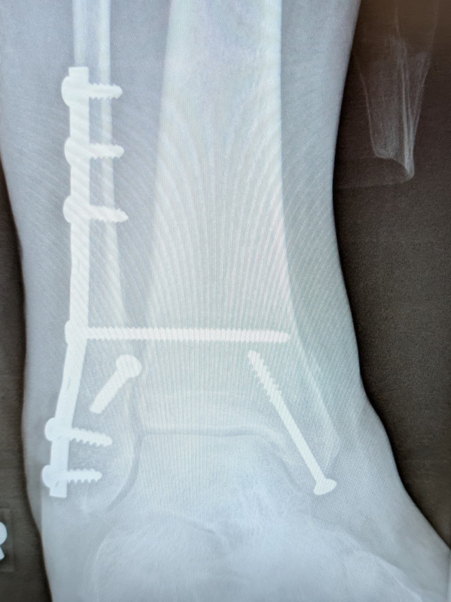
- Surgery (ORIF): open reduction and internal fixation for unstable fractures. The fibula is typically fixed with a plate and screws and the medial malleolus with screws. A torn syndesmosis is held with a screw or a suture button.
- Closed reduction first: a dislocated ankle is pulled back into place right away to protect the skin and blood supply, then splinted until surgery.
The X-ray below shows a right ankle after ORIF for an unstable ankle fracture, with a medical illustration drawn from it: drag the slider to compare them.
Recovery time
The bone heals in about 6 to 8 weeks. After a stable fracture, many people walk in a boot within the first weeks and are back to normal shoes at about 6 to 8 weeks. After surgery, the ankle is usually protected for about 6 weeks, followed by physical therapy for range of motion, strength and balance. Swelling at the end of the day often lasts for months, and full recovery takes 6 to 12 months.
Complications
- Post-traumatic arthritis: when the talus heals out of position or the joint surface is damaged
- Stiffness and chronic swelling
- Wound problems and infection after surgery, more common in smokers and people with diabetes
- Syndesmosis malreduction: the fibula heals out of position in its groove on the tibia
- Blood clots (deep vein thrombosis) during the weeks of reduced walking
- Hardware irritation, sometimes needing plate removal
How an ankle fracture is shown in a personal injury case
Ankle fractures from slips, trips, falls on stairs and car crashes are among the most common injuries in premises liability and auto cases. The X-ray proves the fracture, but jurors rarely understand what a Weber C fracture or a widened medial clear space means. An illustration drawn from the plaintiff's own X-ray, like the one above, shows which bones broke, how the joint shifted, and why surgery was needed. Typical exhibits pair the injury illustration with the hardware placed at surgery and a normal ankle for comparison. For how these images are used at trial, see demonstrative evidence and medical legal illustration.
Related pages
FAQ
How long does a fractured ankle take to heal?+
The bone usually heals in about 6 to 8 weeks. A stable fracture of the fibula alone is often walking in a boot within a few weeks, while fractures that need surgery are usually kept off weight for about 6 weeks. Swelling, stiffness and a limp commonly last 3 to 6 months, and full recovery for sport can take up to a year.
Can you walk on a broken ankle?+
Sometimes. Many people can still take a few steps on a stable fracture of the lateral malleolus, so walking does not rule out a break. Being unable to take four steps, or tenderness over the back edge or tip of either malleolus, are the signs doctors use (the Ottawa ankle rules) to decide an X-ray is needed.
Is it a broken ankle or a sprained ankle?+
A sprain injures the ligaments and a fracture breaks the bone, and they can feel the same at first: pain, swelling and bruising. Bone tenderness over the malleoli, being unable to bear weight, and a visible deformity point toward a fracture. Only an X-ray can tell them apart for certain.
What is a lateral malleolus fracture?+
The lateral malleolus is the bump on the outside of the ankle, formed by the lower end of the fibula. A lateral malleolus fracture is a break of that part of the fibula. It is the most common ankle fracture and is often stable enough to treat in a boot.
What is the difference between a bimalleolar and a trimalleolar fracture?+
A bimalleolar fracture breaks both the lateral malleolus (fibula) and the medial malleolus (tibia). A trimalleolar fracture also breaks the back edge of the tibia, called the posterior malleolus. Both are unstable and usually treated with surgery.
What is a Weber C fracture?+
The Weber classification sorts fibula fractures by height compared with the syndesmosis, the ligaments that hold the tibia and fibula together. Weber A is below it, Weber B is at its level, and Weber C is above it. A Weber C fracture usually means the syndesmosis is torn, so the ankle is unstable and most need surgery.
How long is recovery after ankle surgery with a plate and screws?+
Most people are kept off the foot or in a boot for about 6 weeks after open reduction and internal fixation, then gradually return to walking with physical therapy. Driving is often possible at 6 to 9 weeks for a right ankle. Most return to normal daily activity in 3 to 4 months, and the plate is usually left in unless it causes irritation.
Need an illustration of a patient's own X-ray?
Upload an X-ray, CT or MRI and turn it into a clear, labeled medical illustration for a consult, a chart or a case exhibit.
Try Natomy →