Cervical Spine Anatomy: C1 to C7 Vertebrae Labeled
Cervical spine anatomy labeled: the C1 to C7 vertebrae, atlas and axis, discs, ligaments, nerves, vertebral artery and neck muscles, plus neck injuries.
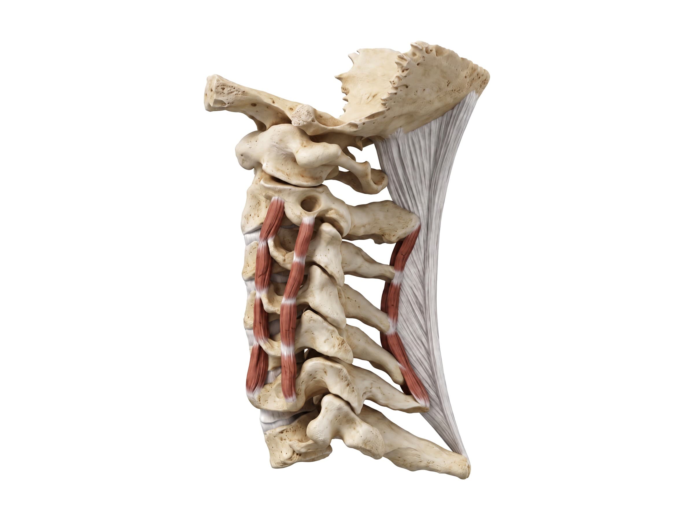
The cervical spine is the neck section of the spine: seven vertebrae, C1 to C7, that run from the base of the skull to the top of the chest. They hold up the head (about 4.5 to 5 kilograms), protect the spinal cord, carry the vertebral arteries to the brain, and give the neck its wide range of movement. Cervical spine anatomy covers the vertebrae themselves, including the specialized atlas (C1) and axis (C2), the intervertebral discs and facet joints between them, the ligaments that hold them together, the eight cervical nerves, the vertebral artery, and the neck muscles that move them. Knowing these parts is the first step to reading a neck X-ray or MRI report, explaining a herniated disc to a patient, or illustrating a whiplash claim.
The seven cervical vertebrae
The labeled side view above shows the cervical spine from the left, with the front on the left. The column curves gently forward in the middle (the cervical lordosis), a curve that can straighten or reverse after injury or with muscle spasm.
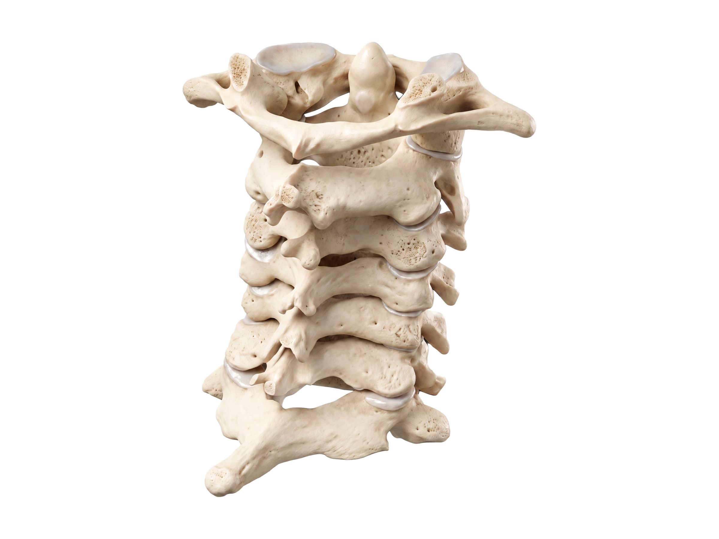
The cervical vertebrae fall into three groups:
- C1 (atlas) and C2 (axis): the upper cervical spine, built for movement of the head rather than weight bearing through a disc. There is no disc between the skull and C1 or between C1 and C2.
- C3 to C6: the typical cervical vertebrae, similar in shape and stacked with a disc between each pair.
- C7 (vertebra prominens): a transition vertebra with a long spinous process, sitting on top of the first thoracic vertebra (T1).
From behind, the spinous processes form the ridge down the back of the neck, and the paired facet joints on each side (the gray joint lines in the figure) guide how each level glides over the next.
Typical cervical vertebra (C3 to C6)
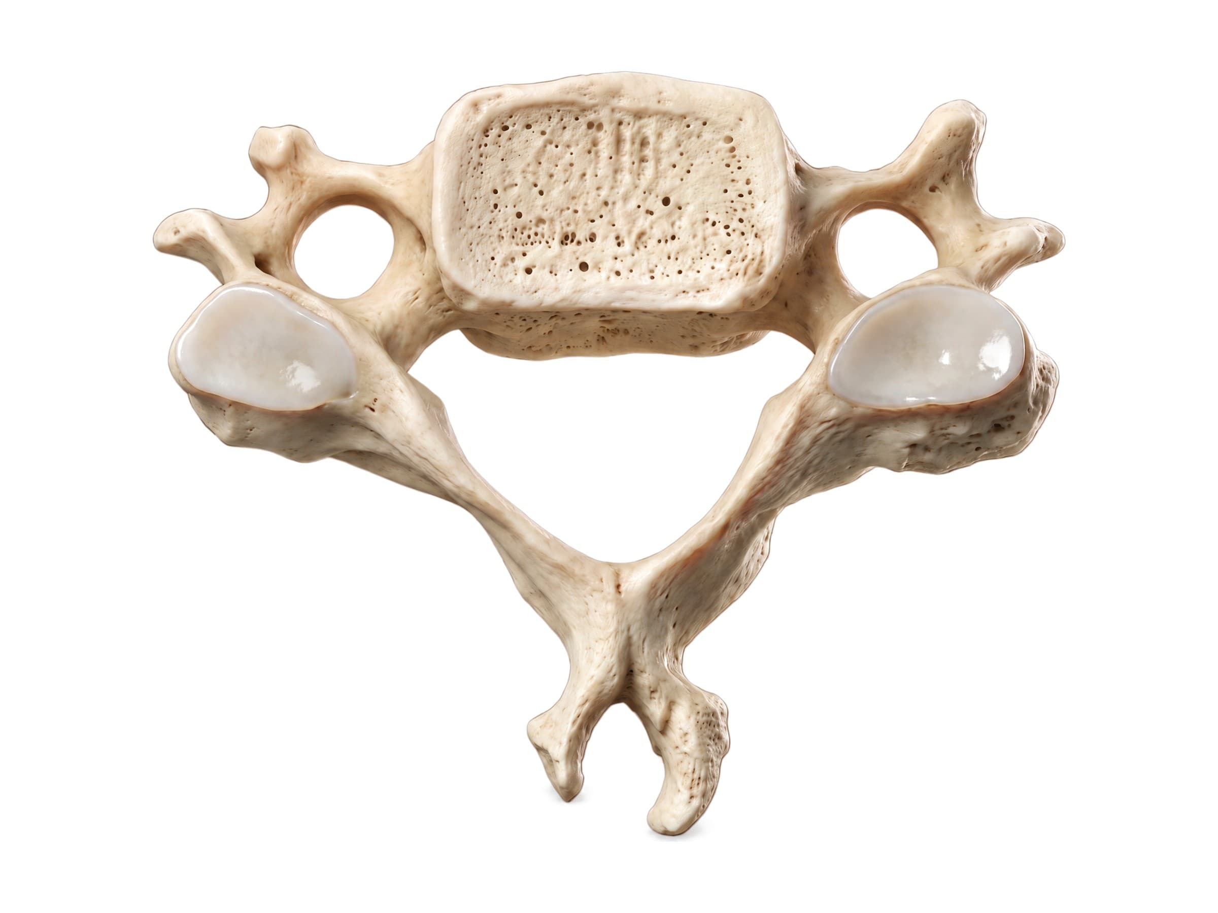
The vertebrae from C3 to C6 share the same layout, seen here from above with the front at the top:
- Vertebral body: a small, wide, oval block in front that carries weight. Its top edges turn up at the sides as the uncinate processes, which form small uncovertebral joints (joints of Luschka) with the vertebra above. Bone spurs from these joints are a common cause of nerve root compression.
- Pedicles and laminae: the short pedicles join the body to the arch, and the thin laminae meet in the midline behind. Together they enclose the vertebral canal (foramen vertebrale), which is large and triangular in the neck because it holds the widest part of the spinal cord.
- Transverse processes and foramen transversarium: each transverse process has a hole, the foramen transversarium, unique to cervical vertebrae. The vertebral artery runs up through these holes from C6 to C1. The front of the C6 transverse process has a large bump, the carotid tubercle, against which the common carotid artery can be pressed.
- Articular processes: flat superior and inferior articular facets form the facet joints with the vertebrae above and below. The bone between them on each side is called the articular pillar or lateral mass.
- Bifid spinous process: the spinous processes of C3 to C6 are short and forked at the tip.
Between each pair of bodies, the intervertebral foramen (foramen intervertebrale) is the opening where a spinal nerve leaves the canal. It is bounded by the disc and uncovertebral joint in front and the facet joint behind, so arthritis or a disc bulge at either can narrow it (foraminal stenosis).
The atlas (C1)
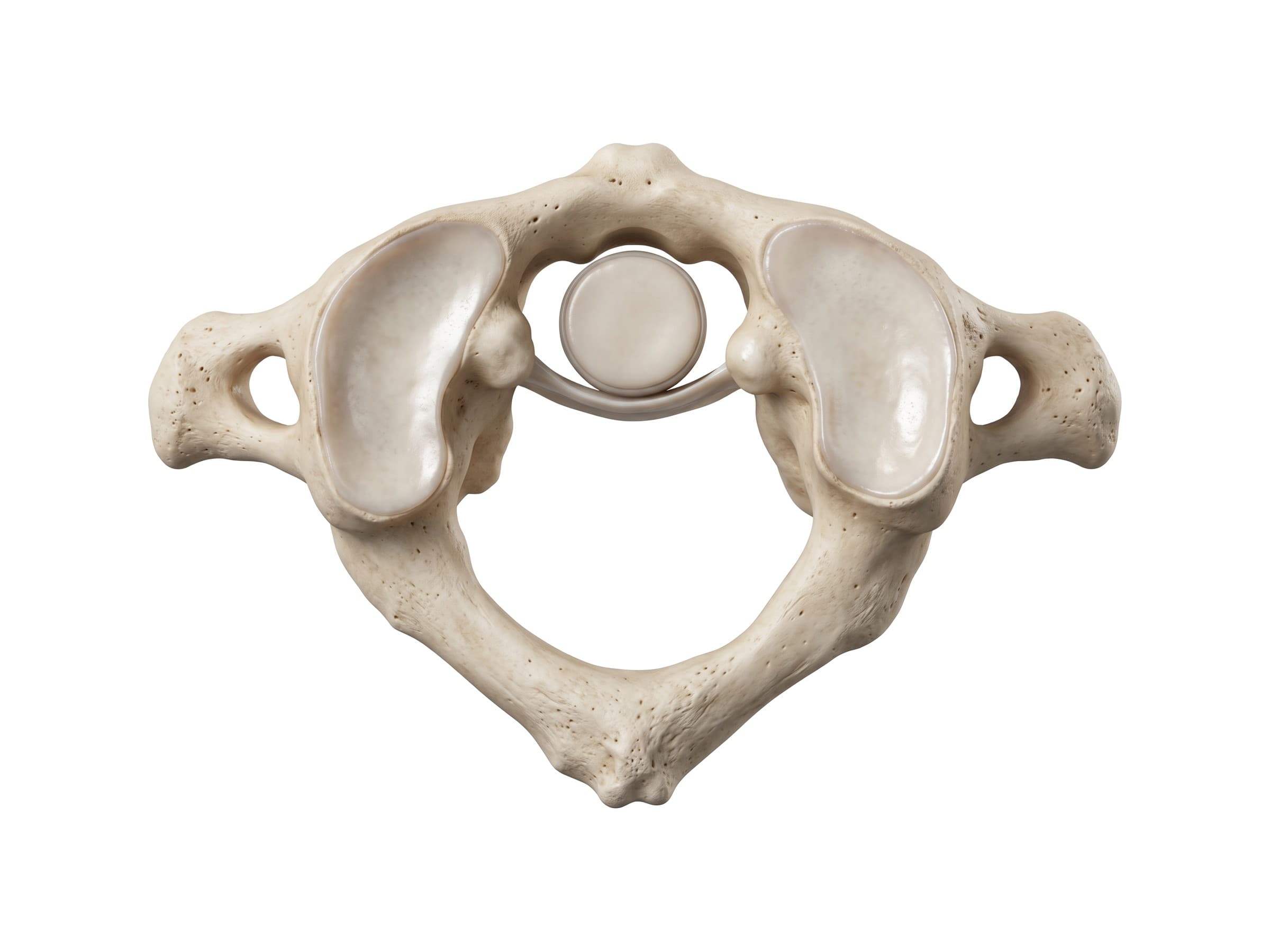
The atlas is the first cervical vertebra, named after the Titan who held up the world, because it holds up the skull. It is a ring of bone with no vertebral body and no spinous process:
- Anterior arch: the short front part of the ring, with a small facet on its inner surface where the dens of C2 rests.
- Lateral masses: the thick sides of the ring. Their large, cup-shaped superior articular facets hold the occipital condyles of the skull, forming the atlanto-occipital joints, which allow nodding (the "yes" movement). Their flat lower facets sit on the axis.
- Transverse ligament: a strong band that stretches across the ring behind the dens, dividing it into a front compartment for the dens and a back compartment for the spinal cord.
- Posterior arch: the longer back part of the ring, with a small posterior tubercle in place of a spinous process. A groove on its top surface carries the vertebral artery as it turns toward the skull.
- Transverse processes: wide and easy to feel just below and in front of the mastoid process behind the ear.
The axis (C2) and the dens
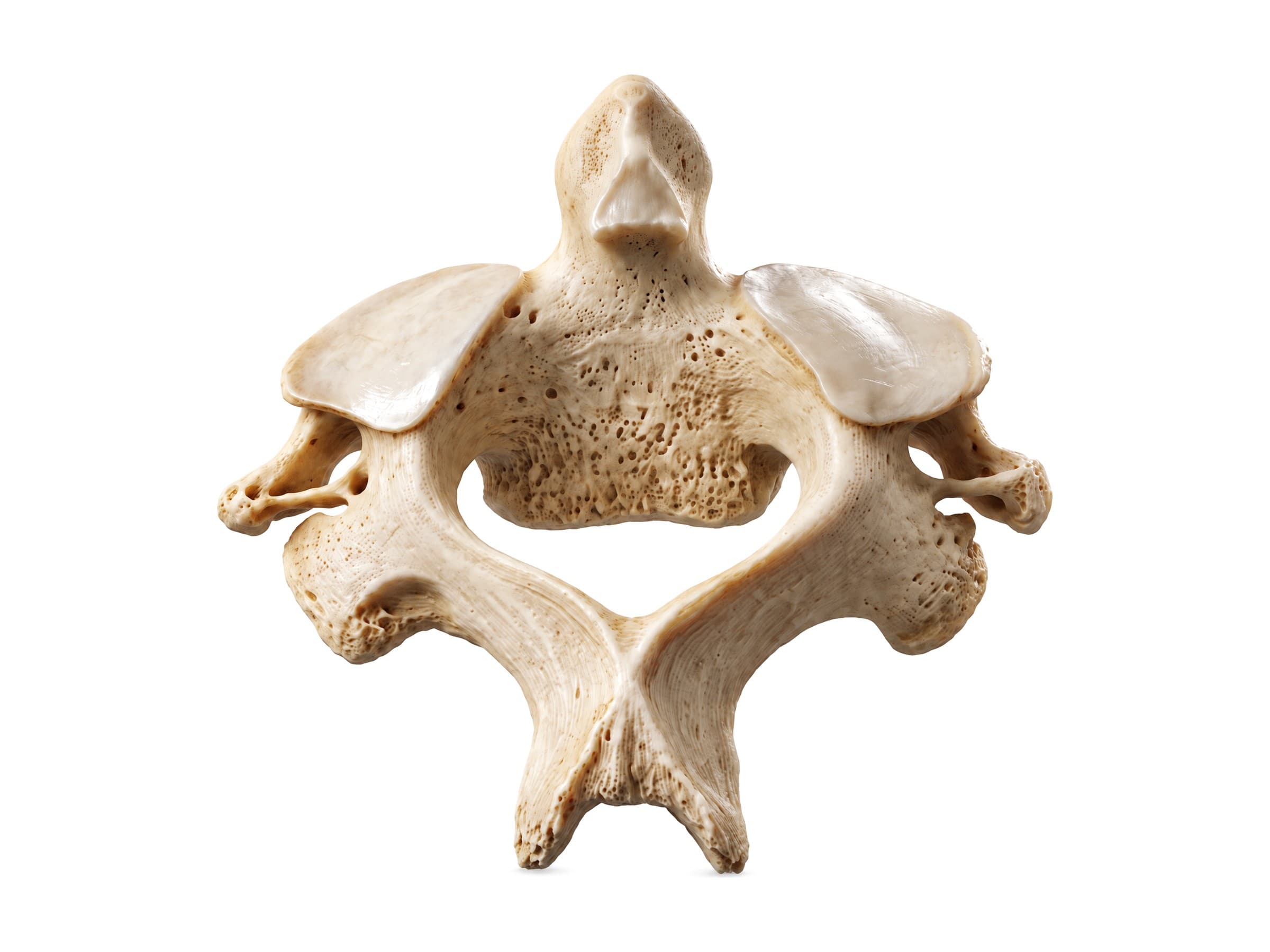
The axis is the second cervical vertebra and the strongest bone in the neck. Its defining feature is the dens, or odontoid process, a tooth-shaped peg that rises from the top of the body into the ring of the atlas. The atlas, carrying the skull, rotates around the dens at the atlantoaxial joint, which provides about half of the neck's rotation (the "no" movement).
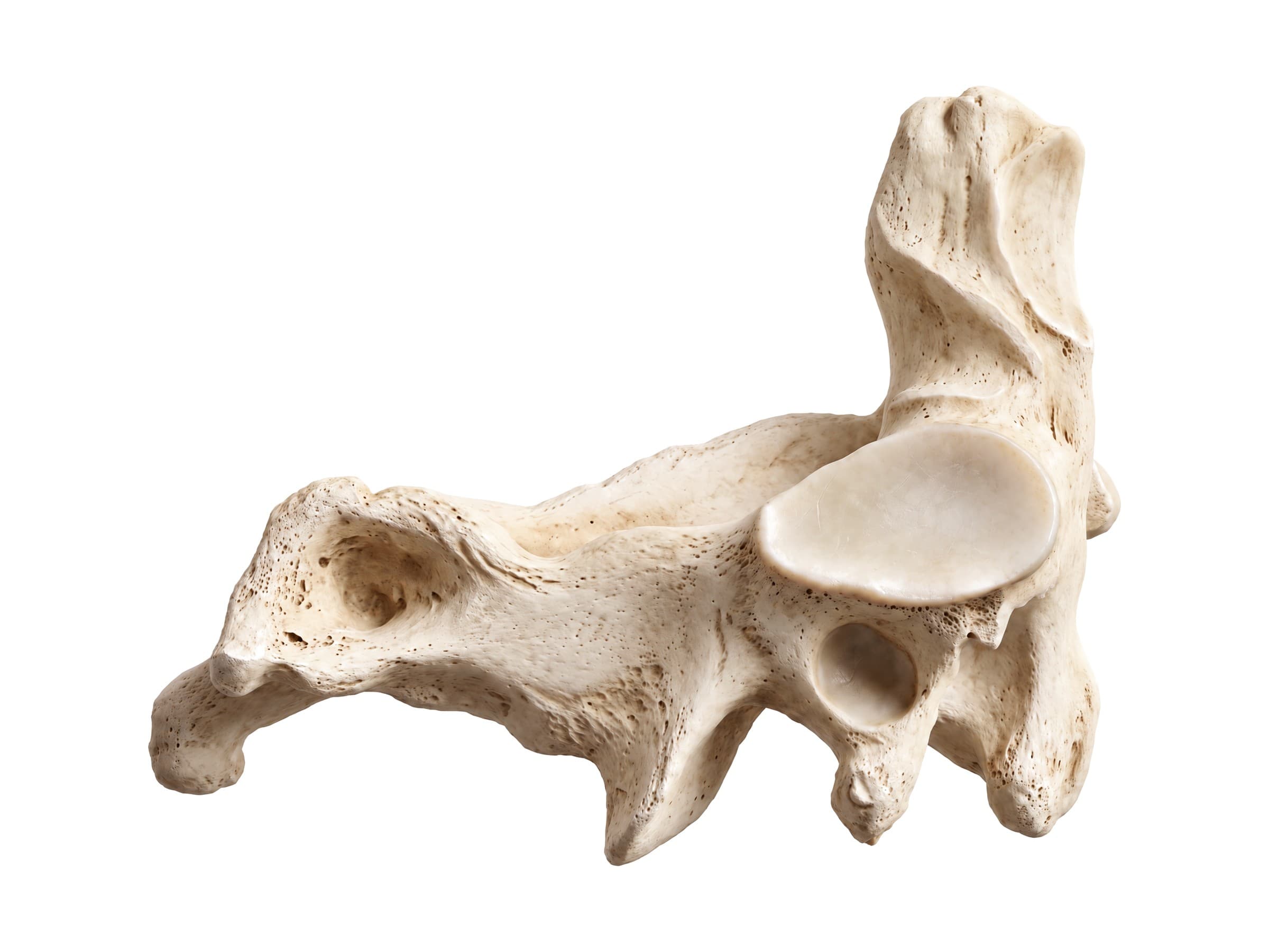
- Dens: its front surface articulates with the anterior arch of the atlas, and its back surface has a groove for the transverse ligament. Its tip is anchored to the skull by the alar and apical ligaments.
- Superior articular surfaces: two broad, flat shoulders on either side of the dens that carry the lateral masses of the atlas.
- Pars interarticularis: the bridge of bone on each side between the upper and lower joint surfaces. It is the site of the hangman's fracture.
- Spinous process: large and bifid, the first spinous process you can feel below the skull.
C7: the vertebra prominens
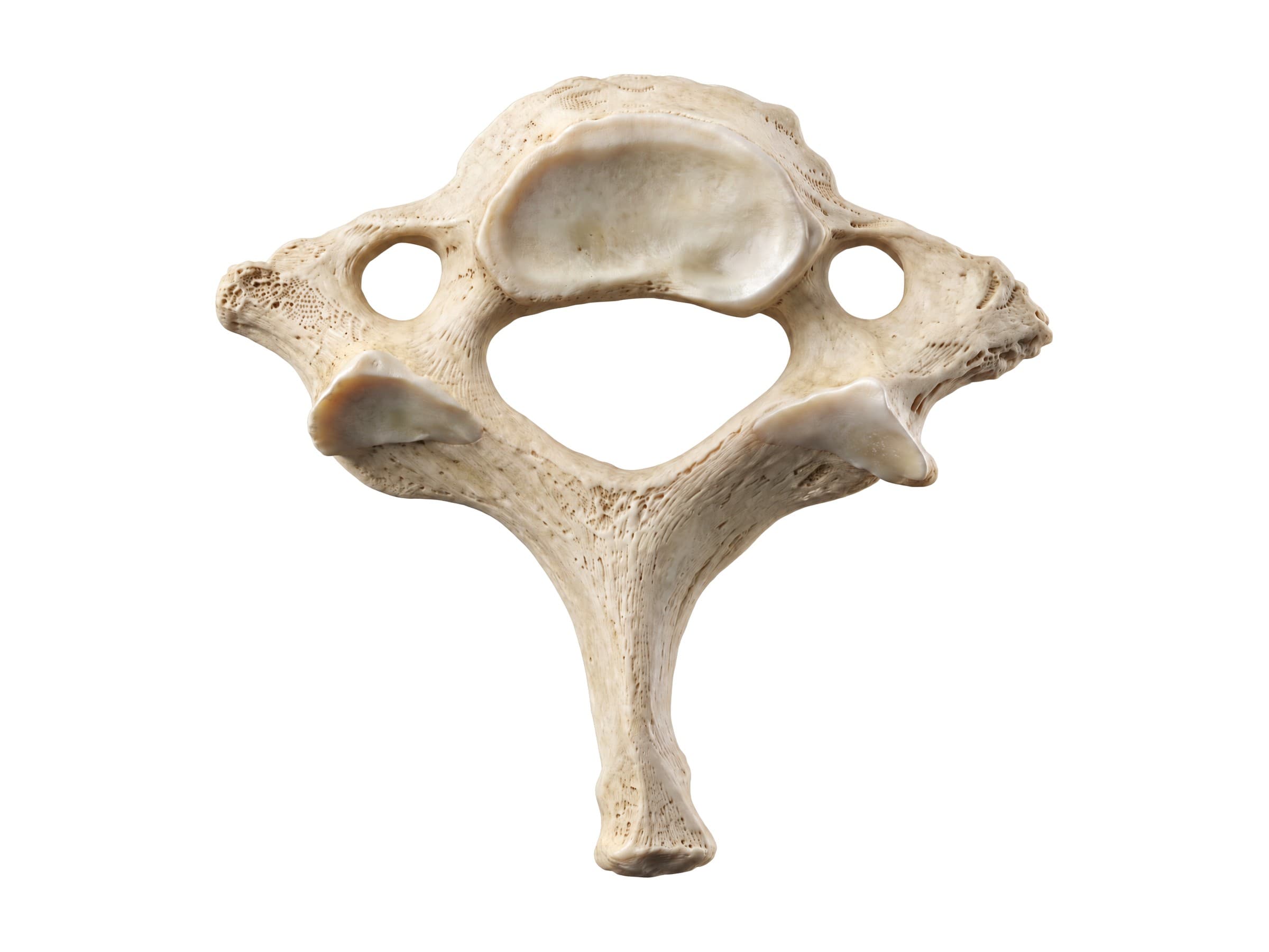
C7 is the largest cervical vertebra and the link to the thoracic spine. Its long, single spinous process is the prominent bump at the base of the neck, used as a landmark to count the vertebrae. Its transverse processes are large, and its foramina transversaria are small, usually carrying only veins, because the vertebral artery normally enters the spine at C6. In some people an extra cervical rib grows from C7 and can press on the brachial plexus or subclavian artery (thoracic outlet syndrome).
Joints, discs and ligaments
The cervical spine has several kinds of joints:
- Atlanto-occipital joints: between the skull and C1, for nodding.
- Atlantoaxial joints: between C1 and C2, one around the dens and two between the lateral masses, for rotation.
- Intervertebral discs: six discs, from C2-C3 down to C7-T1. Each has a tough outer ring, the annulus fibrosus, around a soft gel center, the nucleus pulposus, and acts as a shock absorber between the vertebral bodies.
- Facet (zygapophyseal) joints: small paired joints at the back of each level that guide movement and are a common source of neck pain after whiplash.
- Uncovertebral joints: at the sides of the discs from C3 down.
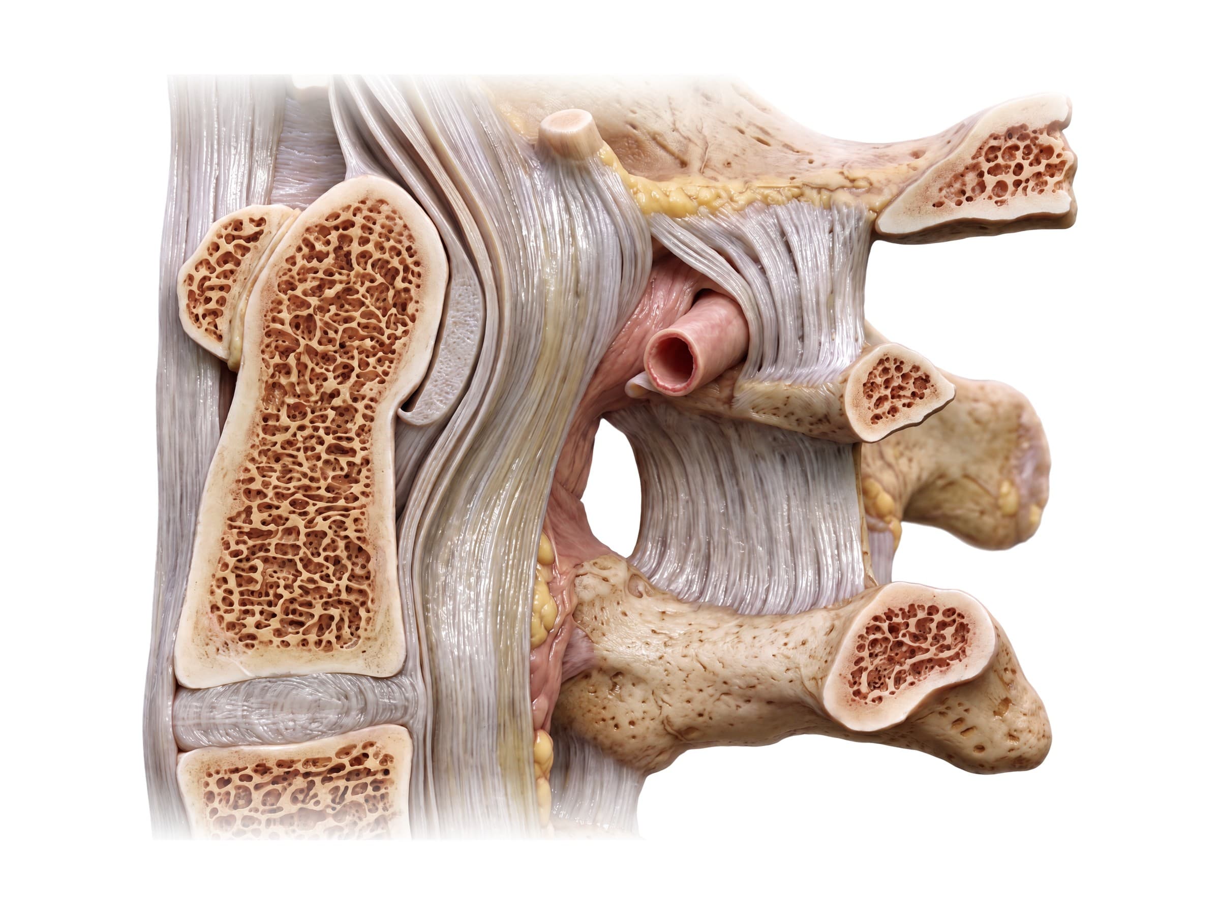
The ligaments of the cervical spine hold the vertebrae together and limit movement:
- Anterior longitudinal ligament: runs down the front of the vertebral bodies and discs; it resists bending backward and is often torn in hyperextension injuries.
- Posterior longitudinal ligament: runs down the back of the bodies inside the canal. Above C2 it continues as the membrana tectoria, which covers the dens and attaches to the base of the skull.
- Ligamentum flavum: elastic yellow ligaments joining the laminae of neighboring vertebrae; when they thicken with age they can narrow the canal.
- Nuchal ligament: the thick midline ligament from the back of the skull to the C7 spinous process, which anchors the neck muscles.
- Transverse, alar and apical ligaments: hold the dens against the atlas and tie it to the skull.
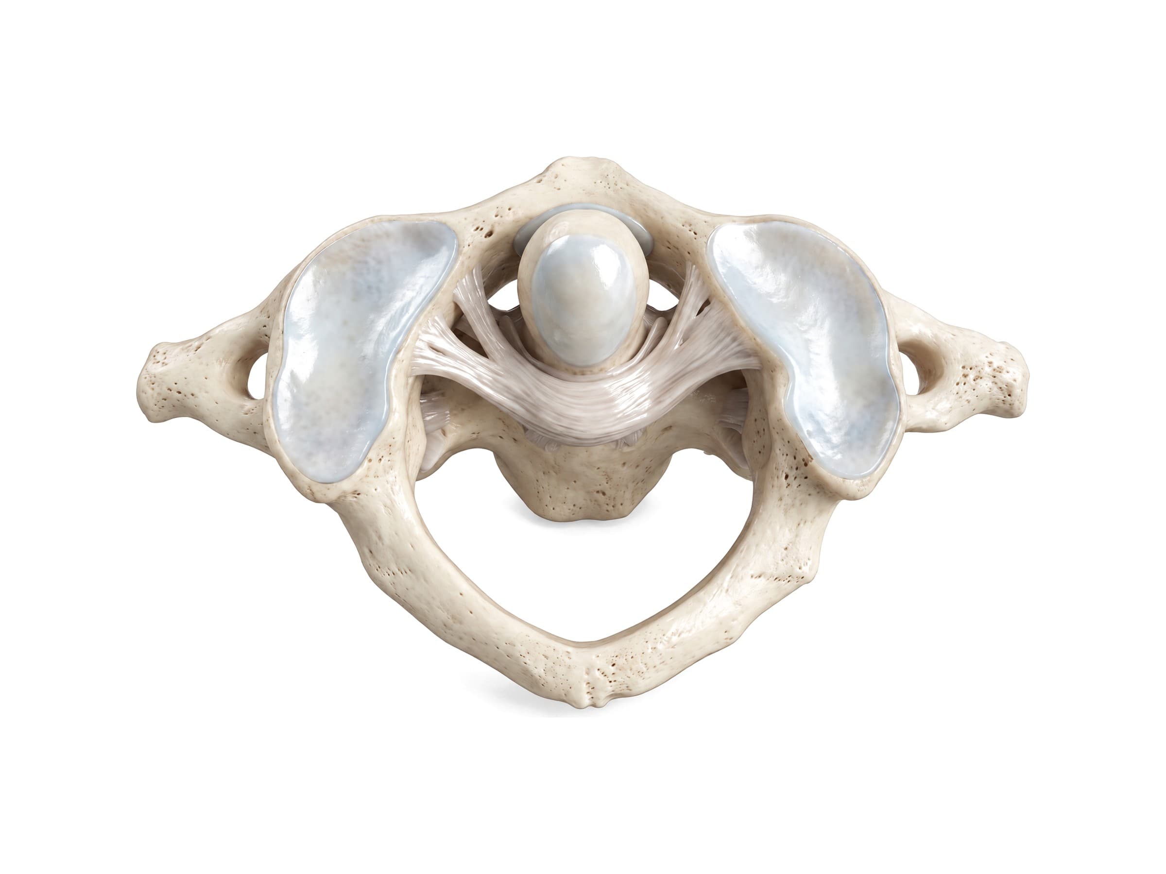
The transverse ligament is the most important stabilizer of the upper neck. If it ruptures, the atlas can slide forward on the axis and the dens can press into the spinal cord, which is why its integrity is checked after any fracture of C1.
Spinal cord and cervical nerves
The cervical spinal cord runs through the vertebral canal from the foramen magnum of the skull. Between C4 and T1 it widens into the cervical enlargement, which holds the nerve cells for the arms. There are eight pairs of cervical nerves but only seven vertebrae: each cervical nerve leaves the canal above the vertebra with the same number (C6 exits between C5 and C6), and C8 exits between C7 and T1. So a disc herniation at C5-C6 usually pinches the C6 root.
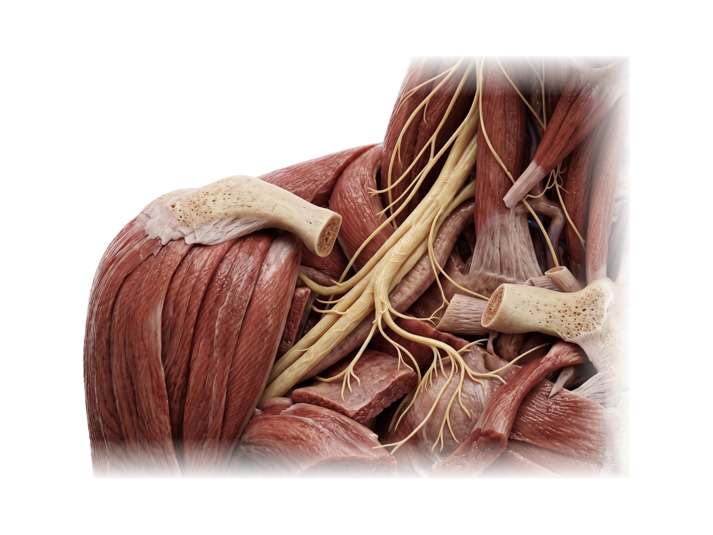
- C1 to C4 (cervical plexus): the back of the head and neck, the skin of the neck, and the small neck muscles. C3, C4 and C5 form the phrenic nerve, which drives the diaphragm, so a cord injury above C5 can stop independent breathing.
- C5 to T1 (brachial plexus): the roots pass between the anterior and middle scalene muscles, run under the clavicle, and divide into the nerves of the arm.
- C5: deltoid and biceps; skin of the outer upper arm; biceps reflex.
- C6: biceps and wrist extensors; thumb and index finger; brachioradialis reflex.
- C7: triceps, wrist flexors and finger extensors; middle finger; triceps reflex.
- C8: finger flexors and small hand muscles; ring and little fingers.
Matching a patient's numbness and weakness to these patterns (the dermatomes and myotomes) is how a clinician tells which root is compressed before the MRI confirms it.
Vertebral artery
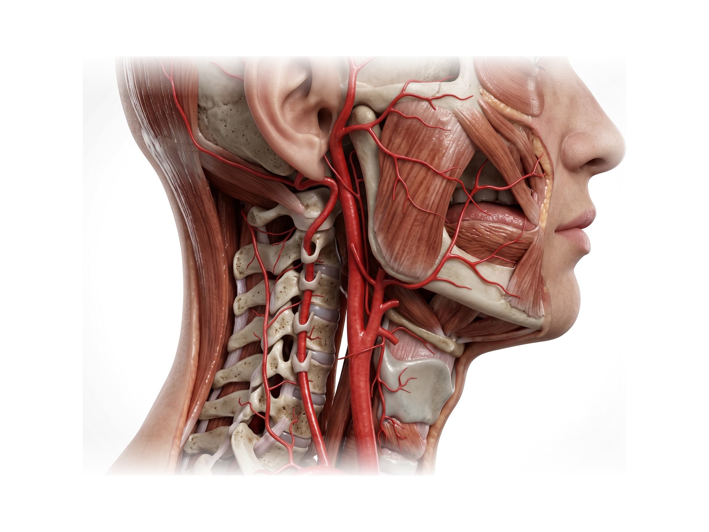
The two vertebral arteries supply the back of the brain, the brainstem and the cerebellum. Each branches from the subclavian artery at the base of the neck, enters the foramen transversarium of C6, climbs through the holes in C6 up to C1, then curves backward over the posterior arch of the atlas and enters the skull through the foramen magnum. Inside the skull the two join to form the basilar artery, part of the circle of Willis.
Because it runs through bone and bends sharply at the top of the neck, the vertebral artery can be torn (vertebral artery dissection) by fractures through the foramen transversarium, sudden neck rotation or extension, or, rarely, neck manipulation. Dissection can cause a stroke, sometimes days after the injury.
Neck muscles
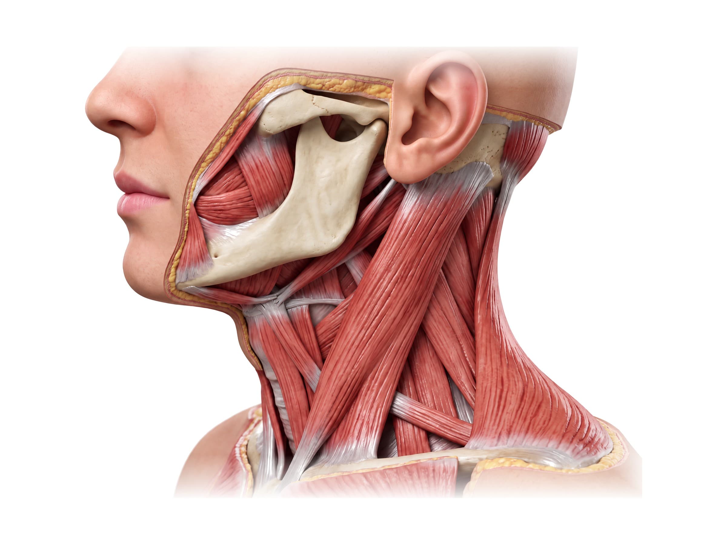
The muscles around the cervical spine hold up the head and move it:
- Sternocleidomastoid: the large strap muscle from the mastoid process behind the ear to the breastbone and collarbone. One side turns the head to the opposite side; both together bend the neck forward. It divides the side of the neck into the anterior and posterior triangles.
- Trapezius: the large muscle at the back of the neck and upper back that extends the head and lifts the shoulder.
- Splenius capitis and cervicis, semispinalis and the small suboccipital muscles: the deep posterior neck muscles that extend and rotate the head. See back muscles anatomy for how they continue down the spine.
- Levator scapulae: from the transverse processes of C1 to C4 down to the shoulder blade; a frequent source of neck and shoulder pain.
- Scalene muscles: on the side of the neck from the transverse processes to the first two ribs; they bend the neck and lift the ribs, and the brachial plexus passes between the anterior and middle scalenes.
- Longus colli and longus capitis: deep muscles on the front of the vertebrae that flex the neck; they are often strained in whiplash.
- Omohyoid, sternohyoid and the other strap muscles in front of the neck move the hyoid bone and larynx rather than the spine. The inferior belly of the omohyoid crosses the lower posterior triangle just above the clavicle.
- Masseter and buccinator: muscles of the face rather than the neck. The masseter runs from the cheekbone (zygomatic arch) to the angle of the jaw and closes the mouth; the buccinator forms the wall of the cheek. They are shown here for orientation.
Range of motion of the cervical spine
The neck is the most mobile part of the spine. Typical normal values are roughly 45 to 50 degrees of flexion (chin to chest), 55 to 70 degrees of extension, 70 to 80 degrees of rotation to each side, and about 40 to 45 degrees of side bending, though range decreases with age. About half of rotation happens at C1-C2 and much of nodding at the skull and C1; most of the remaining flexion and extension happens at C4 to C6. Loss of range of motion, measured with a goniometer or inclinometer, is one of the main ways a neck injury is documented over time.
Common cervical spine injuries and how they appear on imaging
After trauma, doctors use clinical decision rules (the Canadian C-Spine Rule or NEXUS criteria) to decide who needs imaging. CT is the first scan for suspected fractures in adults. X-rays (side, front and open-mouth views of C1-C2) are still used for follow-up and lower-risk cases. MRI shows discs, ligaments, the spinal cord and nerve roots, which neither X-ray nor CT shows well.
- Whiplash (cervical sprain or strain): a rapid back-and-forth movement of the neck, typically in a rear-end collision, that stretches or tears muscles, ligaments and facet joint capsules. X-rays and CT are usually normal or show only a straightened lordosis from muscle spasm; MRI may show ligament or disc injury. Whiplash is graded clinically (Quebec Task Force grades 0 to IV) by symptoms, stiffness, nerve signs and fractures.
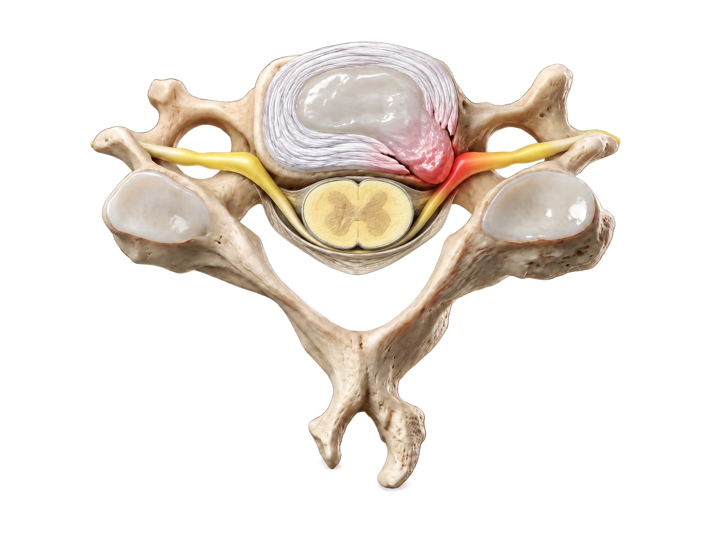
- Cervical disc herniation: the nucleus pulposus pushes through a tear in the annulus, most often at C5-C6 or C6-C7, and presses on a nerve root (radiculopathy) or the cord (myelopathy). MRI shows the disc material and the compressed root. Persistent cases with weakness are often treated with anterior cervical discectomy and fusion (ACDF) or disc replacement.
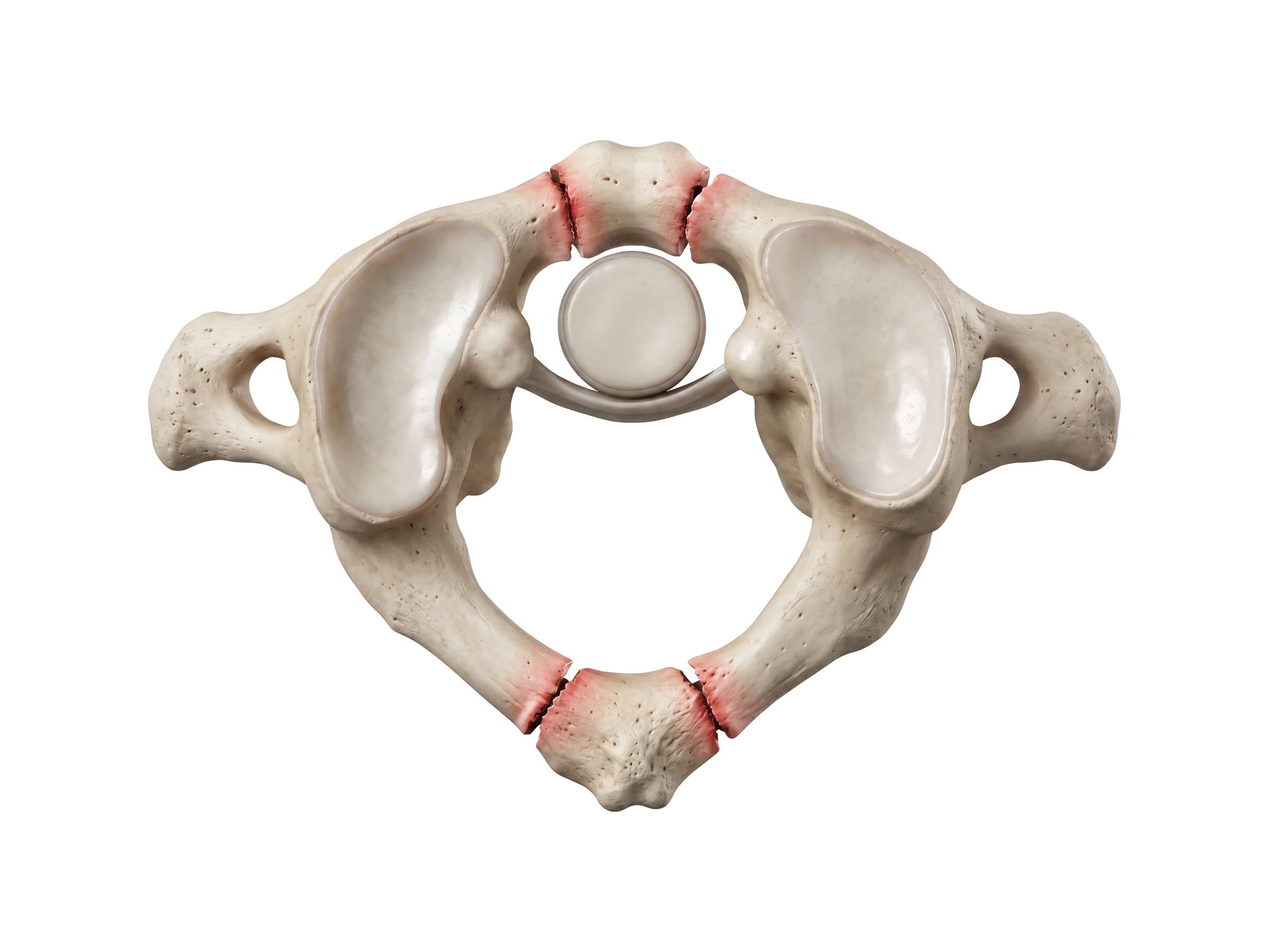
- Jefferson fracture: a burst fracture of the atlas from a load straight down on the top of the head, such as diving into shallow water or the head striking a car roof. The ring breaks in two or more places and the lateral masses spread apart; on the open-mouth X-ray they overhang the edges of C2. CT shows the breaks. Stability depends on whether the transverse ligament is intact.
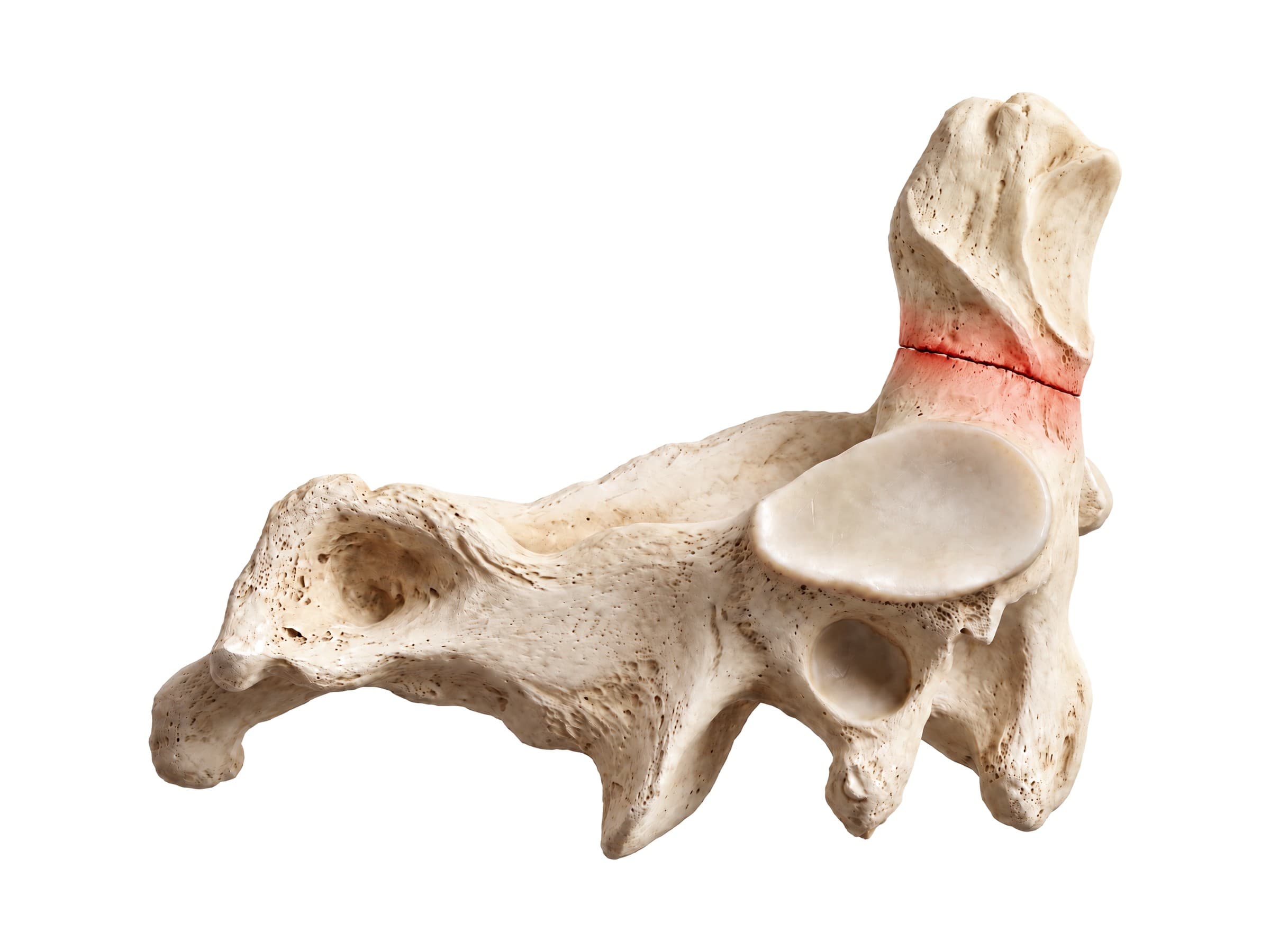
- Odontoid (dens) fracture: classified as type I (the tip), type II (across the base of the dens, the most common and the least likely to heal), or type III (extending down into the body of C2). It is the most common neck fracture in people over 70, often from a simple fall, and in younger people follows high-energy crashes.
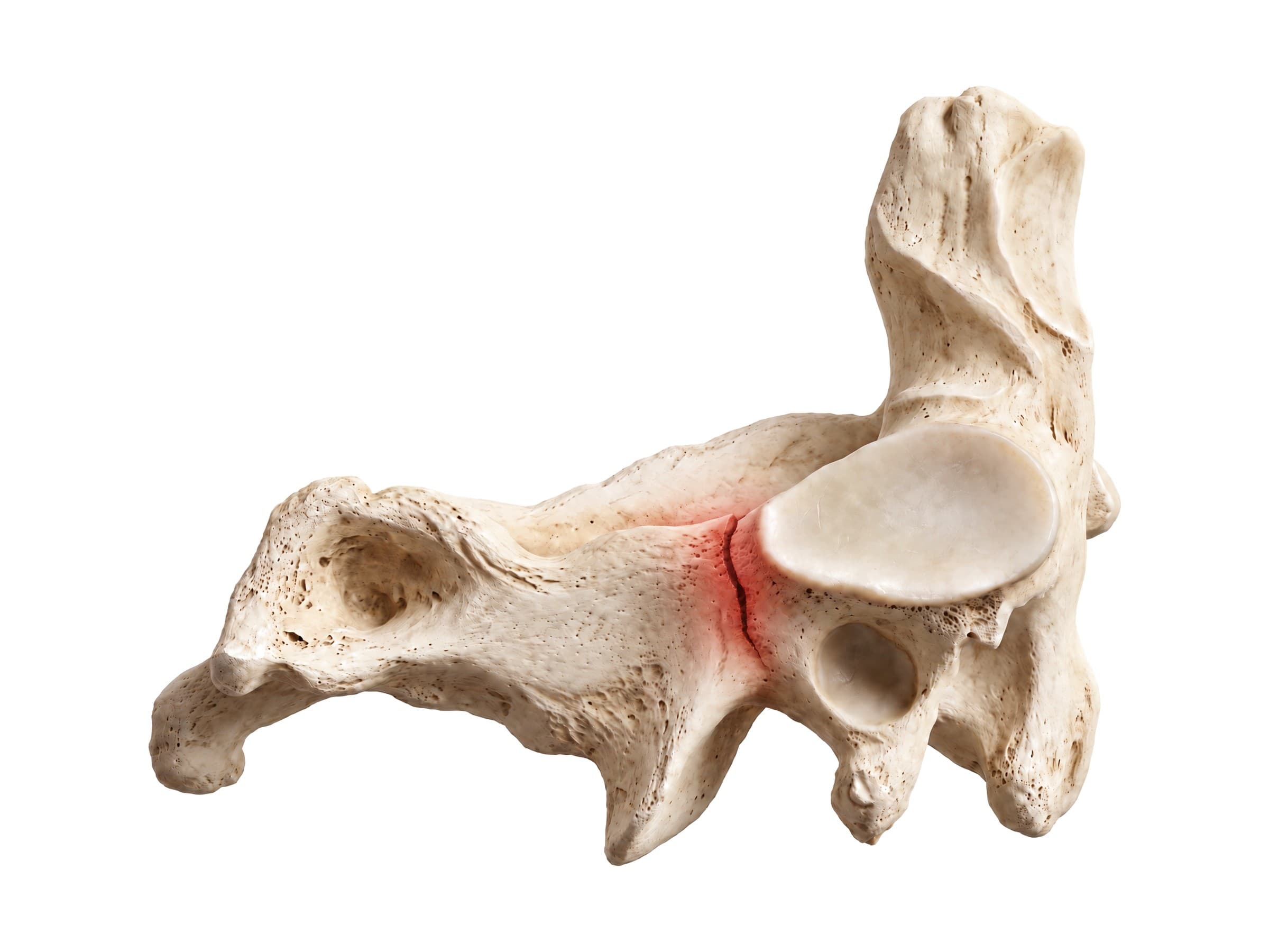
- Hangman's fracture: a break through both pars interarticularis of C2 (traumatic spondylolisthesis of the axis), from forceful hyperextension, classically when the face or forehead strikes the windshield in a car crash. The canal widens with the break, so many patients have no spinal cord injury.
- Facet dislocation and lower cervical fractures: flexion and rotation forces can lock one or both facets of a lower level forward, with a high risk of cord injury. Compression and burst fractures of the vertebral bodies of C5 to C7 follow falls and diving injuries.
- Spinal cord injury: damage to the cord with loss of movement and sensation below the level. Central cord syndrome, with weakness worse in the arms than the legs, typically follows a hyperextension fall in an older person with a narrowed, arthritic canal, even without a fracture.
How the cervical spine is shown in a personal injury case
Neck injuries are among the most common claims after car crashes, falls and workplace accidents, and among the most disputed. The defense often argues that a disc herniation was pre-existing degeneration, that whiplash is a minor strain, or that the symptoms do not match the imaging. Each type of cervical injury follows a recognizable mechanism of injury: whiplash from rear-end acceleration and deceleration, a hangman's fracture from the head striking the windshield, a Jefferson fracture from axial loading of the head. A labeled illustration of the normal cervical spine is the baseline exhibit. It lets an expert show the jury where the C5-C6 disc sits, which nerve root leaves at that level, and why the plaintiff's numbness runs down to the thumb, before showing the injury itself: the herniated disc pressing on the nerve, the fractured vertebra, or the fusion hardware placed in surgery. Illustrations drawn from the plaintiff's own MRI or CT make the connection between the scan, the anatomy and the symptoms concrete.
Natomy creates custom medical legal illustrations of cervical spine injuries for demand packages and trial exhibits, and the rules for using these visuals at trial are covered in demonstrative evidence. For the bones above the neck, see skull anatomy, and for the brain and its blood supply, brain anatomy. Related atlas pages: thoracic spine anatomy, lumbar spine anatomy, back muscles anatomy, shoulder anatomy, elbow anatomy, hand and wrist anatomy and hip anatomy.
FAQ
How many cervical vertebrae are there?+
There are seven cervical vertebrae, numbered C1 to C7 from the top of the neck down. C1 (the atlas) and C2 (the axis) are specialized for nodding and turning the head, C3 to C6 are typical cervical vertebrae, and C7 (the vertebra prominens) is a transition to the thoracic spine. There are eight cervical spinal nerves, because the first one leaves between the skull and C1 and the eighth leaves between C7 and T1.
What nerves do C5, C6 and C7 affect?+
The C5, C6 and C7 nerve roots supply most of the shoulder and arm. C5 powers the deltoid and biceps and carries feeling from the outer upper arm. C6 helps the biceps and wrist extensors and carries feeling from the thumb and index finger; it is tested with the brachioradialis reflex. C7 powers the triceps and the wrist flexors and finger extensors and carries feeling from the middle finger; it is tested with the triceps reflex. C5 to C7 also contribute to the phrenic nerve (C3 to C5) and the brachial plexus.
What nerves are affected by C1, C2 and C3?+
The upper cervical nerves supply the back of the head and the upper neck. C1 is mainly a motor nerve to the small suboccipital muscles under the skull. C2 forms the greater occipital nerve, which carries feeling from the back of the scalp, and contributes to the lesser occipital nerve behind the ear. C3 forms the third occipital nerve and, with C4 and C5, the phrenic nerve to the diaphragm. Irritation of these nerves is a common cause of headaches that start at the base of the skull.
What do C3 to C7 do?+
C3 to C7 carry the weight of the head, protect the spinal cord, and allow the neck to bend forward, backward and to the sides. Their nerve roots control the diaphragm (C3 to C5), the shoulder and elbow (C5 and C6), and the wrist, fingers and hand (C6 to C8). C5-C6 and C6-C7 are the most mobile lower levels, which is why they are where disc herniations and arthritis most often develop.
What is the difference between the atlas and the axis?+
The atlas (C1) is a ring of bone with no vertebral body and no spinous process. It holds up the skull through two cup-shaped facets, and the joint between them allows nodding. The axis (C2) has a peg of bone, the dens or odontoid process, that projects up through the front of the atlas ring. The atlas rotates around the dens, and this atlantoaxial joint provides about half of the neck's ability to turn the head from side to side.
What is the vertebra prominens?+
The vertebra prominens is the seventh cervical vertebra (C7). It is named for its long, single spinous process, which makes the bump you can feel at the base of the back of the neck when you bend your head forward. Clinicians use it as a landmark to count down the spine. Unlike C3 to C6, its spinous process is not forked, and its transverse foramina usually carry only small veins rather than the vertebral artery.
What are common symptoms of cervical spine problems?+
Common symptoms are neck pain and stiffness, headaches that start at the base of the skull, and reduced range of motion. When a disc or bone spur presses on a nerve root (cervical radiculopathy), pain, numbness, tingling or weakness spread into the shoulder, arm or hand in the pattern of that nerve. Pressure on the spinal cord itself (myelopathy) can cause clumsy hands, trouble with balance and walking, and, in severe cases, bladder or bowel problems, which need urgent evaluation.
What is the most commonly injured part of the cervical spine?+
Soft-tissue injury from whiplash, a strain or sprain of the neck muscles and ligaments, is the most common cervical spine injury, typically after a rear-end car collision. Among disc problems, the C5-C6 and C6-C7 levels are affected most often. Among fractures, C2 and the lower levels C5 to C7 are the most common sites; in older adults, odontoid fractures of C2 after a simple fall are especially frequent.
Need a custom illustration of this joint?
Generate a labeled or exhibit-ready figure for a specific patient, injury, or case in minutes.
Try Natomy →