Brain Anatomy: Parts of the Brain Labeled
Parts of the brain labeled: the lobes, cerebellum, brainstem, ventricles, cranial nerves, circle of Willis and meninges, plus epidural and subdural bleeds.
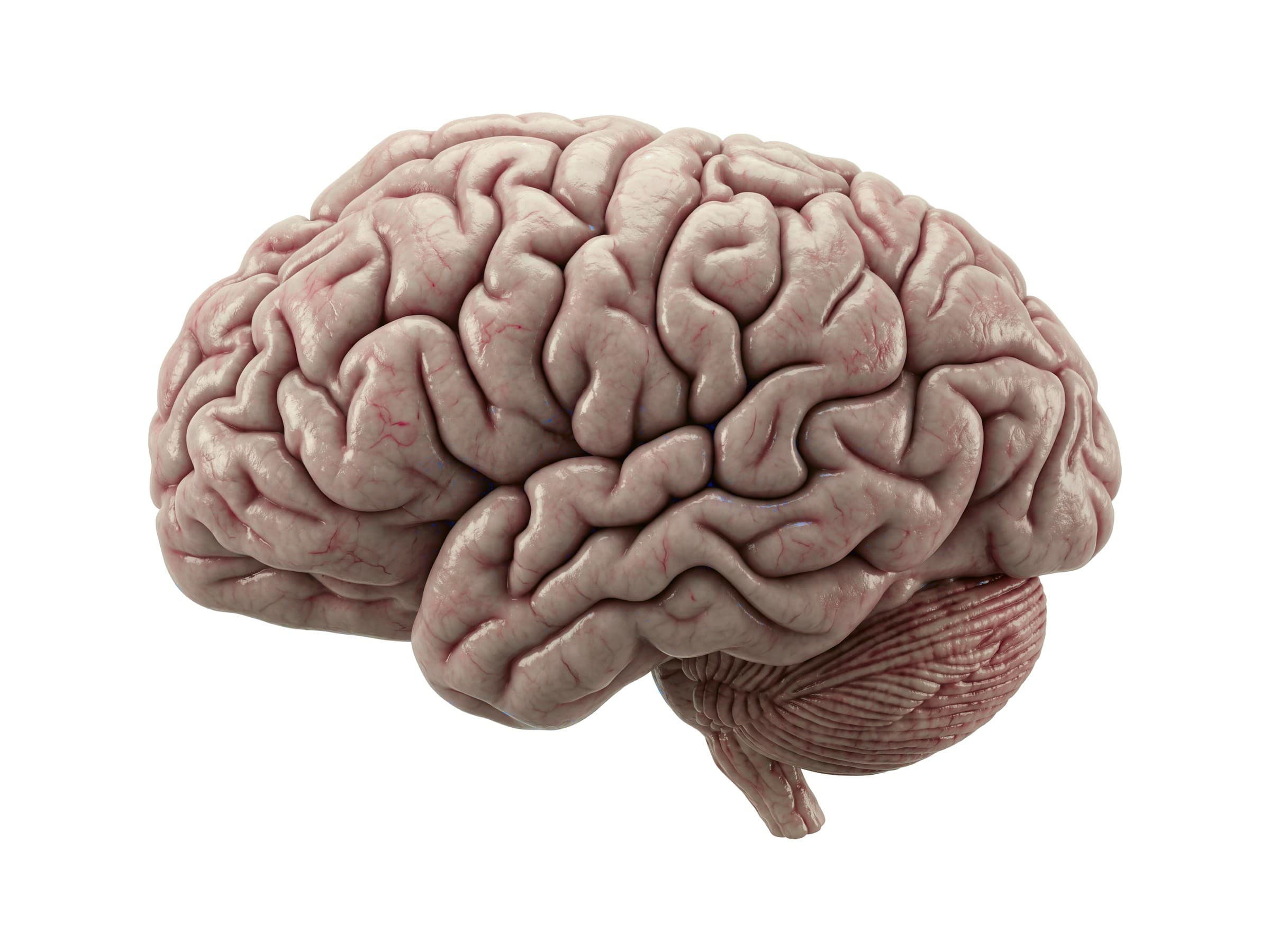
The brain is the control center of the body: about 1.3 to 1.4 kilograms of tissue containing roughly 86 billion neurons. Brain anatomy comes down to three main parts, the cerebrum, the cerebellum and the brainstem, along with the deep structures between them, the fluid-filled ventricles, the cranial nerves, the arteries of the circle of Willis, and the three layers of meninges that wrap it inside the skull. Knowing where each part sits is the first step to reading a CT or MRI report, explaining a brain injury to a patient or family, or illustrating a personal injury claim.
Parts of the brain
The labeled view above shows the left side of the brain, with the front on the left.
- Cerebrum: the largest part, divided by a deep groove into left and right hemispheres. Its folded outer layer, the cerebral cortex, is gray matter; the folds (gyri) and grooves (sulci) pack a large surface area into the skull. The cerebrum handles thinking, memory, language, sensation and voluntary movement. Each hemisphere mainly controls the opposite side of the body.
- Cerebellum: the "little brain" tucked under the back of the cerebrum. It fine-tunes movement, balance, posture and coordination, and it contains more than half of the brain's neurons.
- Brainstem: the stalk that connects the brain to the spinal cord. It is made of the midbrain, pons and medulla oblongata, controls breathing, heart rate, swallowing and wakefulness, and is where ten of the twelve cranial nerves leave the brain.
Lobes of the brain
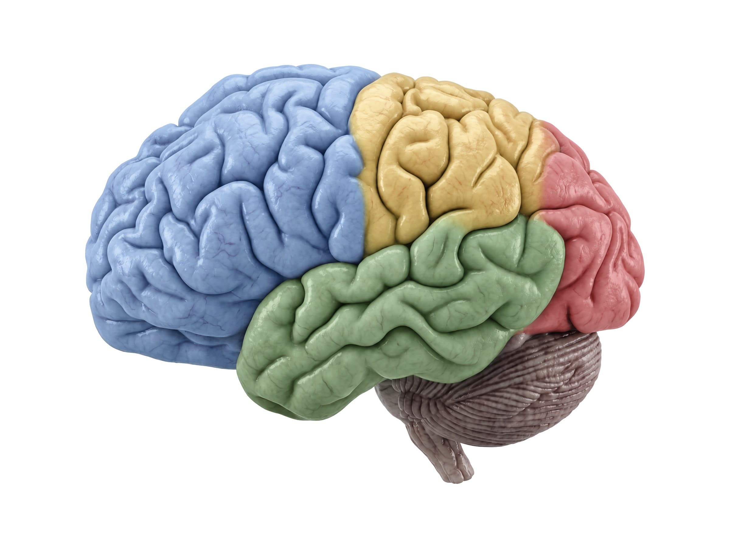
Each cerebral hemisphere is divided into four lobes, named for the skull bones that cover them. Two deep grooves mark the boundaries: the central sulcus, which runs down the side of the brain, and the lateral sulcus (Sylvian fissure), which separates the temporal lobe below.
- Frontal lobe: the front of the brain, ahead of the central sulcus. It controls planning, judgment, personality and behavior. The primary motor cortex (precentral gyrus) at its back edge controls voluntary movement, and Broca's area, usually in the left frontal lobe, produces speech. The frontal lobe is the lobe most often injured in head trauma.
- Parietal lobe: behind the central sulcus. The primary somatosensory cortex (postcentral gyrus) receives touch, pain and temperature from the opposite side of the body, and the parietal lobe combines sensation into spatial awareness.
- Temporal lobe: below the lateral sulcus, on the side of the head. It processes hearing, understands language (Wernicke's area, usually on the left) and, through the hippocampus on its inner side, forms new memories.
- Occipital lobe: the back of the brain. It contains the primary visual cortex, so damage here causes loss of vision in part of the visual field even when the eyes are healthy.
Tucked deep inside the lateral sulcus is a fifth area, the insula, involved in taste, pain and awareness of the body's internal state.
Inside the brain: the midsagittal view
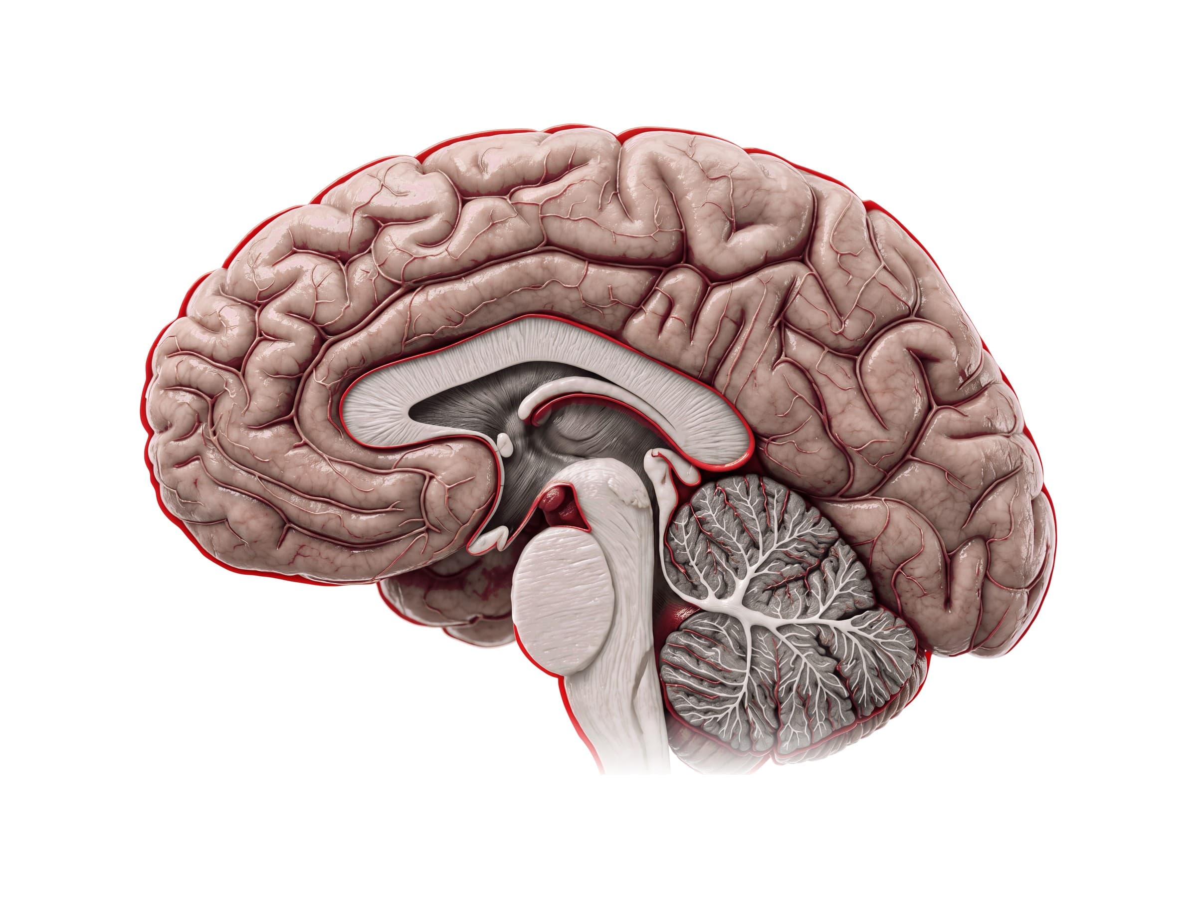
Cutting the brain down the middle shows the structures that sit between the two hemispheres.
- Corpus callosum: a thick C-shaped band of about 200 million nerve fibers that connects the left and right hemispheres.
- Cingulate gyrus: the curved fold just above the corpus callosum, part of the limbic system, involved in emotion and attention.
- Fornix: an arching band under the corpus callosum that carries memory signals from the hippocampus.
- Thalamus: a paired egg-shaped mass in the center of the brain that relays almost all sensory information (except smell) to the cortex.
- Hypothalamus: below the thalamus, above the pituitary gland. It controls hormones, body temperature, hunger, thirst and sleep. The thalamus and hypothalamus together form the diencephalon.
- Brainstem: the midbrain at the top, the bulging pons in the middle and the medulla oblongata below, continuous with the spinal cord.
- Cerebellum: in section, its white matter branches like a tree (the arbor vitae) inside folds of gray matter.
The limbic system, including the hippocampus and amygdala on the inner side of the temporal lobe, sits around these midline structures and drives memory and emotion.
Ventricles of the brain
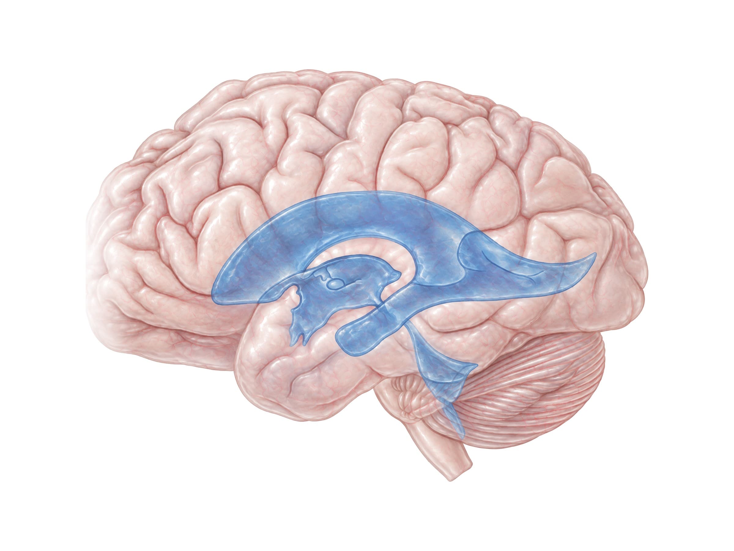
The ventricles are four connected cavities filled with cerebrospinal fluid (CSF), which cushions the brain and carries away waste. CSF is made by the choroid plexus inside the ventricles, about 500 milliliters a day, with roughly 150 milliliters circulating at any one time.
- Lateral ventricles: one C-shaped ventricle in each hemisphere, with an anterior horn in the frontal lobe, a body, a posterior horn in the occipital lobe and an inferior horn in the temporal lobe.
- Third ventricle: a narrow slit in the midline between the two halves of the thalamus, connected to each lateral ventricle by the interventricular foramen.
- Cerebral aqueduct: a thin channel through the midbrain.
- Fourth ventricle: a tent-shaped space between the brainstem and the cerebellum. CSF leaves it through small openings into the subarachnoid space around the brain and spinal cord.
When CSF flow is blocked, most often at the narrow cerebral aqueduct, fluid builds up and the ventricles enlarge (hydrocephalus). On CT, compressed or shifted ventricles are one of the main signs of swelling or bleeding inside the skull.
Base of the brain and cranial nerves
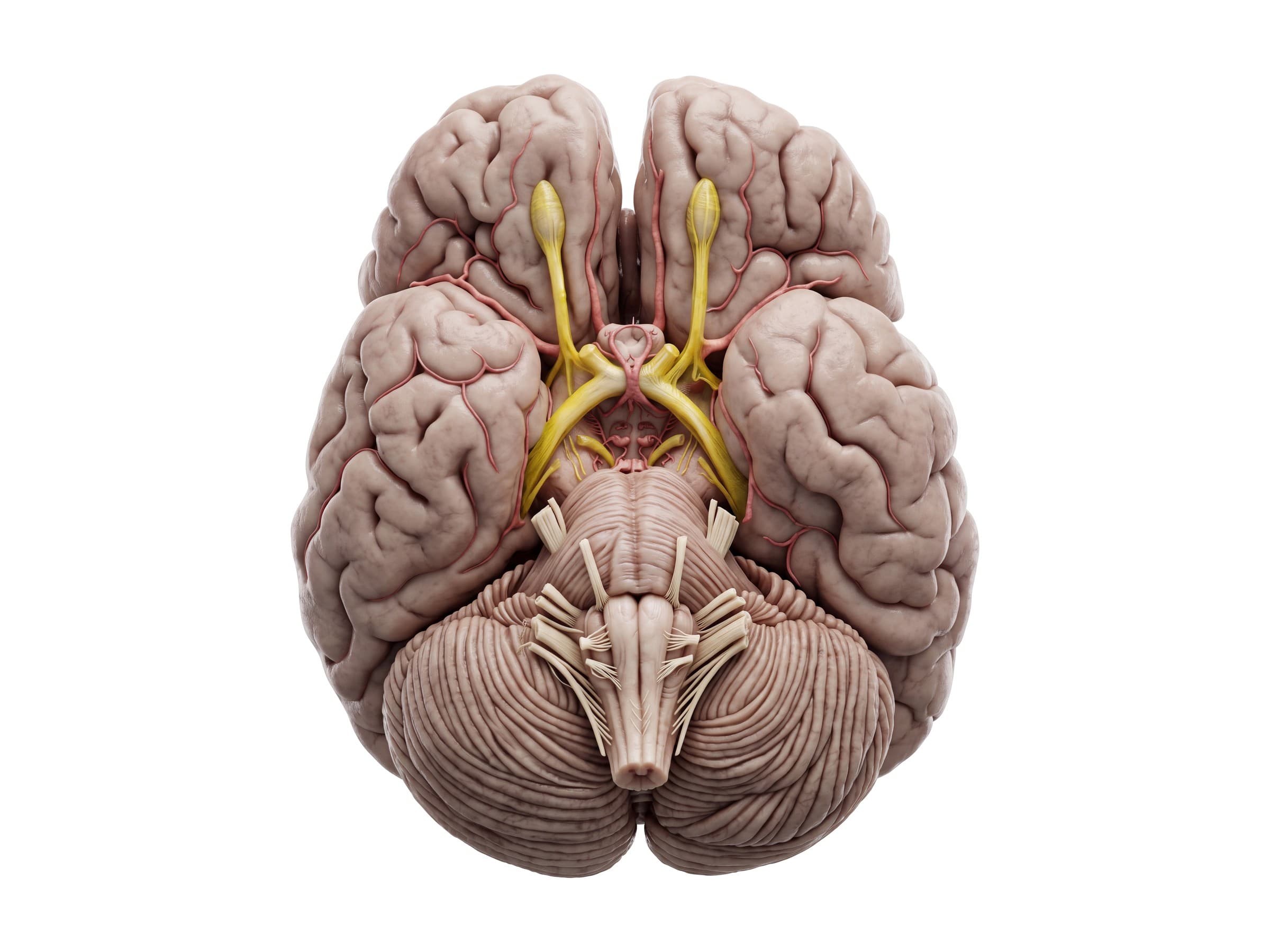
Seen from below, the underside of the frontal lobes carries the olfactory bulbs and tracts, and behind them the two optic nerves meet at the optic chiasm, where fibers from the inner half of each eye cross to the other side. The pons and medulla run down the middle, with the cerebellum on either side.
There are 12 pairs of cranial nerves:
- I. Olfactory: smell.
- II. Optic: vision.
- III. Oculomotor, IV. Trochlear and VI. Abducens: eye movement; the oculomotor nerve also constricts the pupil. A dilated pupil after a head injury can mean the oculomotor nerve is being compressed by swelling or a bleed.
- V. Trigeminal: sensation of the face and the muscles of chewing. It is the thickest cranial nerve, leaving the side of the pons.
- VII. Facial: muscles of facial expression and taste.
- VIII. Vestibulocochlear: hearing and balance.
- IX. Glossopharyngeal and X. Vagus: swallowing, voice, and, for the vagus, the heart, lungs and gut.
- XI. Accessory: the sternocleidomastoid and trapezius muscles.
- XII. Hypoglossal: movement of the tongue.
Blood supply: the circle of Willis
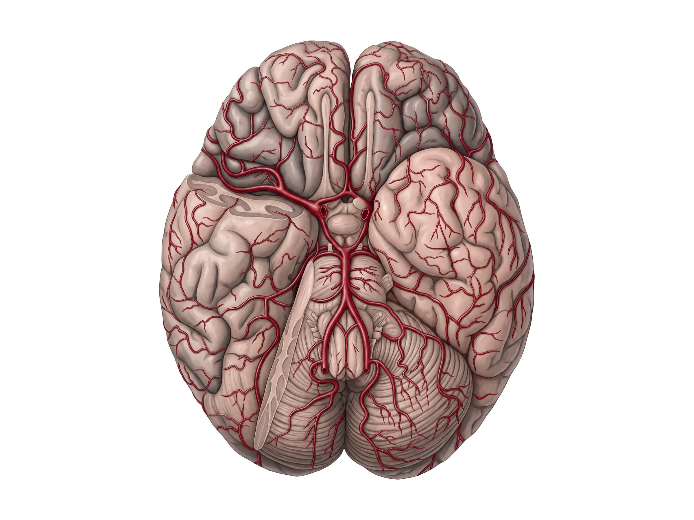
The brain uses about 20 percent of the body's oxygen and is supplied by two pairs of arteries. The two internal carotid arteries come up the front of the neck, and the two vertebral arteries come up through the neck vertebrae and join to form the basilar artery in front of the brainstem. At the base of the brain, these two systems link up in a ring of arteries, the circle of Willis, through the anterior communicating artery, the anterior cerebral arteries, the posterior communicating arteries and the posterior cerebral arteries.
From the circle, three pairs of arteries supply the cerebrum: the anterior cerebral arteries (the inner surface of the frontal and parietal lobes), the middle cerebral arteries (most of the outer surface, including the motor and language areas, and the most common site of strokes) and the posterior cerebral arteries (the occipital lobe and the underside of the temporal lobe). Berry aneurysms form at branch points of the circle of Willis, most often at the anterior communicating artery; when one ruptures, it causes a subarachnoid hemorrhage.
Meninges of the brain
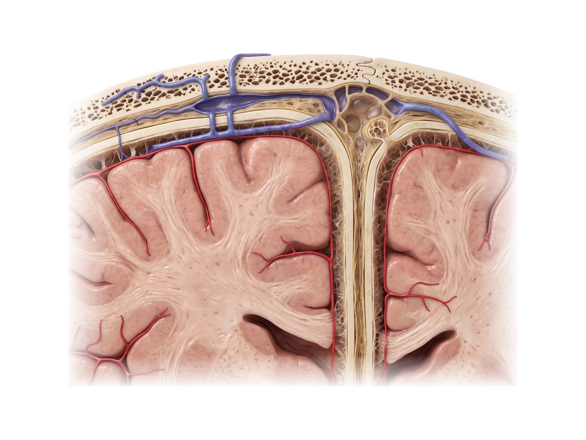
Inside the skull, the brain is wrapped in three layers of membrane, the meninges:
- Dura mater: the tough outer layer, lining the inside of the skull. It folds down between the two hemispheres as the falx cerebri and between the cerebrum and cerebellum as the tentorium. Large veins called dural venous sinuses, such as the superior sagittal sinus along the top, run between its layers and drain blood from the brain.
- Arachnoid mater: a thin middle layer under the dura. Small tufts called arachnoid granulations push into the venous sinuses and return CSF to the blood.
- Pia mater: a delicate layer that clings to every fold of the brain's surface.
The subarachnoid space between the arachnoid and pia is filled with CSF and carries the brain's surface arteries. Bridging veins cross from the brain surface through the subdural space to the sinuses, which is why they tear in a subdural hematoma.
The three potential spaces around these layers explain the three main kinds of bleeding inside the skull: epidural (above the dura), subdural (below the dura) and subarachnoid (in the CSF space).
Common brain injuries and how they appear on imaging
CT without contrast is the first scan after a head injury because it shows fresh blood and skull fractures within minutes. MRI is more sensitive for small contusions and diffuse axonal injury. Severity is graded clinically with the Glasgow Coma Scale.
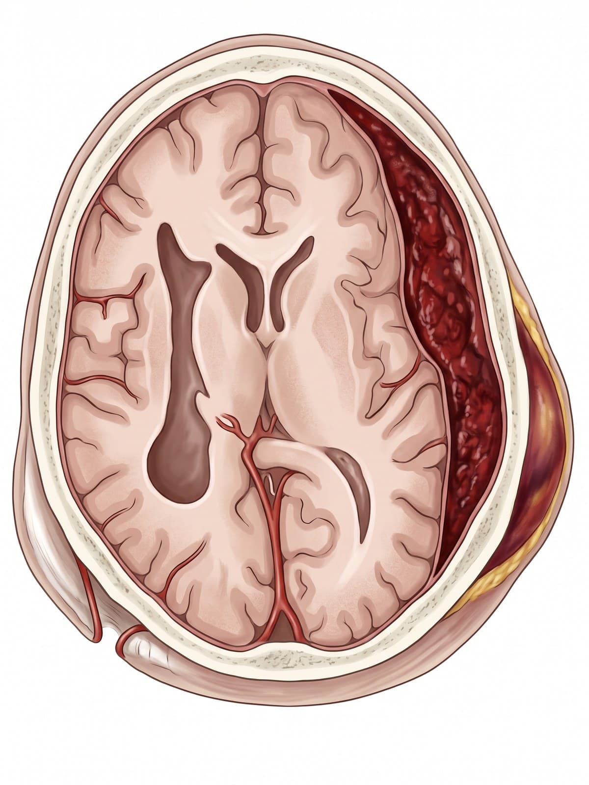
- Epidural hematoma: usually from a skull fracture at the temple that tears the middle meningeal artery. Arterial blood collects between the skull and the dura, which is fixed to the skull at the sutures, so on CT the clot is lens-shaped (biconvex) and does not cross suture lines. Classically, the person may be knocked out, wake up and seem fine (a lucid interval), then deteriorate rapidly as the clot grows. It is a surgical emergency.
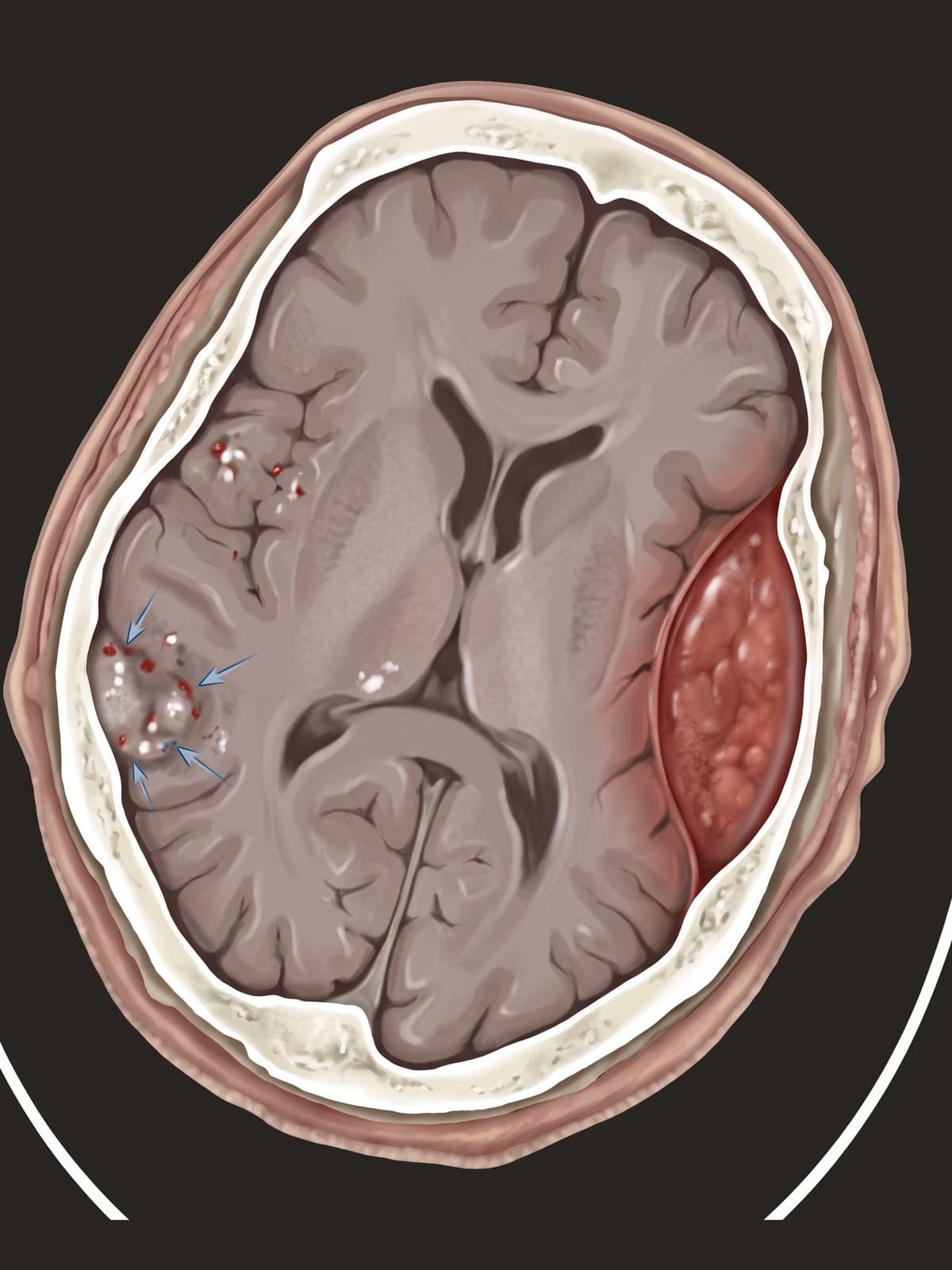
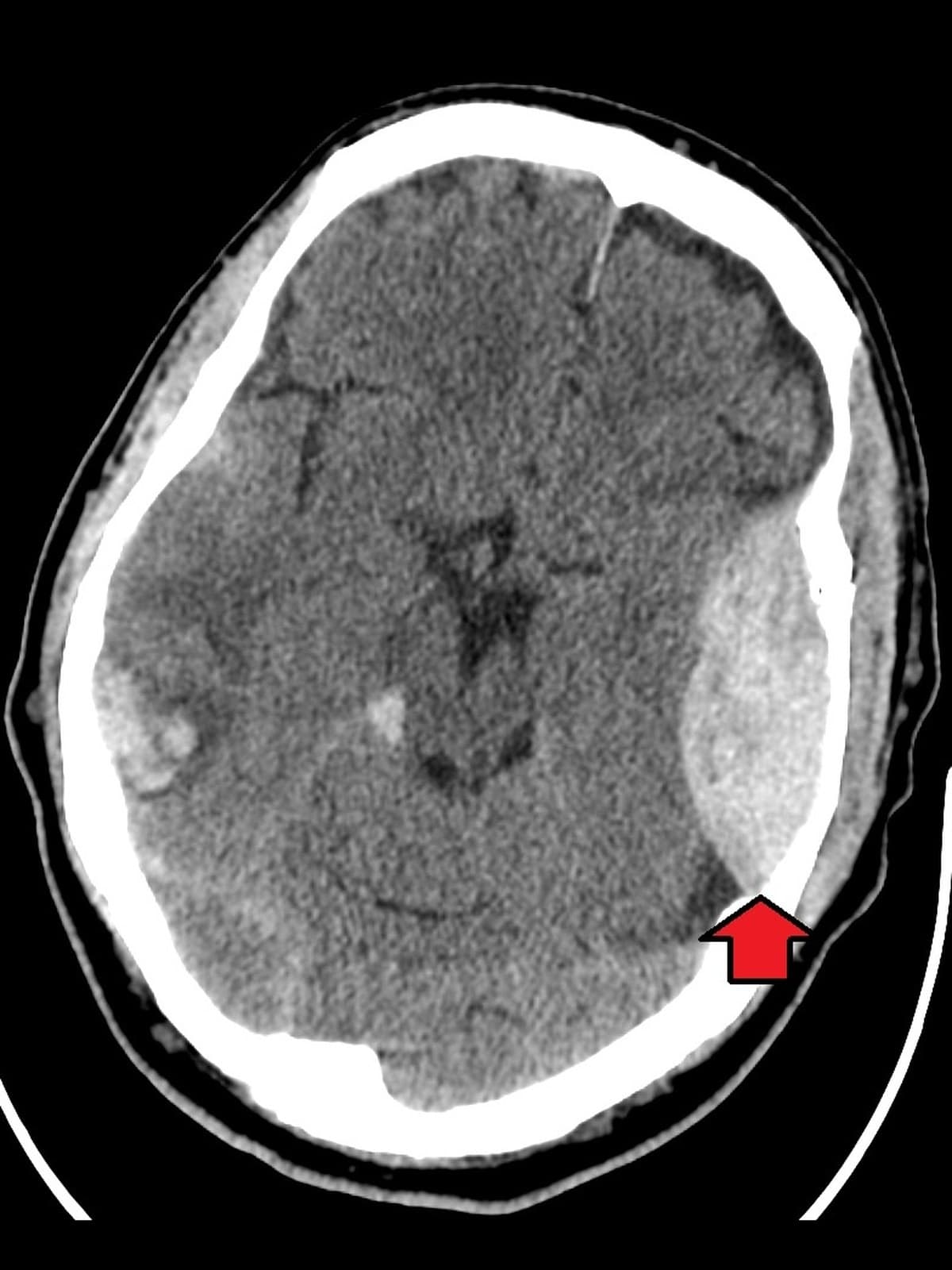
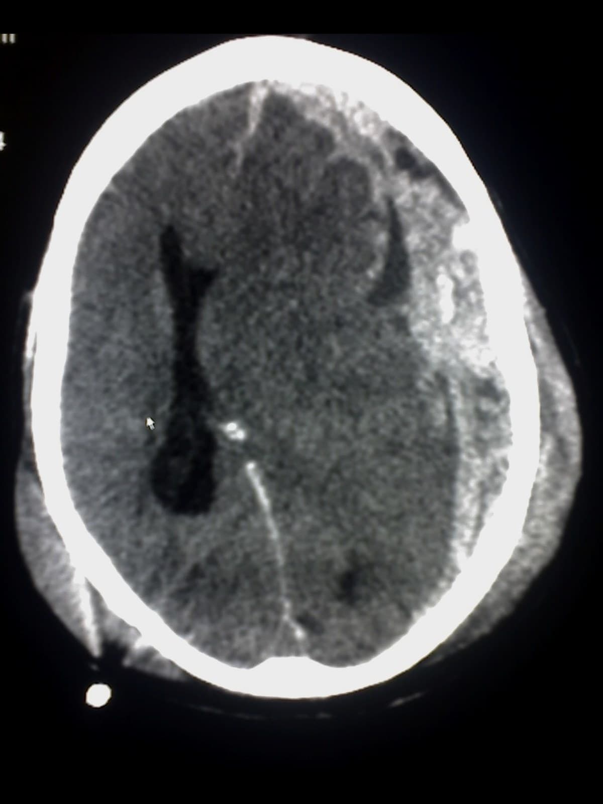
- Subdural hematoma: bleeding from torn bridging veins into the space beneath the dura, often from rapid acceleration and deceleration of the head. On CT it is crescent-shaped, follows the curve of the brain and can cross suture lines but not the midline falx. Acute subdurals follow serious trauma; chronic subdurals build up over weeks in older adults and people on blood thinners, sometimes after a minor fall.
- Subarachnoid hemorrhage: blood in the CSF space around the brain, from trauma or a ruptured aneurysm, seen on CT as blood in the sulci and around the base of the brain.
- Cerebral contusion and coup-contrecoup injury: bruising of the brain surface where it strikes the skull (coup) and on the opposite side as it rebounds (contrecoup). The undersides of the frontal and temporal lobes are most often bruised because they sit against the rough floor of the skull.
- Diffuse axonal injury: shearing of nerve fibers by rotational forces, typically in high-speed crashes. CT may look nearly normal; MRI shows small bleeds at the junction of gray and white matter, in the corpus callosum and in the brainstem.
- Concussion: a mild traumatic brain injury with temporary changes in brain function and normal standard imaging.
How the brain is shown in a personal injury case
Traumatic brain injuries carry some of the highest damages in personal injury and medical malpractice claims, and they are among the hardest injuries for a jury to see. The scans are hard for lay people to read, and the effects (memory loss, personality change, headaches) are invisible. Each type of injury follows a recognizable mechanism of injury: an epidural hematoma from a blow to the side of the head, a subdural hematoma from a fall or a sudden deceleration, diffuse axonal injury from the rotational forces of a high-speed collision. A labeled illustration of the normal brain, its lobes and its coverings is the baseline exhibit. It lets an expert explain which lobe was injured and why that matches the plaintiff's symptoms, for example frontal lobe damage and personality change, before showing the injury exhibit: the clot pressing on the brain, the midline shift, the craniotomy. In delayed-diagnosis malpractice claims, an illustration of a growing epidural or subdural hematoma over time explains why hours mattered.
Natomy creates custom medical legal illustrations of brain injuries for demand packages and trial exhibits, and the role of these visuals at trial is covered in demonstrative evidence. For the bones around the brain, see skull anatomy, and for a closer look at the top of the brain, the superior view of the brain. Related atlas pages: back muscles anatomy, shoulder anatomy, hip anatomy, knee anatomy and foot bones.
FAQ
What are the three main parts of the brain?+
The three main parts of the brain are the cerebrum, the cerebellum and the brainstem. The cerebrum is the large, folded upper part, split into left and right hemispheres, and handles thinking, memory, language, sensation and voluntary movement. The cerebellum sits under the back of the cerebrum and coordinates balance and fine movement. The brainstem connects the brain to the spinal cord and controls breathing, heart rate and consciousness.
What are the 5 main parts of the brain and their functions?+
A common five-part breakdown is: the cerebrum (thinking, memory, language and voluntary movement); the diencephalon, which contains the thalamus (the relay station for sensory signals) and the hypothalamus (hormones, temperature, hunger and thirst); the cerebellum (balance and coordination); the brainstem (breathing, heart rate, swallowing and consciousness); and the limbic system, including the hippocampus and amygdala (memory and emotion).
What are the four lobes of the brain?+
Each cerebral hemisphere has four lobes named after the skull bones above them. The frontal lobe, at the front, controls planning, decision-making, personality, speech production and voluntary movement. The parietal lobe, behind it, processes touch, pain and spatial awareness. The temporal lobe, on the side below the lateral sulcus, handles hearing, language comprehension and memory. The occipital lobe, at the back, processes vision.
What are the 7 major regions of the brain?+
One common way to count seven regions is: the cerebrum (cerebral hemispheres), the diencephalon (thalamus and hypothalamus), the midbrain, the pons, the medulla oblongata, the cerebellum, and the limbic system. The midbrain, pons and medulla together make up the brainstem. Different textbooks group the regions differently, so you will also see counts of three (cerebrum, cerebellum, brainstem) or five.
How many cranial nerves are there?+
There are 12 pairs of cranial nerves, numbered I to XII from front to back: olfactory (I), optic (II), oculomotor (III), trochlear (IV), trigeminal (V), abducens (VI), facial (VII), vestibulocochlear (VIII), glossopharyngeal (IX), vagus (X), accessory (XI) and hypoglossal (XII). The olfactory and optic nerves come from the forebrain; the other ten leave the brainstem. They control smell, vision, eye movement, facial sensation and movement, hearing and balance, swallowing and the tongue.
What is the circle of Willis?+
The circle of Willis is a ring of arteries at the base of the brain, around the pituitary stalk and optic chiasm. It connects the two internal carotid arteries in front with the basilar artery behind through the anterior communicating artery, the anterior cerebral arteries, the posterior communicating arteries and the posterior cerebral arteries. The connections can reroute blood if one supply artery narrows. It is also the most common site of brain aneurysms, especially at the anterior communicating artery.
What is the difference between an epidural and a subdural hematoma?+
An epidural hematoma is bleeding between the skull and the dura mater, usually from a torn middle meningeal artery under a skull fracture at the temple. Because the dura is stuck to the skull at the sutures, the blood forms a lens-shaped clot on CT. A subdural hematoma is bleeding beneath the dura, usually from torn bridging veins, and spreads in a crescent over the brain surface. Subdurals are more common and often occur in older adults and people on blood thinners, sometimes after a minor fall.
What are the meninges?+
The meninges are the three membranes that cover the brain and spinal cord. The tough outer dura mater lines the inside of the skull and folds inward to form the falx cerebri between the hemispheres. The thin middle arachnoid mater sits beneath it, and the delicate pia mater clings to the surface of the brain. Cerebrospinal fluid flows in the subarachnoid space between the arachnoid and the pia, cushioning the brain. Infection of these layers is meningitis.
Need a custom illustration of this joint?
Generate a labeled or exhibit-ready figure for a specific patient, injury, or case in minutes.
Try Natomy →