Skull Anatomy: Bones of the Skull, Sutures & Foramina
Labeled skull anatomy: the 22 bones of the skull, cranial sutures, fontanelles, the skull base and its foramina, the mandible, and common skull fractures.
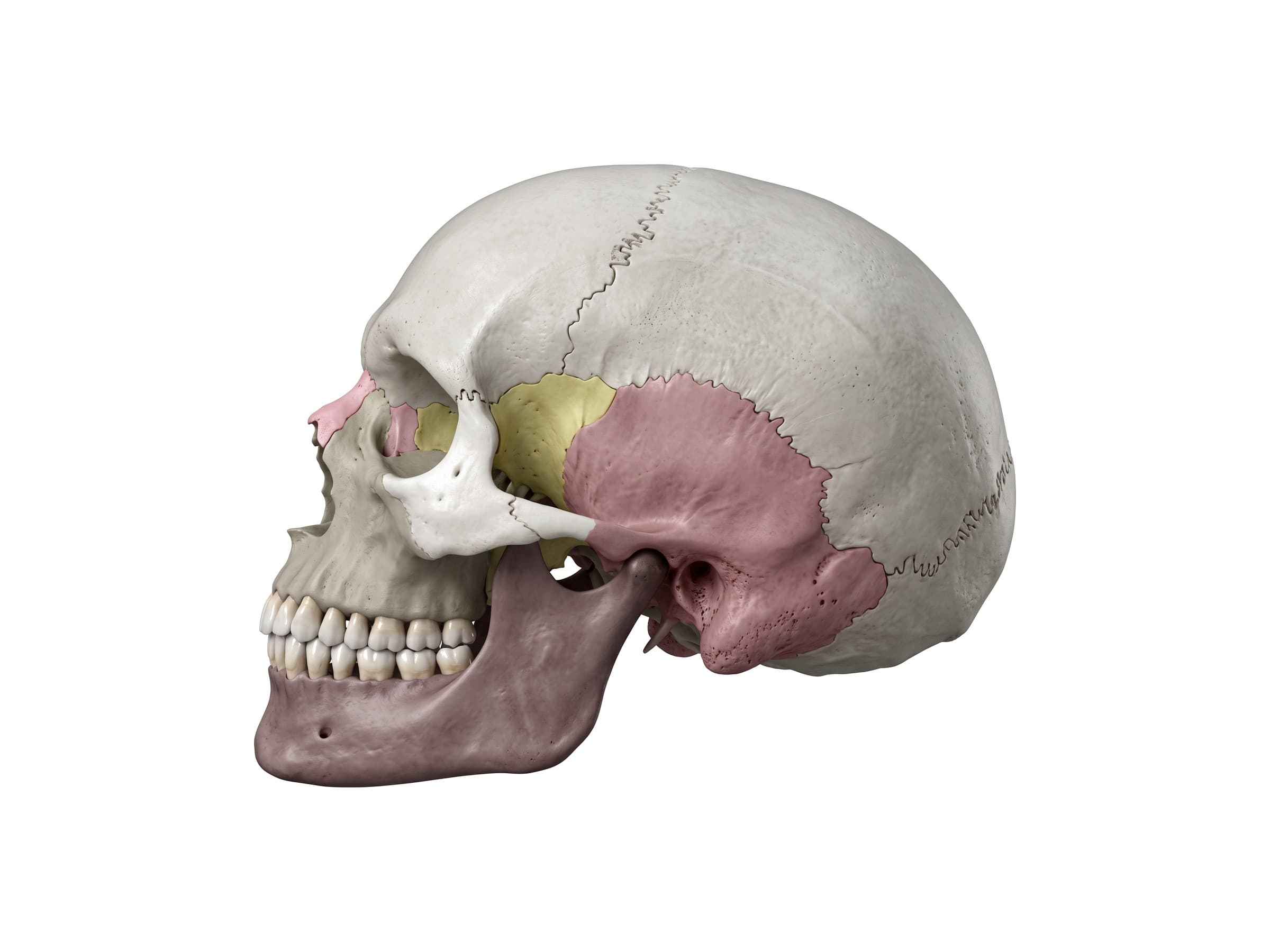
The skull is the bony framework of the head. It has 22 bones in two groups: 8 cranial bones that form the cranium around the brain (the frontal, two parietal, two temporal, occipital, sphenoid and ethmoid bones) and 14 facial bones that shape the face, eye sockets, nose and jaw. All of the skull bones except the mandible are locked together by fibrous joints called sutures, and the base of the skull is pierced by openings (foramina) that carry the cranial nerves, spinal cord and blood vessels. Skull and facial fractures are a common finding after falls, assaults, and motor vehicle collisions, so knowing the bones, sutures and landmarks is the first step to reading a CT report, explaining a head injury to a patient, or illustrating a traumatic brain injury claim.
Bones of the skull
From the side, the skull divides into the rounded cranium above and behind, and the facial skeleton in front and below. The frontal bone forms the forehead and the roof of the eye sockets. Behind it, the two parietal bones form most of the top and upper sides of the skull, and the occipital bone forms the back. Low on each side, the temporal bone holds the ear canal (the external acoustic meatus) and the mastoid process behind it. In front of the temporal bone, the greater wing of the sphenoid fills the temple.
Cranial bones
- Frontal bone: the forehead, the upper rims and roofs of the orbits, and the frontal sinuses.
- Parietal bones (2): the top and upper sides of the cranium, meeting each other at the sagittal suture.
- Temporal bones (2): the lower sides of the cranium. Each has a flat squamous part, the zygomatic process that forms the back half of the zygomatic arch, the mastoid process, and the dense petrous part in the skull base that houses the inner and middle ear.
- Occipital bone: the back of the skull and the back of the skull base, surrounding the foramen magnum.
- Sphenoid bone: a butterfly-shaped bone that runs across the middle of the skull base. Its body holds the sella turcica, the seat of the pituitary gland, and its greater wings appear on the side of the skull at the temple.
- Ethmoid bone: a light, air-filled bone between the orbits. Its cribriform plate forms the roof of the nasal cavity and lets the olfactory nerves pass up to the brain.
Facial bones
From the front, the face is built around the two orbits and the nasal cavity. The two nasal bones form the bridge of the nose. The maxillae (upper jaw bones) form the middle of the face, the floor of each orbit, the sides of the nasal cavity and the upper tooth row, and each holds a large maxillary sinus. The zygomatic bones (cheekbones) form the outer rims of the orbits and the prominence of the cheek. The mandible is the lower jaw.
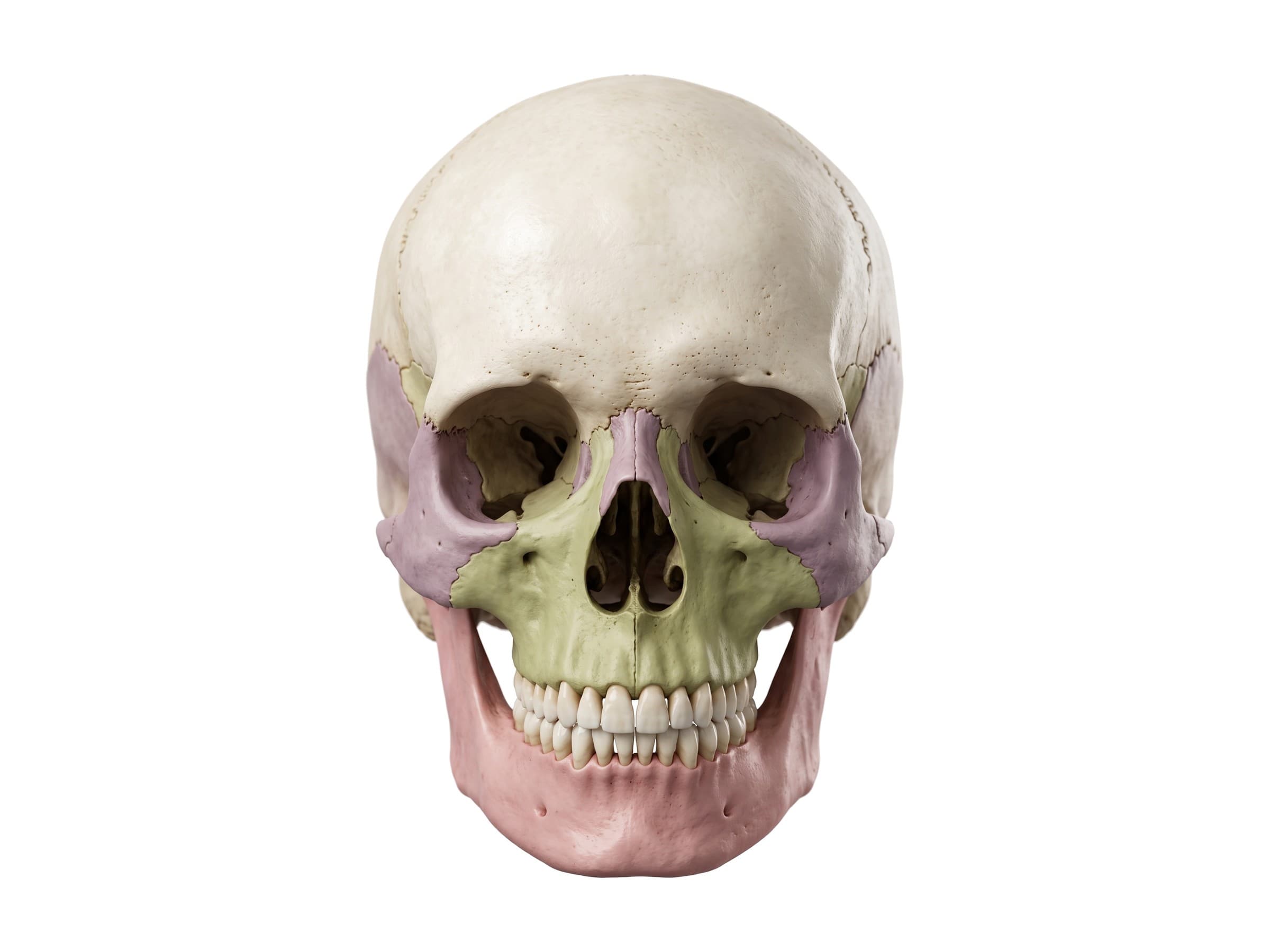
- Nasal bones (2): the bony bridge of the nose; the most commonly fractured facial bones.
- Maxillae (2): the upper jaw, the floor of the orbit, and the hard palate in front.
- Zygomatic bones (2): the cheekbones, joined to the temporal bone by the zygomatic arch.
- Lacrimal bones (2): tiny bones in the inner wall of each orbit, holding the tear sac.
- Palatine bones (2): the back of the hard palate and part of the nasal cavity walls.
- Inferior nasal conchae (2): scroll-shaped bones on the side walls of the nasal cavity.
- Vomer: a thin plate forming the lower back of the nasal septum.
- Mandible: the lower jaw, the only movable bone of the skull.
Sutures of the skull
The skull bones are joined by sutures, fibrous joints with interlocking, saw-tooth edges that allow almost no movement in adults. From the side, the coronal suture runs across the top of the skull between the frontal and parietal bones, the squamous suture arches over the temporal bone where it overlaps the parietal, and the lambdoid suture runs down the back between the parietal and occipital bones. From above, the sagittal suture runs along the midline between the two parietal bones. For a dedicated suture diagram with and without labels, see the skull sutures labelled diagram.
Where the coronal and sagittal sutures meet is called the bregma, and where the sagittal and lambdoid sutures meet is the lambda. On the side of the skull, the pterion is the H-shaped junction of the frontal, parietal, sphenoid and temporal bones, the thinnest part of the skull wall.
Fontanelles of the newborn skull
At birth, the skull bones are not yet joined. The sutures are wide, and where several of them meet, the gaps are covered only by membrane: the fontanelles, or soft spots. The anterior fontanelle is the diamond-shaped soft spot on top of the head where the two halves of the frontal bone meet the two parietal bones; it usually closes between 12 and 18 months. The posterior fontanelle is a smaller triangle where the parietal bones meet the occipital bone; it closes by about 2 months. Smaller sphenoidal and mastoid fontanelles on each side close in the first year or two. In a newborn, the frontal bone is still in two halves, separated by the metopic suture.
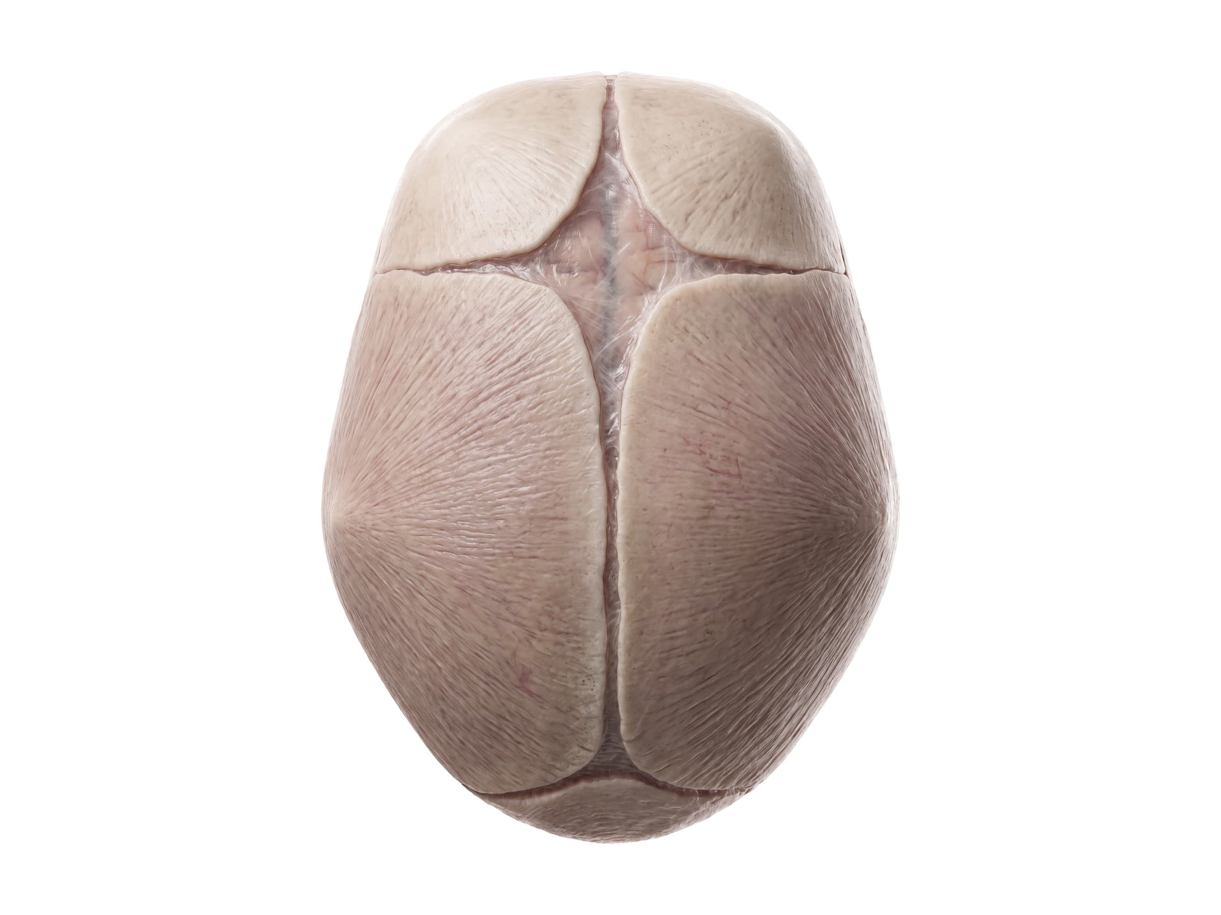
Fontanelles let the skull bones overlap during delivery and expand as the brain grows. A tense, bulging fontanelle can be a sign of raised pressure inside the skull, and a sunken one a sign of dehydration. Early fusion of a suture (craniosynostosis) changes the shape of the head, depending on which suture closes.
Base of the skull and its foramina
With the top of the skull removed, the floor of the cranial cavity steps down from front to back in three levels:
- Anterior cranial fossa: formed by the frontal bone, the cribriform plate of the ethmoid in the middle, and the lesser wings of the sphenoid at the back. It holds the frontal lobes of the brain.
- Middle cranial fossa: butterfly-shaped, formed by the sphenoid and the temporal bones. In the middle sits the sella turcica, which holds the pituitary gland. The lateral parts hold the temporal lobes.
- Posterior cranial fossa: the deepest, formed mainly by the occipital bone and the back of the petrous part of the temporal bone. It holds the cerebellum and brainstem.
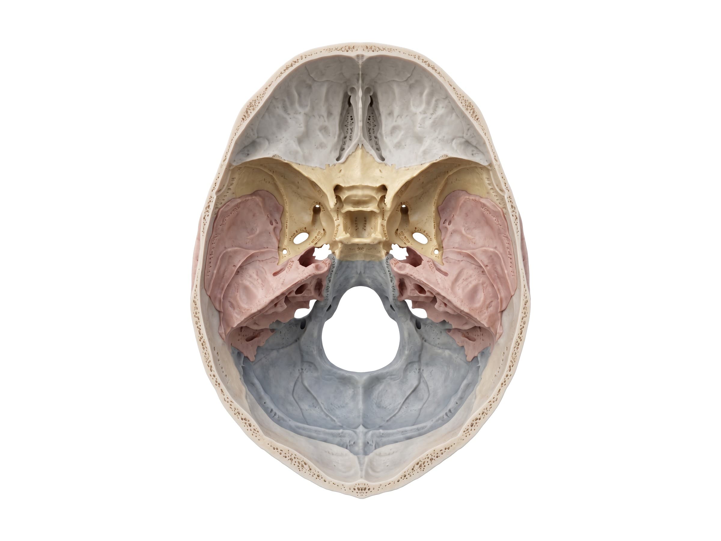
The skull base is pierced by openings that carry nerves and vessels. In the middle cranial fossa, the foramen rotundum carries the maxillary nerve, the foramen ovale carries the mandibular nerve, and the small foramen spinosum carries the middle meningeal artery up into the skull. In the posterior fossa, the foramen magnum carries the brainstem down into the spinal canal, and the internal acoustic meatus and jugular foramen carry the facial, vestibulocochlear, glossopharyngeal, vagus and accessory nerves and the internal jugular vein.
| Opening | Bone | What passes through |
|---|---|---|
| Cribriform plate | Ethmoid | Olfactory nerves (CN I) |
| Optic canal | Sphenoid | Optic nerve (CN II), ophthalmic artery |
| Superior orbital fissure | Sphenoid | CN III, IV, V1 and VI |
| Foramen rotundum | Sphenoid | Maxillary nerve (V2) |
| Foramen ovale | Sphenoid | Mandibular nerve (V3) |
| Foramen spinosum | Sphenoid | Middle meningeal artery |
| Internal acoustic meatus | Temporal | Facial (VII) and vestibulocochlear (VIII) nerves |
| Jugular foramen | Temporal and occipital | CN IX, X and XI, internal jugular vein |
| Hypoglossal canal | Occipital | Hypoglossal nerve (CN XII) |
| Foramen magnum | Occipital | Medulla and spinal cord, vertebral arteries |
The orbit
Each orbit (eye socket) is a four-sided bony cone built from seven bones: the frontal bone above, the zygomatic bone and greater wing of the sphenoid on the outside, the maxilla below, and the ethmoid, lacrimal and sphenoid on the inside wall. The rim of the orbit is thick and strong, but the floor (the maxilla, over the maxillary sinus) and the inner wall (the paper-thin lamina papyracea of the ethmoid) are among the thinnest bones in the body. That is why a blow to the eye often breaks the floor or inner wall while the rim stays intact.
The mandible
The mandible is the largest and strongest facial bone and the only skull bone that moves. Its horseshoe-shaped body carries the lower teeth in the alveolar process and meets in the midline at the chin, the mental protuberance. Below the second premolar, the mental foramen lets the mental nerve out to supply the skin of the lower lip and chin. At the back, the body turns up at the angle of the mandible into the vertical ramus, which ends in two processes separated by the mandibular notch: the coronoid process in front, where the temporalis muscle attaches, and the condyle behind, which sits on its neck and forms the temporomandibular joint with the temporal bone.
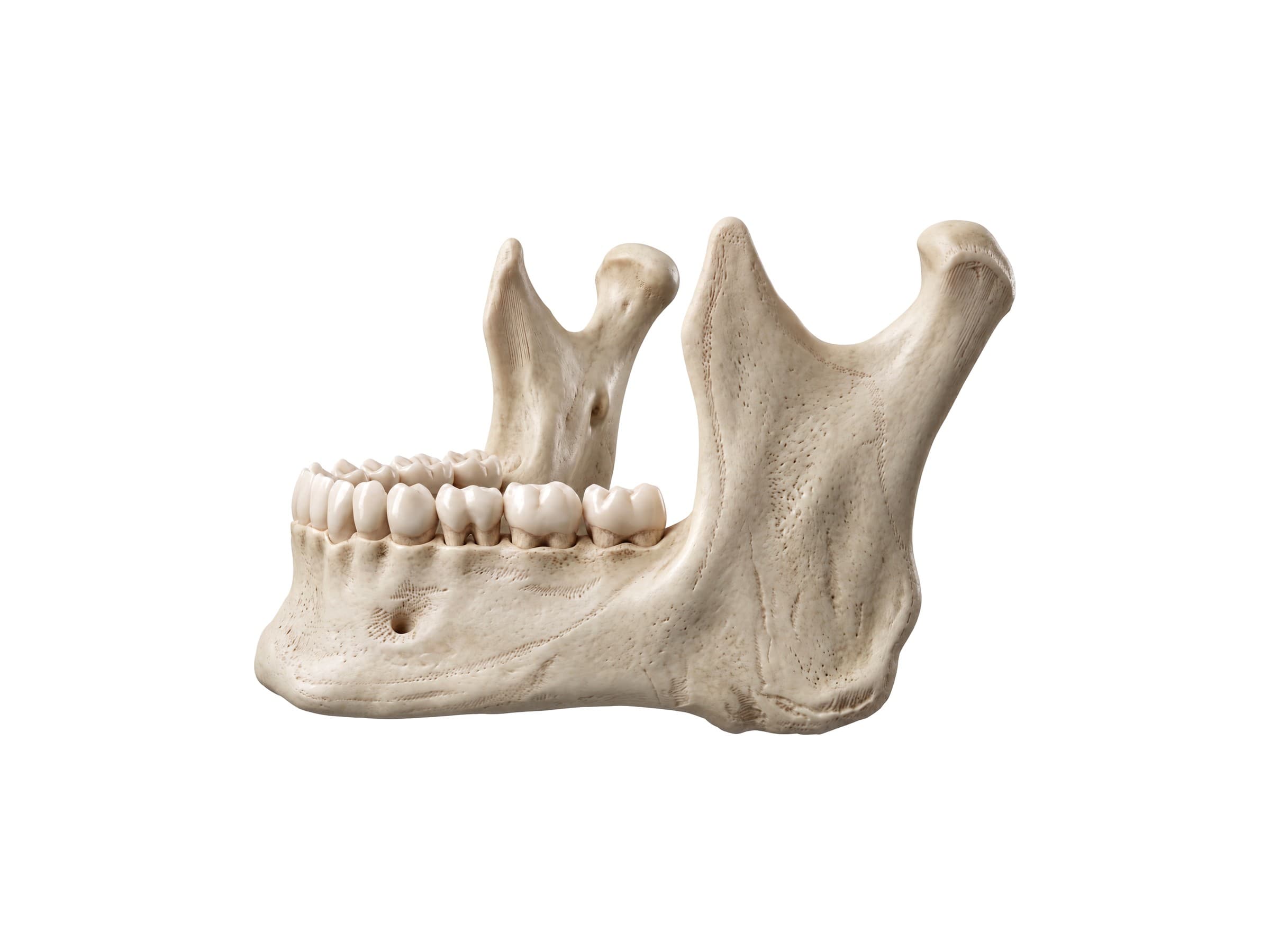
Because it is a ring with the skull, the mandible often breaks in two places, typically at the point of impact and on the opposite side at the angle or condyle.
Common skull fractures and how they appear on imaging
Skull fractures are diagnosed on CT, which shows both the bone and any bleeding inside the skull. Most are described by pattern and location:
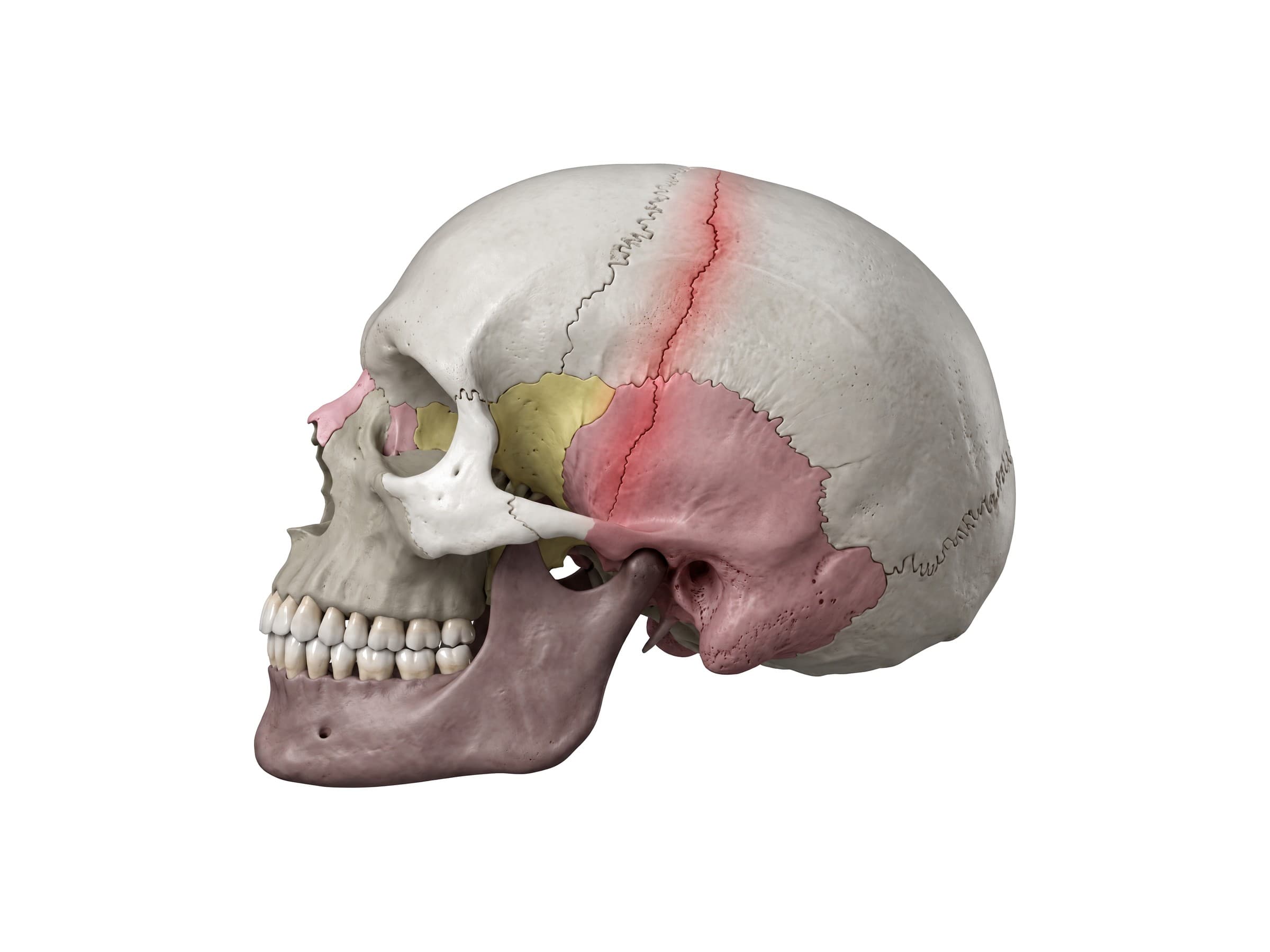
- Linear skull fracture: a straight break through the full thickness of the bone without displacement, the most common type. On its own it usually heals without treatment, but a linear fracture across the temporal bone near the pterion can tear the middle meningeal artery and cause an epidural hematoma, a lens-shaped collection of blood between the skull and dura that can become life-threatening within hours.
- Depressed skull fracture: a fragment of bone pushed inward, usually from a blow with a small, hard object. A fragment depressed more than the thickness of the skull, an open wound, or injury to the brain beneath often needs surgery to lift the bone.
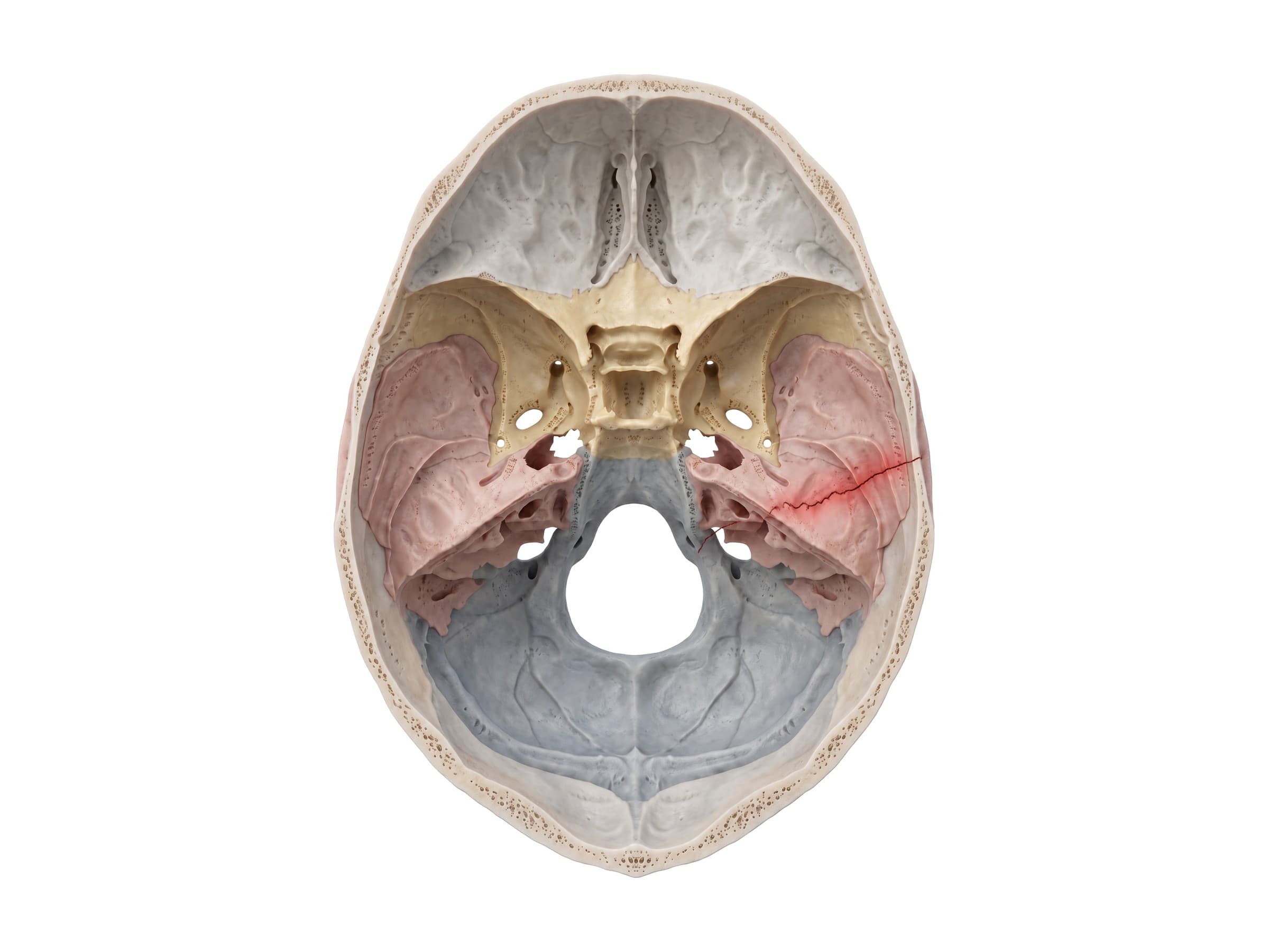
- Basilar skull fracture: a break in the bones of the skull base, most often the petrous temporal bone, the sphenoid, or the occipital bone. Clinical signs include bruising behind the ear (Battle sign), bruising around the eyes (raccoon eyes), blood behind the eardrum, and cerebrospinal fluid leaking from the nose or ear. It is best seen on thin-slice CT of the skull base and can injure the facial and hearing nerves.
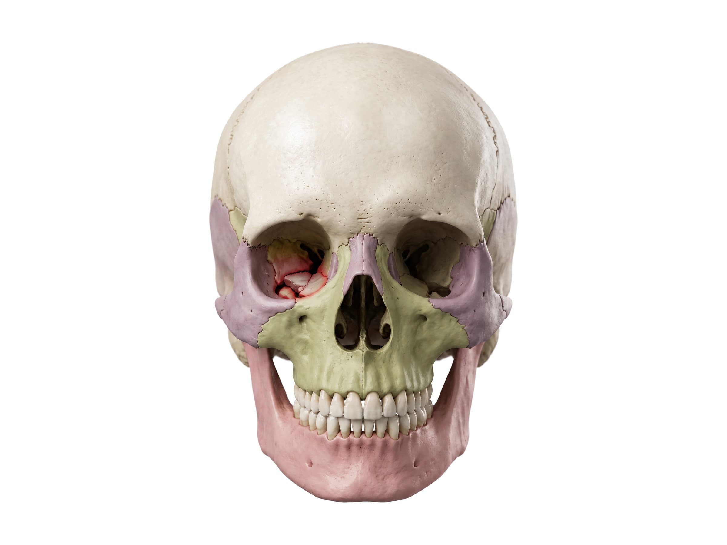
- Orbital blowout fracture: a blow to the eye from an object larger than the orbit (a fist, a ball, a dashboard) raises the pressure inside the orbit and breaks the thin floor or inner wall, while the rim stays intact. Orbital fat or the inferior rectus muscle can herniate through the floor into the maxillary sinus and trap the eye's upward movement, causing double vision. Coronal CT shows the defect and the herniated tissue.
- Facial fractures: nasal bone fractures are the most common; zygomaticomaxillary complex (tripod) fractures separate the cheekbone at its three connections; and Le Fort I, II and III fractures separate the maxilla or the whole midface from the skull at progressively higher levels, typically in high-energy motor vehicle collisions.
- Mandible fractures: most often at the condyle, the angle, and the body, frequently in two places at once. A panoramic X-ray (Panorex) or CT shows them.
Epidural vs subdural hematoma: an epidural hematoma lies between the skull and the dura, usually from an arterial bleed under a fracture, and stops at the sutures, so it looks lens-shaped (biconvex) on CT. A subdural hematoma lies between the dura and the brain, usually from torn bridging veins after rapid acceleration and deceleration, often without a fracture; it crosses sutures and looks crescent-shaped.
Skull anatomy for artists
For drawing the head, the skull sets the proportions everything else sits on. From the side, the cranium is roughly a ball, and the face hangs off its front lower quarter. The brow ridge (the supraorbital margin of the frontal bone) sits about halfway down the head, and the eye sockets sit just below it. The zygomatic arch lines up roughly with the bottom of the nose and the top of the ear, and the ear opening sits just behind the jaw joint. The angle of the mandible lines up with the ear lobe, and the cheekbones mark the widest point of the face. From the front, the skull is about two-thirds as wide as it is tall, and the orbits are about one eye-width apart. For flat reference views to trace or study, see the labelled lateral view of the skull and anterior view of the skull.
How the skull is shown in a personal injury case
A labeled illustration of normal skull anatomy is the baseline exhibit in many head-injury claims: falls, assaults, motor vehicle collisions, and workplace injuries. It shows a jury where the temporal bone, the pterion, and the skull base sit before the injury-specific exhibit is introduced, such as the fracture line on CT, the epidural hematoma beneath it, or the plates and screws from a facial fracture repair. Because the brain injury itself is often what the case is about, a skull exhibit that traces the mechanism of injury, the point of impact, the fracture, and the bleed that followed, connects the force of the event to the medical findings in a way a CT slice alone does not.
Natomy creates custom medical legal illustrations of skull fractures and traumatic brain injuries for demand packages and trial exhibits. For more on how normal anatomy is used in court, see demonstrative evidence. For the brain itself, see the superior view of the brain. Related atlas pages: shoulder anatomy, back muscles anatomy, hand and wrist anatomy, elbow anatomy, hip anatomy, knee anatomy and ankle anatomy.
FAQ
How many bones are in the human skull?+
The adult human skull has 22 bones: 8 cranial bones that enclose the brain and 14 facial bones. The cranial bones are the frontal, the two parietal, the two temporal, the occipital, the sphenoid, and the ethmoid. The facial bones are the two nasal, two maxillae, two zygomatic, two lacrimal, two palatine, two inferior nasal conchae, the vomer, and the mandible. Counting the six ear ossicles and the hyoid bone, which are sometimes included, brings the total to 29.
What are the 8 cranial bones?+
The eight cranial bones are the frontal bone (forehead), the two parietal bones (the top and upper sides), the two temporal bones (the lower sides, containing the ear), the occipital bone (the back and base, around the foramen magnum), the sphenoid bone (a butterfly-shaped bone across the middle of the skull base), and the ethmoid bone (between the eye sockets, forming the roof of the nasal cavity).
What are the sutures of the skull?+
Sutures are the fibrous joints between the skull bones. The four main ones are the coronal suture (between the frontal and the two parietal bones, running across the top of the head from ear to ear), the sagittal suture (between the two parietal bones, along the midline), the lambdoid suture (between the parietal bones and the occipital bone, at the back), and the squamous suture (between the parietal and temporal bones on each side). In infants, the metopic suture also runs down the middle of the forehead; it usually fuses in the first year.
What is the pterion and why is it important?+
The pterion is the H-shaped junction on the side of the skull where four bones meet: the frontal, parietal, sphenoid (greater wing), and temporal. It sits about two finger-widths above the zygomatic arch and a thumb's width behind the eye socket. The bone is thinnest here, and the anterior branch of the middle meningeal artery runs just underneath it, so a blow to the side of the head can fracture the pterion, tear the artery, and cause an epidural hematoma.
What is a basilar skull fracture?+
A basilar skull fracture is a break in the bones of the skull base, most often the temporal bone (including its petrous part), the sphenoid, or the occipital bone around the foramen magnum. It usually results from significant head trauma. Signs include bruising behind the ear (Battle sign), bruising around both eyes (raccoon eyes), blood behind the eardrum, and clear cerebrospinal fluid leaking from the nose or ear. It is diagnosed with a thin-slice CT of the skull base.
What is the difference between an epidural and a subdural hematoma?+
An epidural hematoma is bleeding between the skull and the dura mater, usually from a torn middle meningeal artery under a temporal bone fracture. It is limited by the sutures, where the dura is firmly attached, so on CT it looks lens-shaped (biconvex). A subdural hematoma is bleeding between the dura and the brain, usually from torn bridging veins, often without a fracture. It can spread across sutures, so on CT it looks crescent-shaped and follows the curve of the brain.
What are fontanelles?+
Fontanelles are the soft, membrane-covered gaps in a newborn's skull where the sutures meet before the bones have fully grown together. The anterior fontanelle, the diamond-shaped soft spot on top of the head where the frontal and parietal bones meet, usually closes between 12 and 18 months. The smaller posterior fontanelle, where the parietal bones meet the occipital bone, closes by about 2 months. Fontanelles let the skull compress during birth and expand as the brain grows.
What is the foramen magnum?+
The foramen magnum is the large opening in the occipital bone at the base of the skull. The lower end of the brainstem (the medulla oblongata) passes through it to become the spinal cord, along with the vertebral arteries and the spinal roots of the accessory nerve. It is the largest foramen in the skull and sits just above the first cervical vertebra (the atlas), which articulates with the occipital condyles on either side of it.
Need a custom illustration of this joint?
Generate a labeled or exhibit-ready figure for a specific patient, injury, or case in minutes.
Try Natomy →