Facial Muscles Labeled: Muscles of the Face, Facial Nerve & Masseter
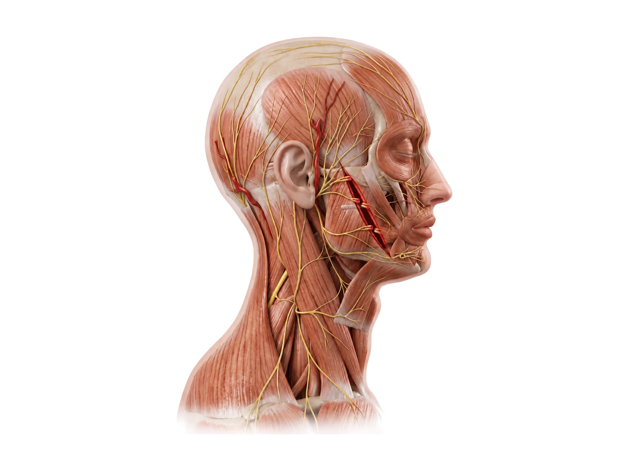
Labeled facial muscles: the muscles of facial expression, the masseter and other chewing muscles, the facial nerve and its branches, and the parotid gland.
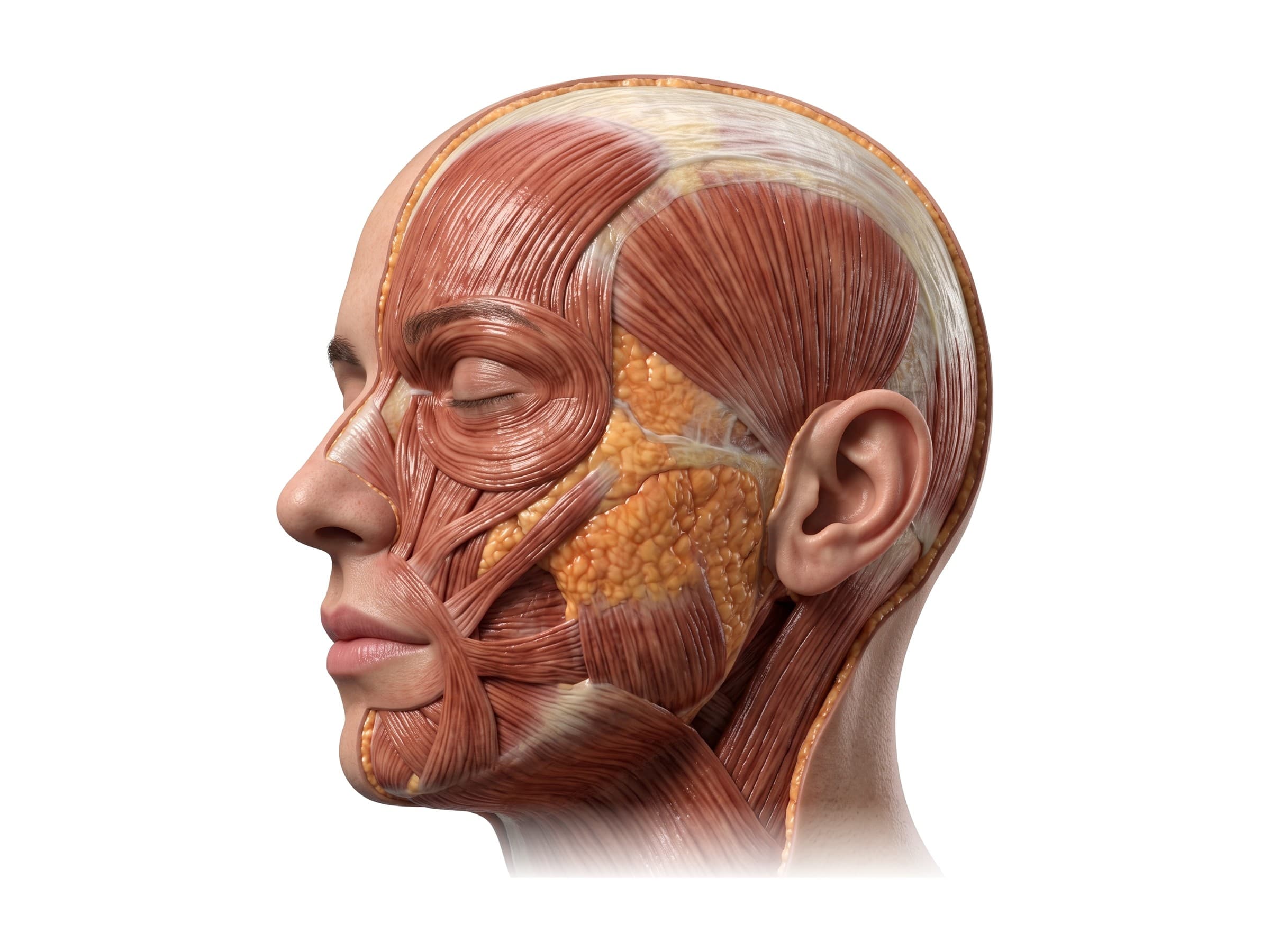
The facial muscles are the thin muscles under the skin of the face that open and close the eyes, nose and mouth and create every facial expression, together with the larger chewing muscles that move the jaw. Facial muscle anatomy comes down to three groups: the muscles of facial expression (more than 20 on each side, all supplied by the facial nerve), the four muscles of mastication (the masseter, temporalis and two pterygoids, supplied by the trigeminal nerve), and the platysma in the neck. Knowing where each one lies, and which branch of the facial nerve reaches it, is the first step to understanding facial nerve palsy, a cheek laceration, or a scar that changes how a face moves.
Facial muscles labeled
The hero illustration above shows the superficial layer of the left side of the face with the skin removed. Unlike most muscles, the muscles of facial expression attach to the skin, so when they contract they pull the skin into lines and folds. They are grouped around the openings of the face:
- Around the eye: orbicularis oculi, corrugator supercilii and procerus.
- Around the nose: nasalis and the small depressor septi.
- Around the mouth: orbicularis oris, the muscles that lift the lip (levator labii superioris, levator anguli oris, zygomaticus major and minor), the muscles that lower it (depressor anguli oris, depressor labii inferioris, mentalis), risorius and buccinator.
- Scalp and ear: frontalis and occipitalis, joined by the epicranial aponeurosis, plus the small auricular muscles.
- Neck: platysma.
Muscles of the forehead and scalp
- Frontalis: the broad muscle of the forehead. It raises the eyebrows and wrinkles the forehead into horizontal lines. It is supplied by the temporal branch of the facial nerve, so injury to that branch leaves the eyebrow drooping on one side.
- Occipitalis: the muscle at the back of the scalp. With the frontalis it forms the occipitofrontalis, two muscle bellies joined by a tough sheet of tendon over the top of the head called the epicranial aponeurosis (galea aponeurotica).
- Corrugator supercilii: a small muscle under the inner end of the eyebrow that pulls the brows together and down, producing the vertical frown lines between them.
- Procerus: a small muscle over the bridge of the nose that pulls the inner eyebrows down, making horizontal lines at the top of the nose. The corrugator and procerus are the muscles treated for frown (glabellar) lines.
Orbicularis oculi: the muscles around the eye
The orbicularis oculi is a ring of muscle around the eye that closes the eyelids. It has three parts:
- Orbital part: the thick outer ring over the bone around the eye socket. It squeezes the eye tightly shut and causes crow's feet when you squint or smile.
- Palpebral part: the thin inner part inside the eyelids, used for gentle closing and blinking.
- Lacrimal part: a small deep slip behind the tear sac that helps drain tears from the eye into the nose.
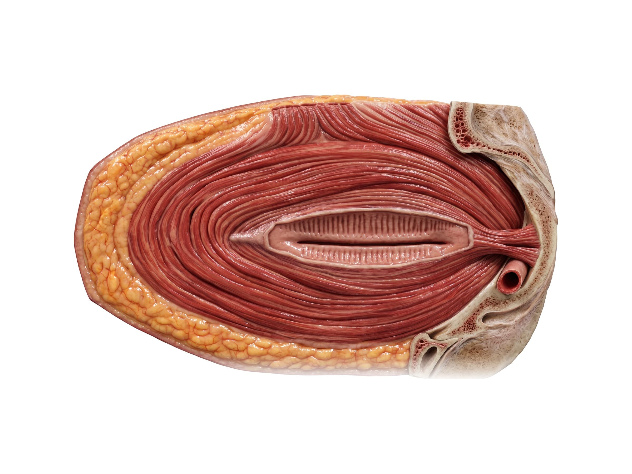
The orbicularis oculi is supplied by the temporal and zygomatic branches of the facial nerve. When these branches stop working, as in Bell's palsy, the eye cannot close fully and the cornea is at risk of drying out. The muscle that lifts the upper eyelid, levator palpebrae superioris, is not a facial muscle: it sits inside the eye socket and is supplied by the oculomotor nerve, so a drooping eyelid (ptosis) points to a different nerve than an eye that will not close.
Muscles of the nose
- Nasalis: the main muscle of the nose. Its transverse part runs across the bridge and compresses the nostril; its alar part flares the nostril open during deep breathing.
- Depressor septi: a small muscle under the nose that pulls the tip of the nose down.
- Levator labii superioris alaeque nasi: a slip that runs down the side of the nose to the nostril and upper lip, lifting both (the snarl).
Muscles of the mouth
Most facial muscles insert around the mouth, where they meet at a dense knot of fibers just outside each corner called the modiolus.
- Orbicularis oris: the ring of muscle inside the lips that closes and purses the mouth. It is essential for speech, eating and drinking without leaking.
- Zygomaticus major: runs from the cheekbone to the corner of the mouth and pulls it up and out in a smile. Zygomaticus minor lies just in front of it and lifts the upper lip.
- Levator labii superioris: lifts the upper lip and deepens the fold between the nose and mouth (the nasolabial fold).
- Levator anguli oris: lies deep to the zygomaticus and lifts the corner of the mouth.
- Risorius: a thin strip that pulls the corner of the mouth sideways. It is absent in some people.
- Depressor anguli oris: pulls the corner of the mouth down in a frown.
- Depressor labii inferioris: pulls the lower lip down.
- Mentalis: the muscle of the chin. It raises and pushes out the lower lip (pouting) and wrinkles the skin of the chin.
- Buccinator: the muscle that forms the wall of the cheek. It holds the cheek against the teeth so food stays between them while chewing, and it is used to blow out air. Although it lies deep, it is a muscle of facial expression and is supplied by the facial nerve.
Platysma
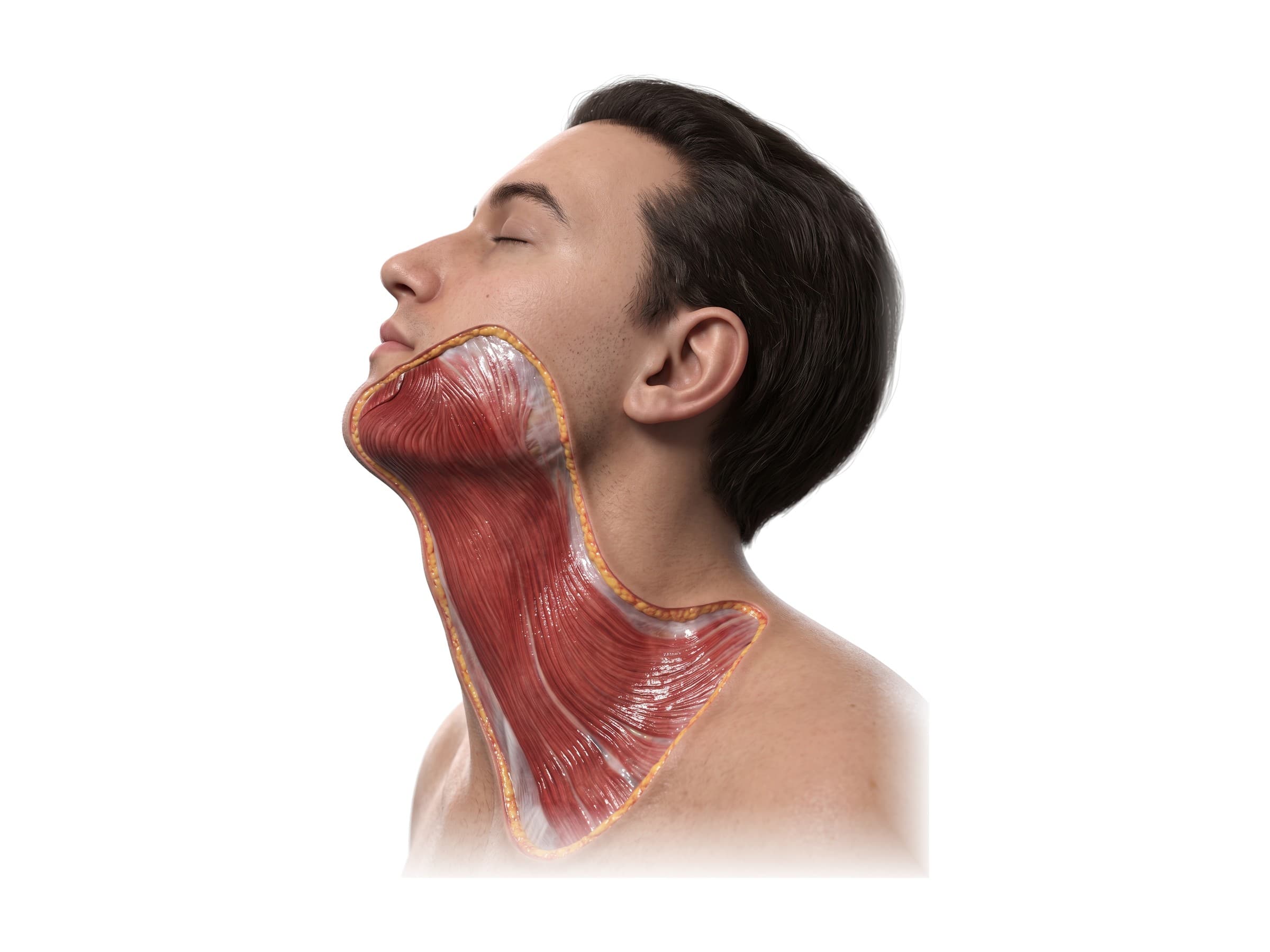
The platysma is a thin, broad sheet of muscle just under the skin of the neck. It starts over the upper chest and collarbone and runs up over the jaw, where its fibers blend with the muscles of the lower lip and chin. It tenses the skin of the neck and helps pull the corners of the mouth down. It is supplied by the cervical branch of the facial nerve.
The SMAS
The muscles of facial expression are linked by a continuous layer of fibrous tissue called the superficial musculoaponeurotic system (SMAS). It connects the platysma below to the fascia over the temple and the epicranial aponeurosis above, and it carries the facial muscles and the fat above them as a single layer. The branches of the facial nerve run just underneath it, which is why surgeons who lift or tighten the SMAS in a facelift must work in a plane that protects them.
Muscles of mastication: masseter, temporalis and pterygoids
The four chewing muscles lie deeper than the muscles of facial expression and move the lower jaw (mandible) at the jaw joint (temporomandibular joint). All four are supplied by the mandibular branch of the trigeminal nerve, not the facial nerve.
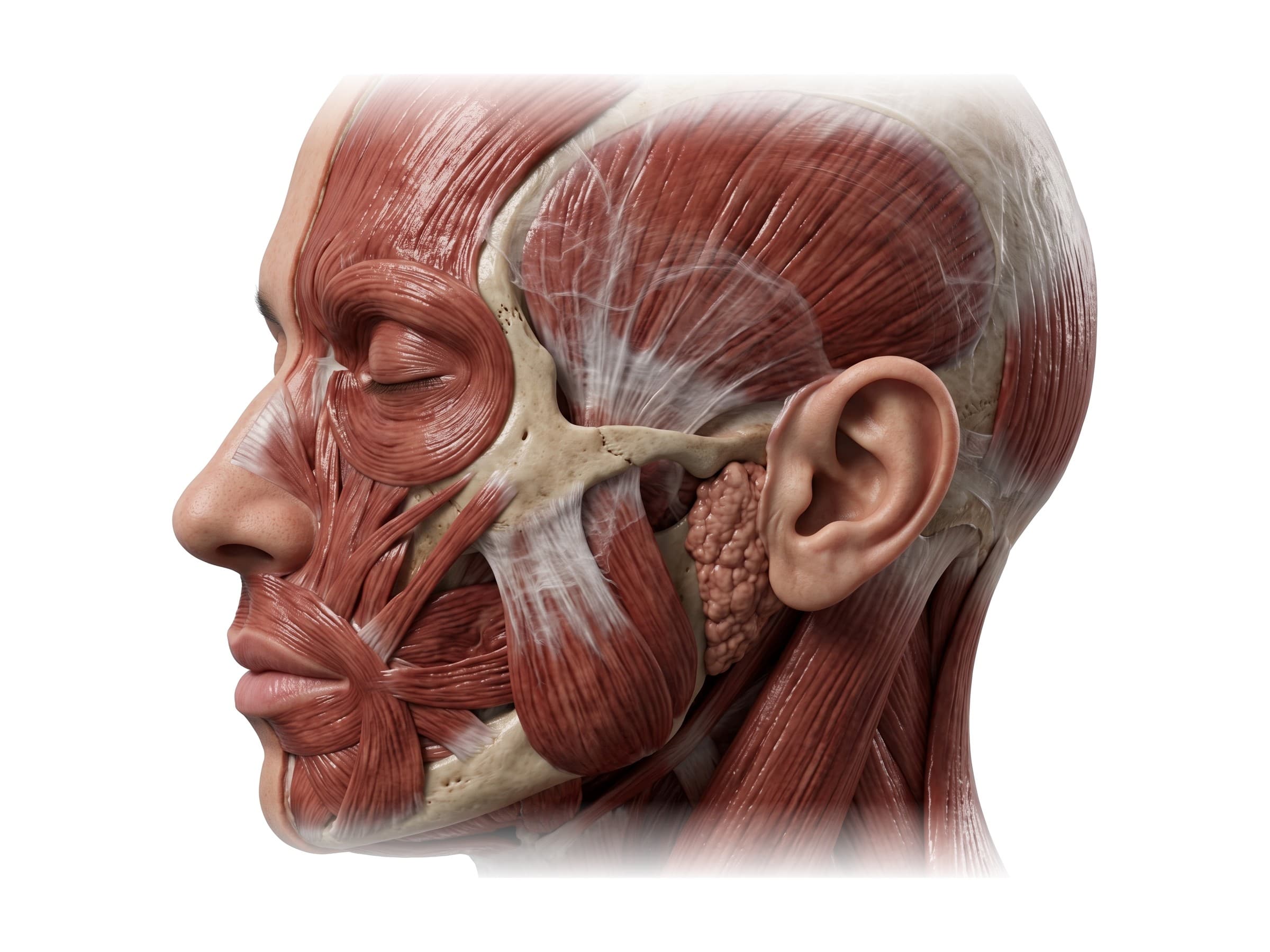
- Masseter: the thick, rectangular muscle at the side of the jaw. It runs from the zygomatic arch (the cheekbone) down to the outer surface of the angle of the mandible and closes the jaw with great force. It bulges when you clench your teeth, and it enlarges in people who grind their teeth (bruxism).
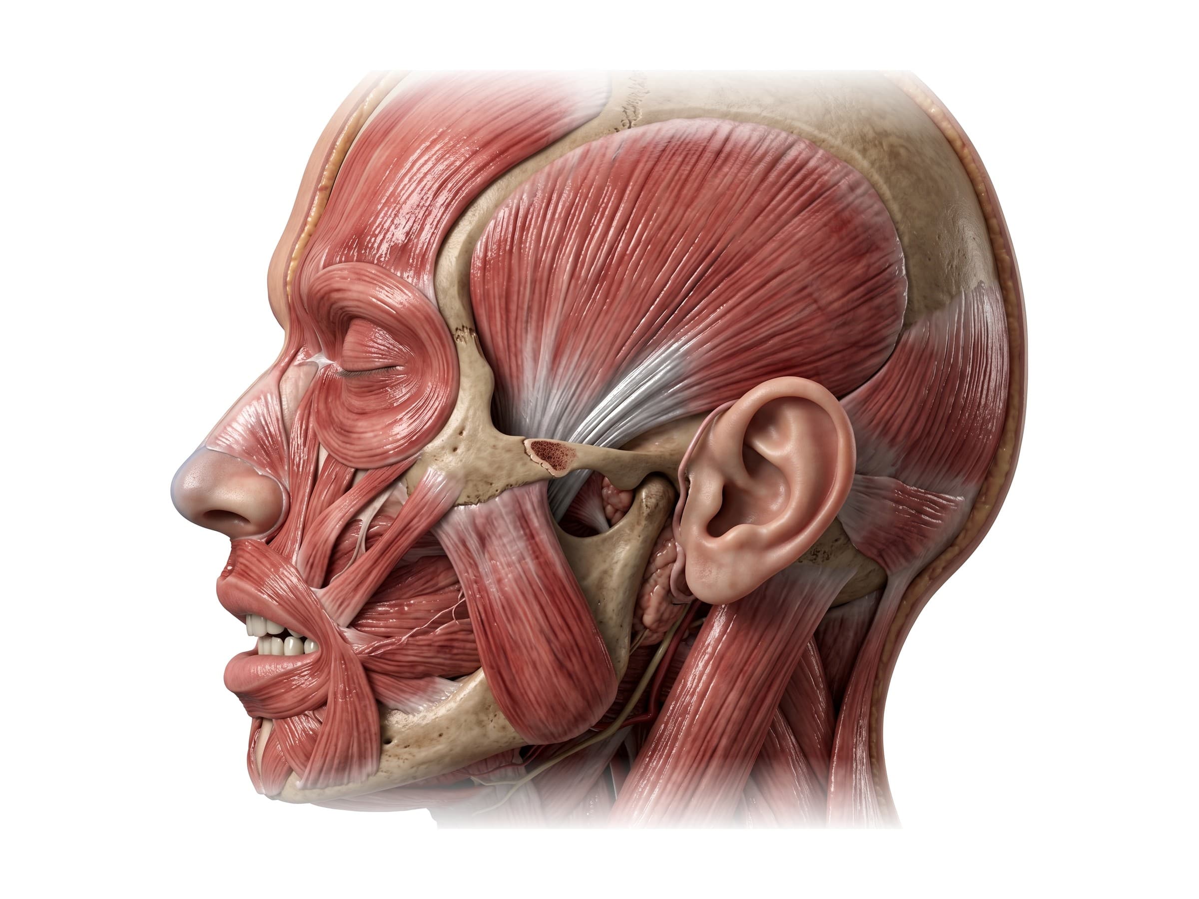
- Temporalis: a fan-shaped muscle that fills the hollow at the side of the skull (the temporal fossa). Its fibers converge into a tendon that passes under the zygomatic arch to the coronoid process of the mandible. It closes the jaw and pulls it backward, and you can feel it contract at the temple when you clench.
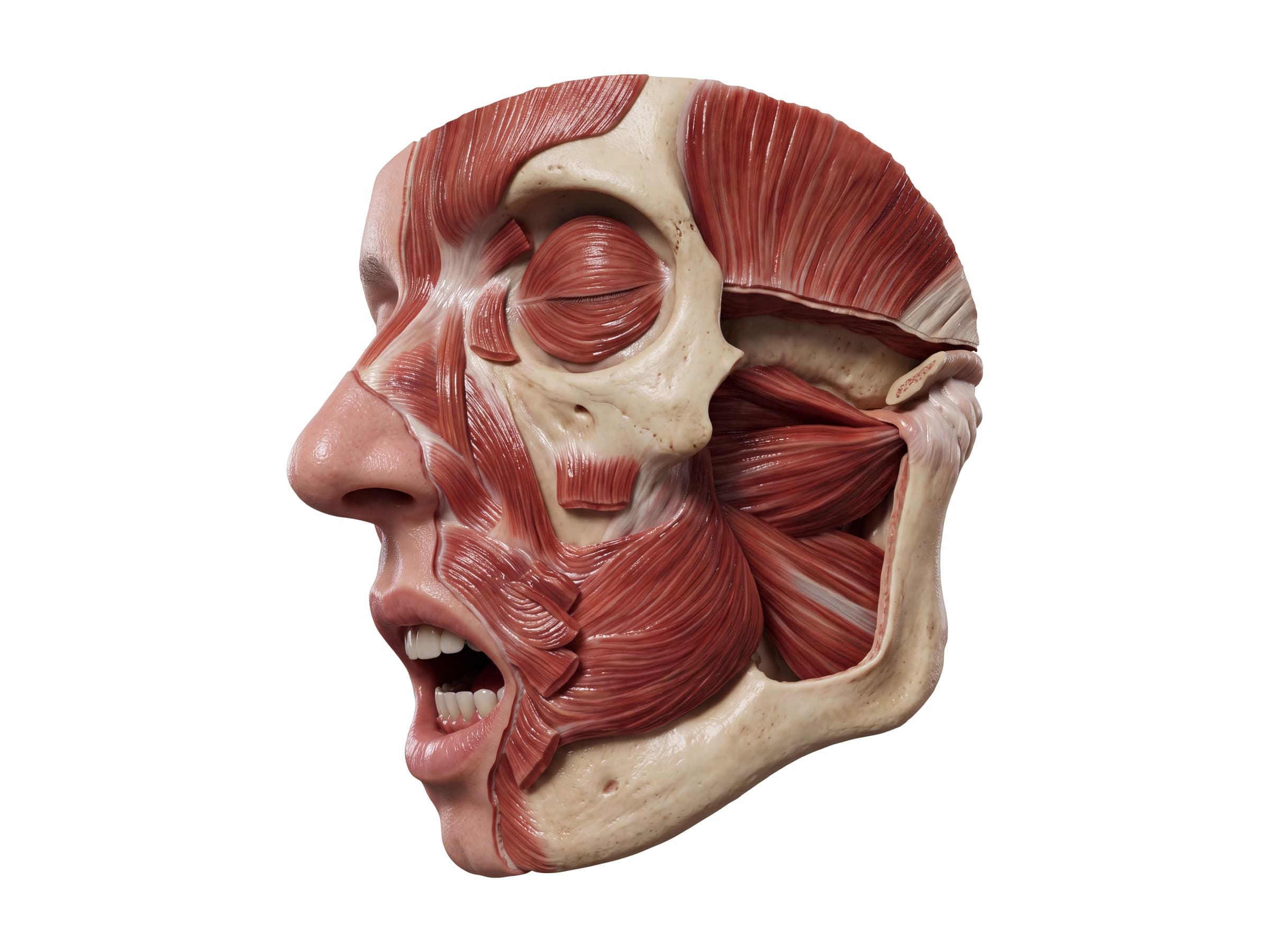
- Lateral pterygoid: a short, nearly horizontal muscle deep to the jaw that pulls the head of the mandible and the joint disc forward. It is the main muscle that opens the mouth and moves the jaw from side to side.
- Medial pterygoid: a thick muscle on the inner side of the jaw, running from the skull base down to the inner surface of the angle of the mandible. It mirrors the masseter on the inside and helps close the jaw.
Facial nerve branches
The facial nerve (cranial nerve VII) leaves the skull through a small opening behind the ear (the stylomastoid foramen), enters the parotid gland and divides inside it into five branches that fan out across the face:
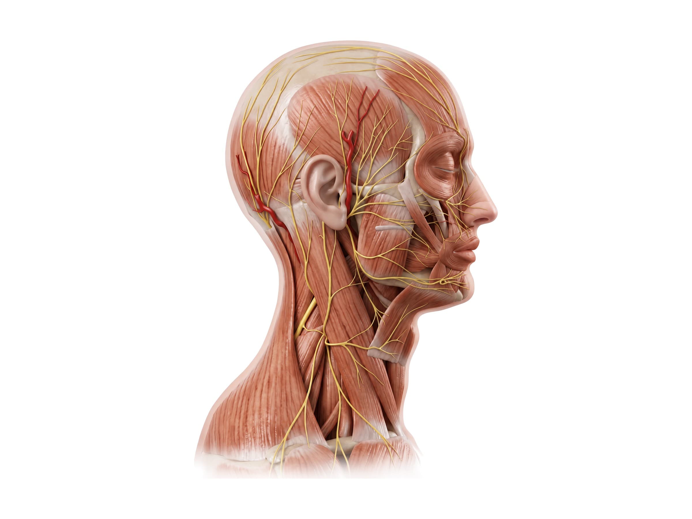
- Temporal branch: crosses the zygomatic arch to the frontalis, the upper orbicularis oculi and the corrugator. It runs close to the skin over the arch, where it is easily injured.
- Zygomatic branches: run along the cheekbone to the orbicularis oculi and help close the eye.
- Buccal branches: cross the masseter alongside the parotid duct to the muscles of the cheek, the upper lip and the nose, including the buccinator and zygomaticus.
- Marginal mandibular branch: runs along the lower border of the jaw to the muscles of the lower lip and chin. Injury leaves the lower lip unable to move down on one side, producing a crooked smile.
- Cervical branch: passes down into the neck to supply the platysma.
Before it enters the parotid gland, the nerve also gives off the posterior auricular branch to the occipitalis and the muscles behind the ear. Higher up, inside the temporal bone, other fibers carry taste from the front of the tongue and control tear and saliva production. The facial nerve controls movement only: sensation in the skin of the face comes from the trigeminal nerve. For where the facial nerve leaves the brainstem, see the cranial nerves on the brain anatomy page.
The face is also supplied by the facial artery, which crosses the lower border of the mandible just in front of the masseter and winds up toward the corner of the mouth and the side of the nose, and by the superficial temporal artery, which runs up in front of the ear to the scalp.
Parotid gland and parotid duct
The parotid gland is the largest salivary gland. It sits in front of and below the ear, wrapped around the back of the mandible and overlapping the masseter. Saliva drains forward through the parotid duct (Stensen's duct), which runs across the outer surface of the masseter, turns inward through the buccinator and opens inside the cheek opposite the upper second molar. A small accessory parotid gland often lies along the duct.
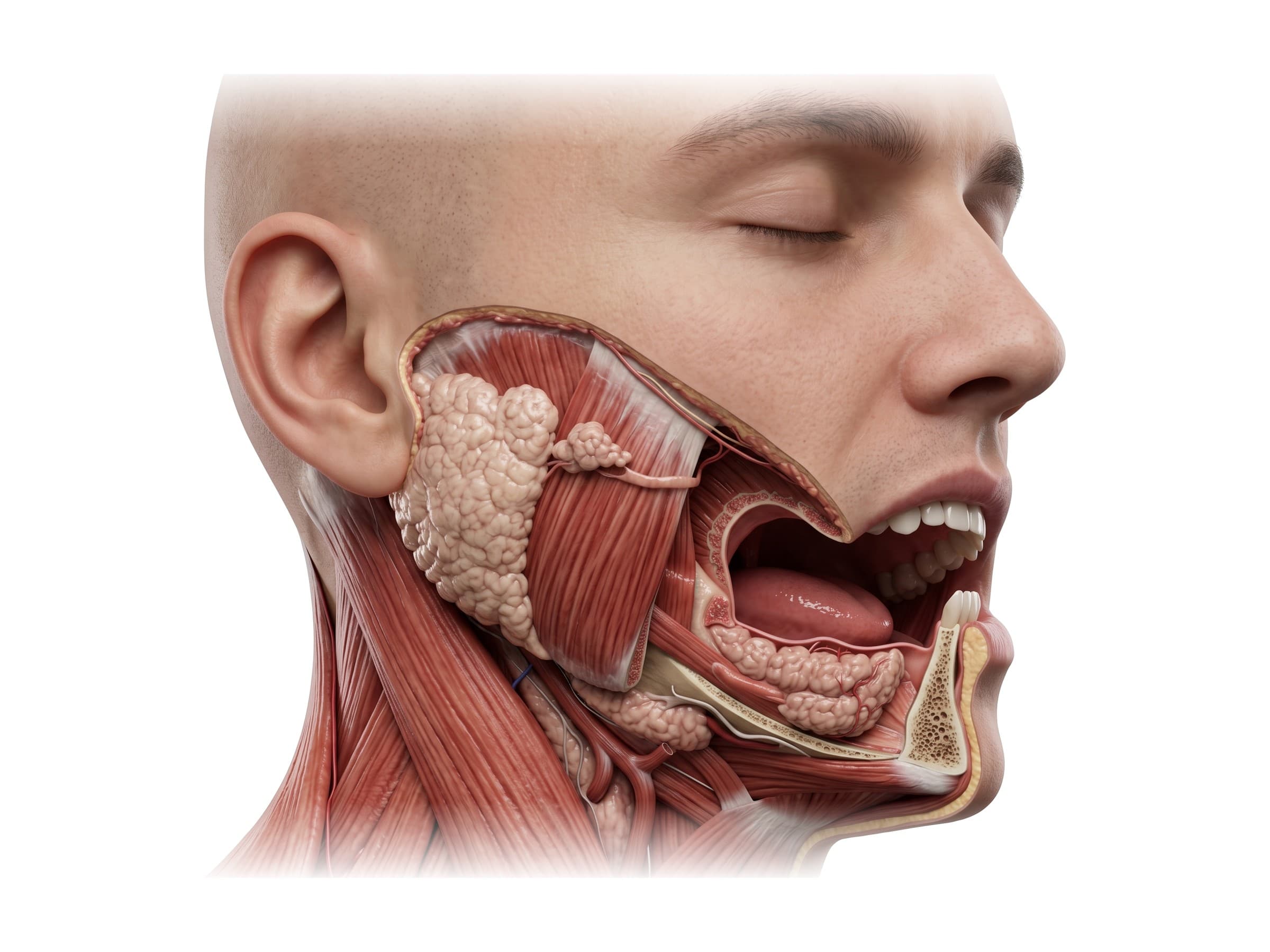
The two smaller salivary glands sit below the jaw: the submandibular gland under the angle of the mandible and the sublingual gland under the tongue. The facial nerve passes through the parotid gland and divides inside it, which is why parotid tumors and parotid surgery carry a risk of facial weakness, and why a cheek wound deep enough to cut the parotid duct can also cut the nerve branches that run with it.
Common facial muscle and facial nerve injuries
- Facial nerve injury from lacerations: a deep cut across the cheek can sever the zygomatic or buccal branches, weakening eye closure or the smile on that side. Cuts close to the ear, where the branches are larger and fewer, cause the most weakness and are usually repaired with microsurgery. Facial nerve function is graded on scales such as House-Brackmann, from normal (I) to total paralysis (VI).
- Bell's palsy: sudden paralysis of one side of the face from inflammation of the facial nerve. The forehead is involved, which helps separate it from a stroke. Imaging is usually not needed unless the weakness is atypical or does not improve.
- Facial nerve injury in skull fractures: a fracture through the temporal bone (a basilar skull fracture) can injure the nerve inside its bony canal; see skull anatomy. CT shows the fracture line.
- Parotid duct injury: a cheek laceration that cuts the duct can leak saliva into the tissues (a sialocele) or through the skin (a fistula). The duct runs along a line from the ear to the middle of the upper lip, so any deep wound crossing that line is checked for it.
- Dog bites and facial lacerations: dog bites to the face, most common in children, often involve the lips, nose and cheeks, where they can damage the orbicularis oris, the lip elevators and the nerve branches beneath them. Scarring that tethers the skin to the muscle can change facial expression permanently.
- Zygomatic arch and mandible fractures: a depressed zygomatic arch fracture can press on the temporalis and the coronoid process and limit mouth opening (trismus); in mandible fractures, the pull of the masseter and pterygoids displaces the broken fragments.
- Temporomandibular joint (TMJ) disorders: pain and clicking at the jaw joint, often with tender, overworked masseter and temporalis muscles from clenching or grinding.
- Marginal mandibular nerve injury: weakness of the lower lip on one side after surgery or injury near the lower border of the jaw.
How facial injuries are shown in a personal injury case
A labeled illustration of normal facial muscle anatomy is the baseline exhibit in facial injury claims: dog bites, motor vehicle collisions, assaults, and falls. It shows a jury which muscles produce a smile, close the eye or hold the lips together before the injury-specific exhibit is introduced: the laceration, the severed nerve branch, the scar that tethers the muscle. In facial nerve cases, a figure of the five branches explains why a cut in one place weakens the eye and a cut in another weakens the lower lip, and it ties the injury to the patient's own mechanism of injury. In medical malpractice claims following parotid, facelift or neck surgery, the same branch-by-branch figure shows where the nerve runs relative to the surgical field.
Natomy creates custom medical legal illustrations of facial injuries for demand packages and trial exhibits, and you can read more about how illustrations are used as demonstrative evidence. For the bones beneath these muscles, including Le Fort and orbital blowout fractures, see skull anatomy; for the cranial nerves and the brain, brain anatomy; and for the neck muscles below the platysma, cervical spine anatomy.
FAQ
How many muscles are in the face?+
There are more than 20 muscles of facial expression on each side of the face, depending on how small muscles and muscle slips are counted, plus four pairs of chewing muscles (the masseter, temporalis, and medial and lateral pterygoids). The muscles of facial expression are unusual because most of them attach to the skin rather than only to bone, which is why they can move the eyebrows, eyelids, nose and lips so finely.
What nerve controls the facial muscles?+
The facial nerve, the seventh cranial nerve (CN VII), supplies every muscle of facial expression, including the platysma in the neck and the buccinator in the cheek. The chewing muscles are different: the masseter, temporalis and pterygoids are supplied by the mandibular branch of the trigeminal nerve (CN V). The trigeminal nerve also carries sensation from the skin of the face, so numbness and weakness of the face point to different nerves.
What are the 5 branches of the facial nerve?+
After leaving the skull and entering the parotid gland, the facial nerve divides into five branches, from top to bottom: temporal, zygomatic, buccal, marginal mandibular and cervical. A common mnemonic is "To Zanzibar By Motor Car". The temporal branch raises the eyebrow, the zygomatic branch closes the eye, the buccal branch moves the cheek and upper lip, the marginal mandibular branch moves the lower lip, and the cervical branch supplies the platysma.
What are the muscles of mastication?+
The four muscles of mastication (chewing) are the masseter, temporalis, medial pterygoid and lateral pterygoid. The masseter, temporalis and medial pterygoid close the jaw and grind the teeth together; the lateral pterygoid pulls the jaw forward and helps open the mouth and move the jaw from side to side. All four are supplied by the mandibular branch of the trigeminal nerve.
Which muscles make you smile?+
The main smiling muscle is the zygomaticus major, which runs from the cheekbone to the corner of the mouth and pulls it up and out. It is helped by the zygomaticus minor, the levator labii superioris and the levator anguli oris, which lift the upper lip, and the risorius, which pulls the corner of the mouth sideways. A genuine smile also contracts the orbicularis oculi around the eyes, which produces crow's feet.
What is the strongest muscle in the face?+
The masseter is usually described as the strongest muscle in the body for its size. It runs from the zygomatic arch (cheekbone) to the angle of the jaw and, together with the temporalis and medial pterygoid, can produce a biting force of several hundred newtons at the back teeth. You can feel it bulge at the side of the jaw when you clench your teeth.
What is Bell's palsy?+
Bell's palsy is a sudden weakness or paralysis of the facial muscles on one side, caused by inflammation of the facial nerve with no other identified cause. Because the nerve itself is affected, the whole half of the face droops, including the forehead, and the eye may not close. A stroke usually spares the forehead, because the part of the brain that controls the forehead muscles is supplied from both sides. Most people with Bell's palsy recover over weeks to months.
What does the parotid gland do?+
The parotid gland is the largest salivary gland. It sits in front of and below the ear, wrapped around the back of the jaw, and makes watery saliva that drains through the parotid duct (Stensen's duct) across the masseter and into the mouth opposite the upper second molar. The facial nerve runs through the gland and divides inside it, which is why tumors, surgery and deep injuries in the parotid region can weaken the face.
Need a custom illustration of this joint?
Generate a labeled or exhibit-ready figure for a specific patient, injury, or case in minutes.
Try Natomy →