Lumbar Spine Anatomy: L1 to S1 Vertebrae, Sacrum and Nerves
Lumbar spine anatomy labeled: L1 to L5, the lumbosacral joint and sacrum, discs, ligaments, cauda equina and nerves, plus common lower back injuries.
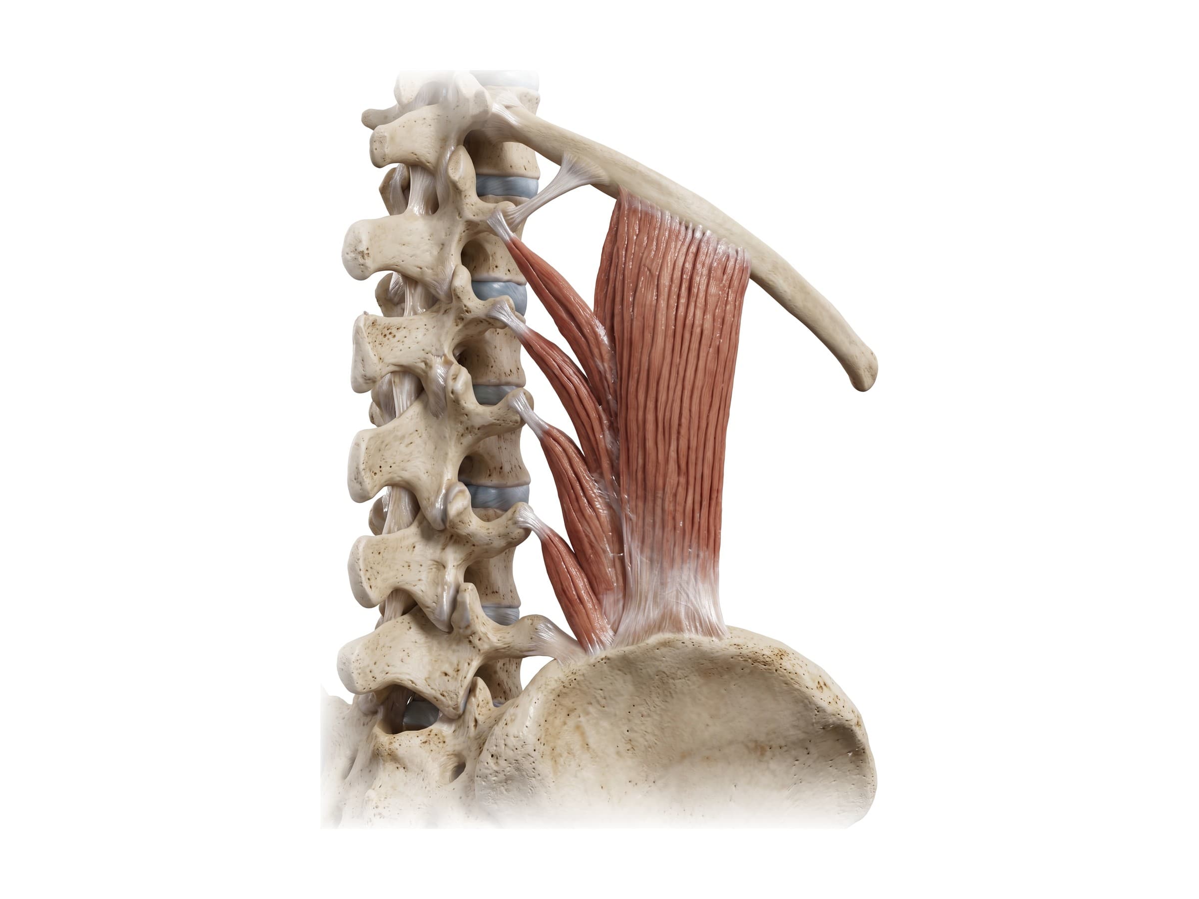
The lumbar spine is the lower back section of the spine: five large vertebrae, L1 to L5, between the rib cage and the pelvis. Below them sits the sacrum, a triangle of five fused vertebrae (S1 to S5) wedged between the hip bones, and the small coccyx (tailbone). Together they form the lumbosacral spine. The lumbar vertebrae carry most of the weight of the upper body, bend and straighten the trunk, and protect the cauda equina, the bundle of nerve roots that runs to the legs, bladder and bowel. Lumbar spine anatomy covers the vertebrae, the intervertebral discs and facet joints, the ligaments, the lumbosacral joint at L5-S1, the sacrum, and the nerves of the lumbar plexus. Knowing these parts is the first step to reading a lumbar MRI report, explaining sciatica to a patient, or illustrating a back injury claim.
The five lumbar vertebrae
The labeled view above shows the lumbar spine from T12 down to L5, with the twelfth rib and the quadratus lumborum muscle beside it. From the side, the lumbar spine curves gently forward, a curve called the lumbar lordosis, typically about 40 to 60 degrees between L1 and S1. Too much curve (hyperlordosis) or a flattened curve, often from muscle spasm after an injury, both show on a side X-ray.
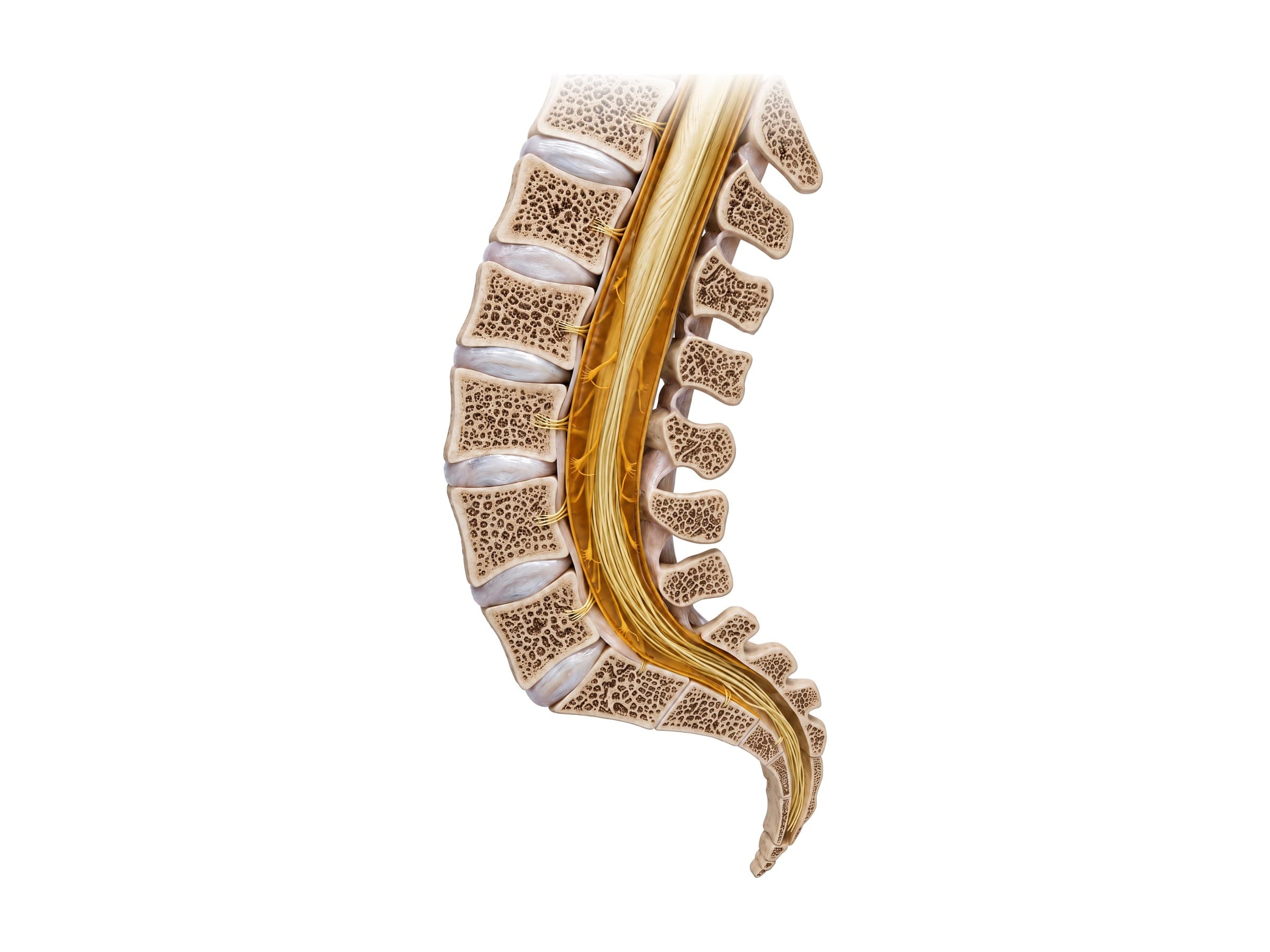
Cut down the midline, the lumbar spine is a column of five vertebral bodies separated by thick discs, sitting on the sacrum. The L4-L5 and L5-S1 levels at the bottom carry the most load and movement, so they are where most disc herniations, arthritis and slips develop. The spinal cord itself ends at about L1-L2; below that, the canal holds the cauda equina.
Anatomy of a lumbar vertebra
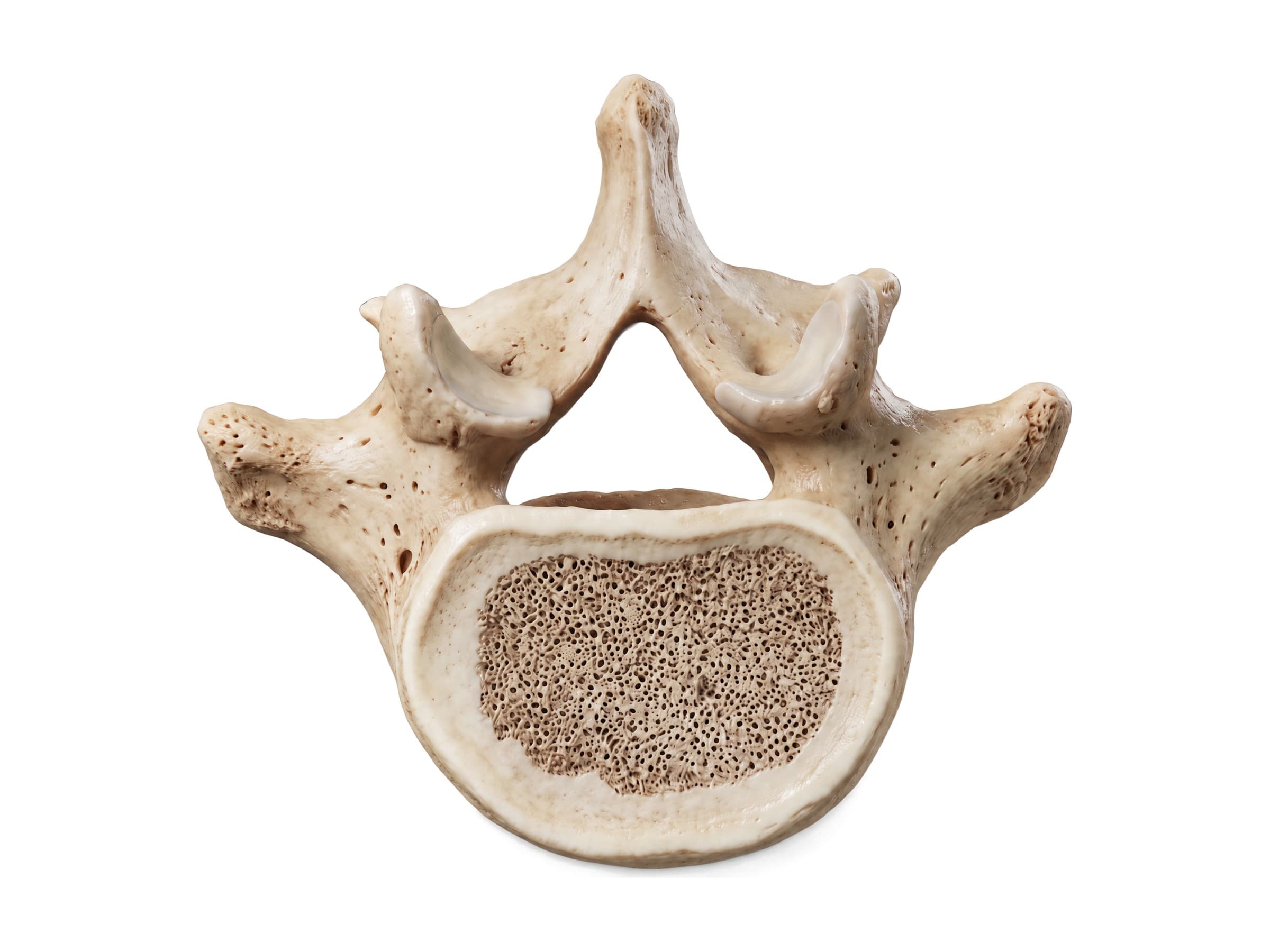
Each lumbar vertebra has the same parts, seen here from above with the front at the bottom:
- Vertebral body: the large, kidney-shaped block in front that carries weight. Lumbar bodies are the largest in the spine, and they get bigger from L1 to L5.
- Pedicles: two short, thick pillars that join the body to the arch behind. Surgeons place pedicle screws through them in a lumbar fusion.
- Laminae: flat plates that meet in the midline and form the roof of the canal. Removing part of them (laminectomy) makes room for compressed nerves.
- Vertebral canal: the triangular opening that holds the dural sac and cauda equina. Narrowing of the canal is lumbar spinal stenosis.
- Transverse processes: long, thin projections to the sides that anchor the quadratus lumborum, psoas and deep back muscles. Unlike cervical vertebrae, lumbar vertebrae have no holes in them, and unlike thoracic vertebrae, no rib facets.
- Spinous process: short, thick and square (hatchet-shaped), pointing straight back. You can feel the lumbar spinous processes down the middle of the lower back.
- Superior and inferior articular processes: paired joint processes that form the facet joints with the vertebrae above and below. The upper facets face inward and the lower facets face outward, which locks neighboring vertebrae together and allows bending forward and backward while limiting twisting. Small knobs on the back of the superior articular processes (mammillary processes) anchor the multifidus muscle.
- Pars interarticularis: the narrow bridge of bone in each side of the arch between the upper and lower articular processes, the weak point that breaks in spondylolysis.
Between each pair of vertebrae, the intervertebral foramen (foramen intervertebrale) is the opening where a spinal nerve leaves the canal. It is bounded by the disc and vertebral bodies in front, the pedicles above and below, and the facet joint behind, so a disc bulge, bone spur or thickened ligament can narrow it (foraminal stenosis).
Intervertebral discs and ligaments
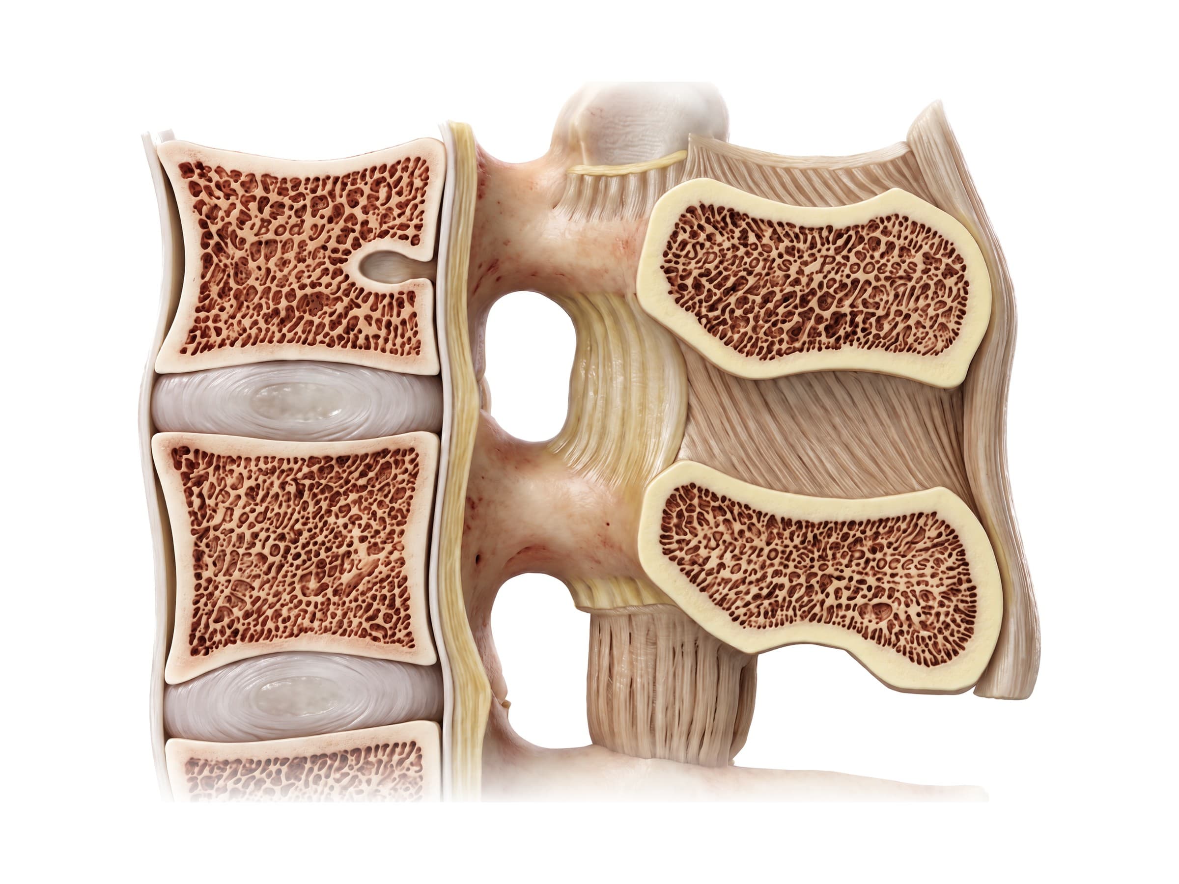
Each lumbar intervertebral disc has a tough outer ring, the annulus fibrosus, made of layers of crossing collagen fibers, around a soft gel center, the nucleus pulposus. The lumbar discs are the thickest in the spine and act as shock absorbers. With age the nucleus dries out and the disc loses height, which is why degenerative disc disease is so common on lumbar MRI even in people without pain.
The ligaments hold the lumbar spine together:
- Anterior longitudinal ligament: a broad band down the front of the vertebral bodies and discs that limits bending backward.
- Posterior longitudinal ligament: runs down the back of the bodies inside the canal. It narrows in the lumbar spine, leaving the back corners of each disc less supported, which is why most disc herniations go back and to one side (posterolateral), toward a nerve root.
- Ligamentum flavum: elastic yellow ligaments joining the laminae. When they thicken and buckle with age they narrow the canal, a major cause of lumbar spinal stenosis.
- Interspinous and supraspinous ligaments: join neighboring spinous processes and limit bending forward; they can tear in flexion injuries such as seatbelt fractures.
- Facet joint capsules: thin capsules around each facet joint, a common source of lower back pain.
The lumbosacral joint (L5-S1)
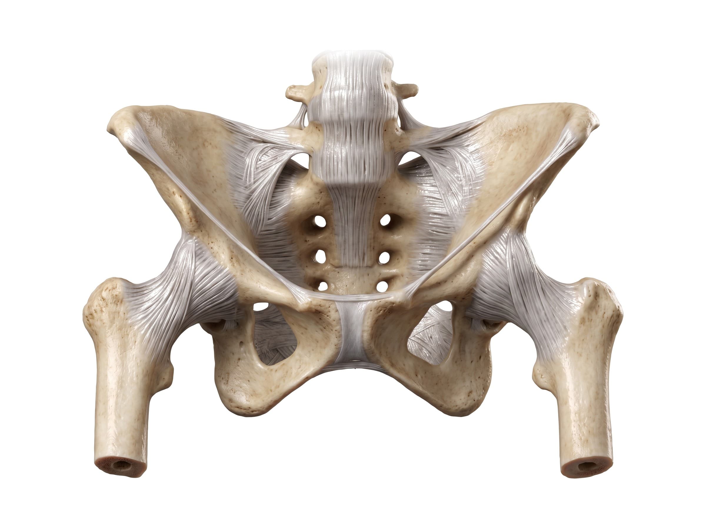
The lumbosacral joint is where the last lumbar vertebra, L5, meets the top of the sacrum, S1. It includes the L5-S1 disc in front and two facet joints behind. Because the top of the sacrum tilts forward (the lumbosacral angle), L5 constantly tends to slide forward on S1. The L5-S1 disc is wedge shaped, taller in front, and the joint is braced by the iliolumbar ligaments, strong bands from the transverse processes of L5 to the iliac crest on each side. This shear load is why L5-S1 is a common site of disc herniation and of isthmic spondylolisthesis. The level of the iliac crests, the tops of the hip bones, usually crosses L4 or the L4-L5 disc, a landmark used to count the levels.
The sacrum and coccyx
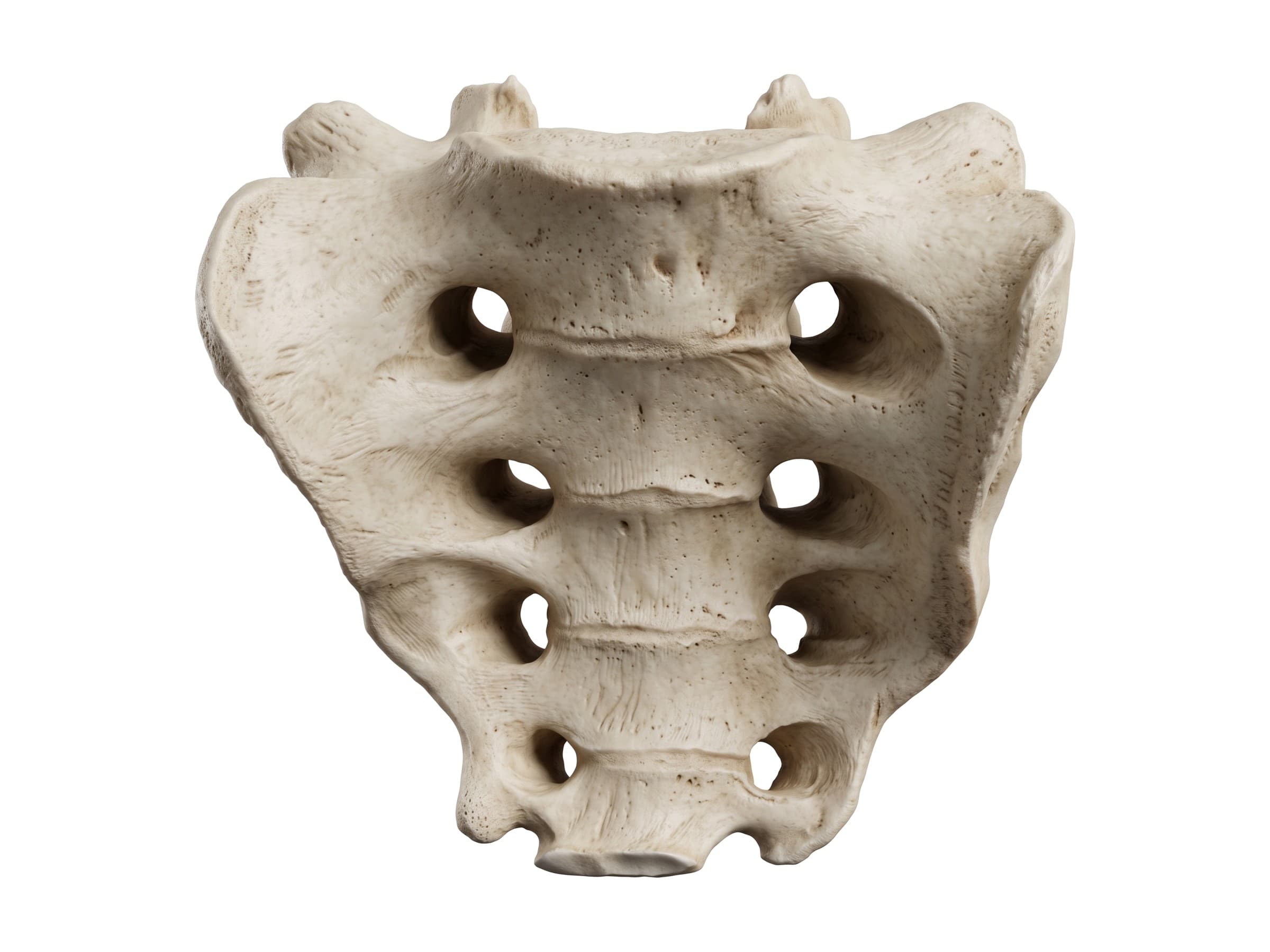
The sacrum is a large triangular bone formed by five vertebrae (S1 to S5) that fuse in early adulthood. It forms the back wall of the pelvis and joins the hip bones at the sacroiliac joints (covered in hip anatomy).
- Sacral promontory: the projecting front edge of the top of S1, an important landmark in childbirth and surgery.
- Body of S1: the top of the sacrum, which carries the L5-S1 disc.
- Sacral ala: the two wing-like masses on either side of S1. The L5 nerve root runs across the ala, so ala fractures in pelvic trauma can injure it.
- Anterior sacral foramina: four pairs of openings on the front surface where the front branches of the sacral nerves leave to form the sacral plexus and sciatic nerve.
- Apex: the narrow lower end, which joins the coccyx.
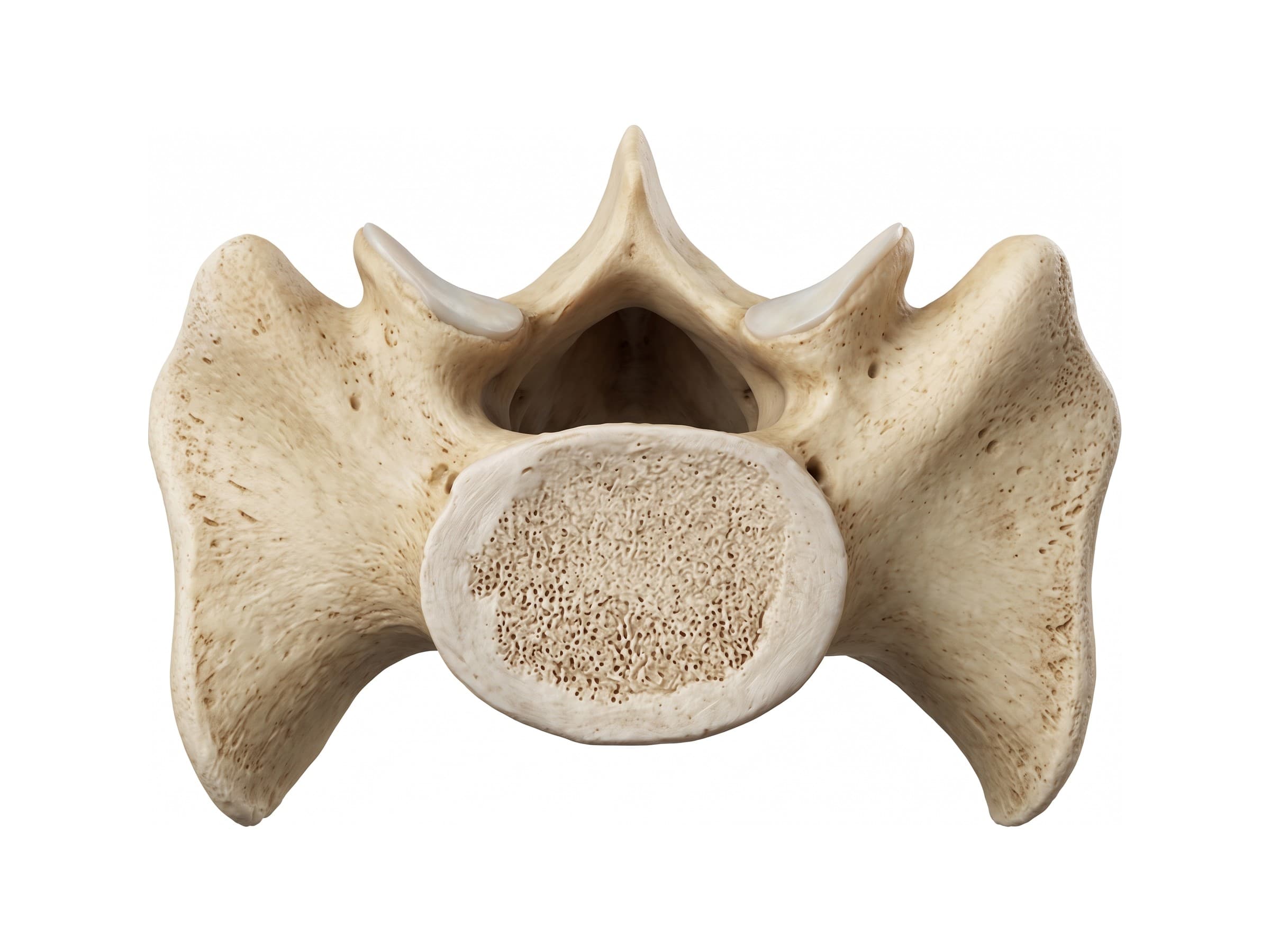
Seen from above, the base of the sacrum shows the oval body of S1, the triangular sacral canal behind it (the continuation of the vertebral canal), the superior articular processes that form the L5-S1 facet joints, and the broad ala on each side.
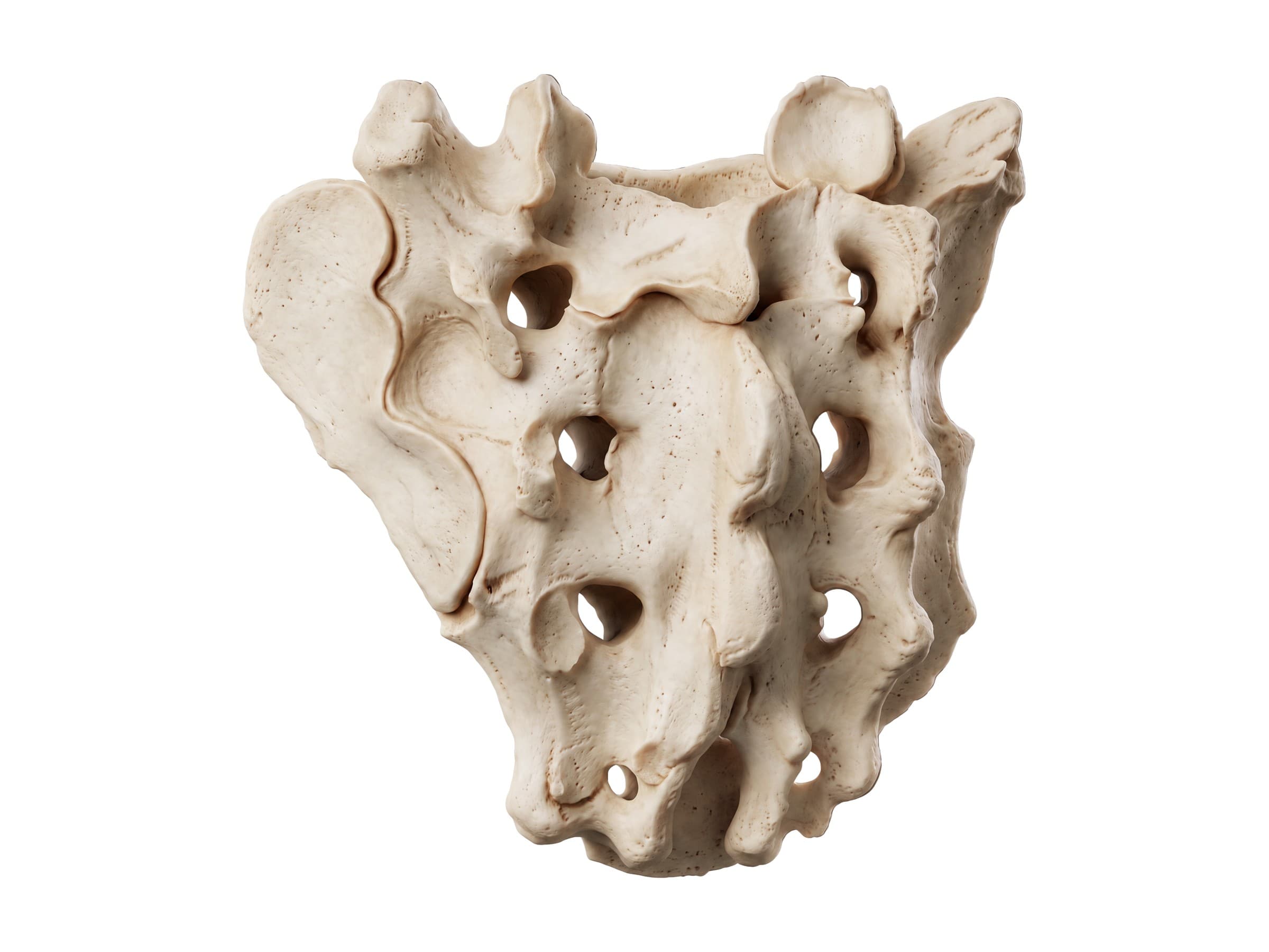
The back of the sacrum is rough. The median sacral crest runs down the middle (the fused spinous processes), with the posterior sacral foramina on either side for the back branches of the sacral nerves. The ear-shaped auricular surface on each side forms the sacroiliac joint with the ilium. At the bottom, the sacral canal opens as the sacral hiatus, the entry point for a caudal epidural injection.
The coccyx, or tailbone, is three to five small fused vertebrae below the sacrum. It anchors the gluteus maximus and pelvic floor muscles and is often bruised or fractured in a fall onto the buttocks.
Spinal cord, cauda equina and lumbar nerves
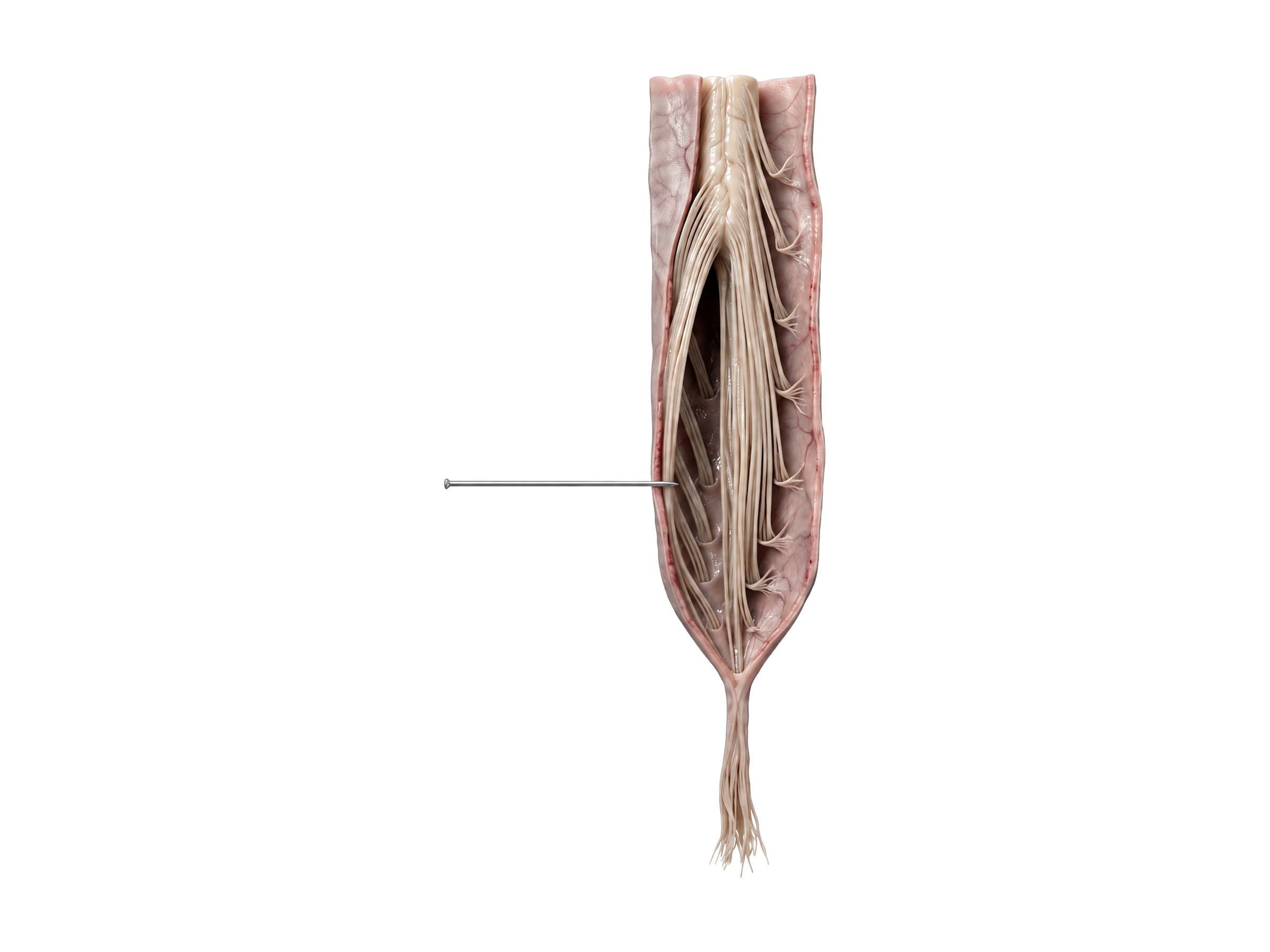
In adults the spinal cord ends at about the L1-L2 disc, where it tapers into the conus medullaris. Below that, the lumbar and sacral nerve roots hang down the canal inside the dural sac as the cauda equina (Latin for horse's tail). A thin strand, the filum terminale, continues from the conus to anchor the cord to the coccyx. The dural sac ends at about S2, so lumbar punctures and spinal anesthesia are done at L3-L4 or L4-L5, below the end of the cord.
There are five pairs of lumbar nerves. Each lumbar nerve leaves below the vertebra with the same number: the L4 nerve exits between L4 and L5, the L5 nerve between L5 and S1. A typical posterolateral disc herniation catches the nerve root heading down to the next level, so an L4-L5 herniation usually affects L5, and an L5-S1 herniation affects S1.
- L1 to L3: the groin and front of the thigh; hip flexion.
- L4: the knee straightener (quadriceps), the inner shin, and the knee reflex.
- L5: lifting the big toe and the front of the foot; the outer shin and top of the foot. Severe L5 weakness causes foot drop.
- S1: pushing off on the toes (calf); the sole and outer foot; the ankle reflex.
- S2 to S4: the bladder, bowel and sexual function, and the skin of the saddle area. Compression of these roots is cauda equina syndrome.
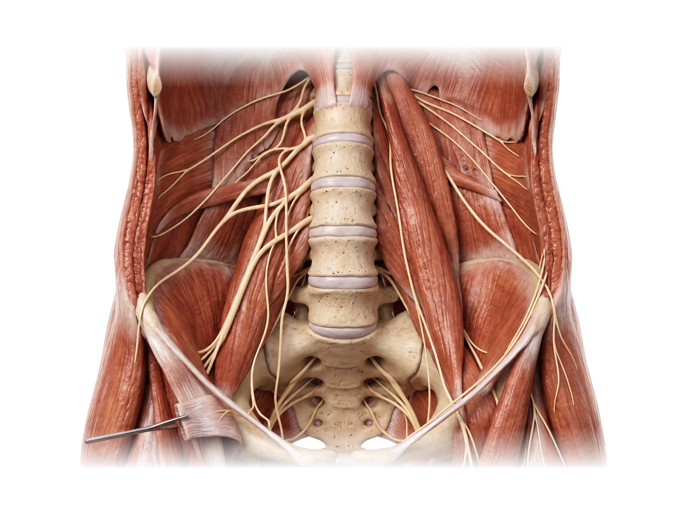
The front branches of L1 to L4 form the lumbar plexus inside the psoas major muscle. Its main branches are the iliohypogastric and ilioinguinal nerves (the lower abdominal wall and groin), the genitofemoral nerve, the lateral femoral cutaneous nerve (the skin of the outer thigh, compressed in meralgia paresthetica), the femoral nerve (the quadriceps and front of the thigh) and the obturator nerve (the inner thigh muscles). L4 and L5 also join the sacral nerves as the lumbosacral trunk to form the sacral plexus and the sciatic nerve (L4 to S3), the nerve of sciatica.
Muscles of the lower back
The muscles around the lumbar spine hold it upright and move it. The erector spinae and multifidus run along the back of the spine and extend it, the quadratus lumborum runs from the twelfth rib to the iliac crest on each side, and the psoas major runs along the front of the lumbar vertebrae to the thigh. All are covered in detail in back muscles anatomy, and the psoas continues as the main hip flexor in hip anatomy. Lumbar muscle strain is the most common cause of acute lower back pain.
Range of motion of the lumbar spine
The lumbar spine moves mostly forward and backward. Typical normal values are roughly 40 to 60 degrees of flexion (bending forward), 20 to 30 degrees of extension (bending back) and 20 to 30 degrees of side bending each way, with very little rotation because the facet joints lock against twisting. Much of forward bending also comes from the hips tilting the pelvis. Loss of lumbar range of motion, measured with an inclinometer, is one of the main ways a lower back injury is documented over time.
Common lumbar spine injuries and how they appear on imaging
X-rays (front, side and sometimes bending views) show alignment, fractures, slips and disc height. CT shows fractures in detail after trauma. MRI shows the discs, nerve roots, ligaments and the cauda equina, and is the test for sciatica that does not settle or for any red flag symptoms.
- Lumbar strain and sprain: overstretching or tearing of the muscles, ligaments or facet capsules, the most common lower back injury after lifting, falls and car crashes. Imaging is usually normal or shows only straightening of the lordosis from spasm.
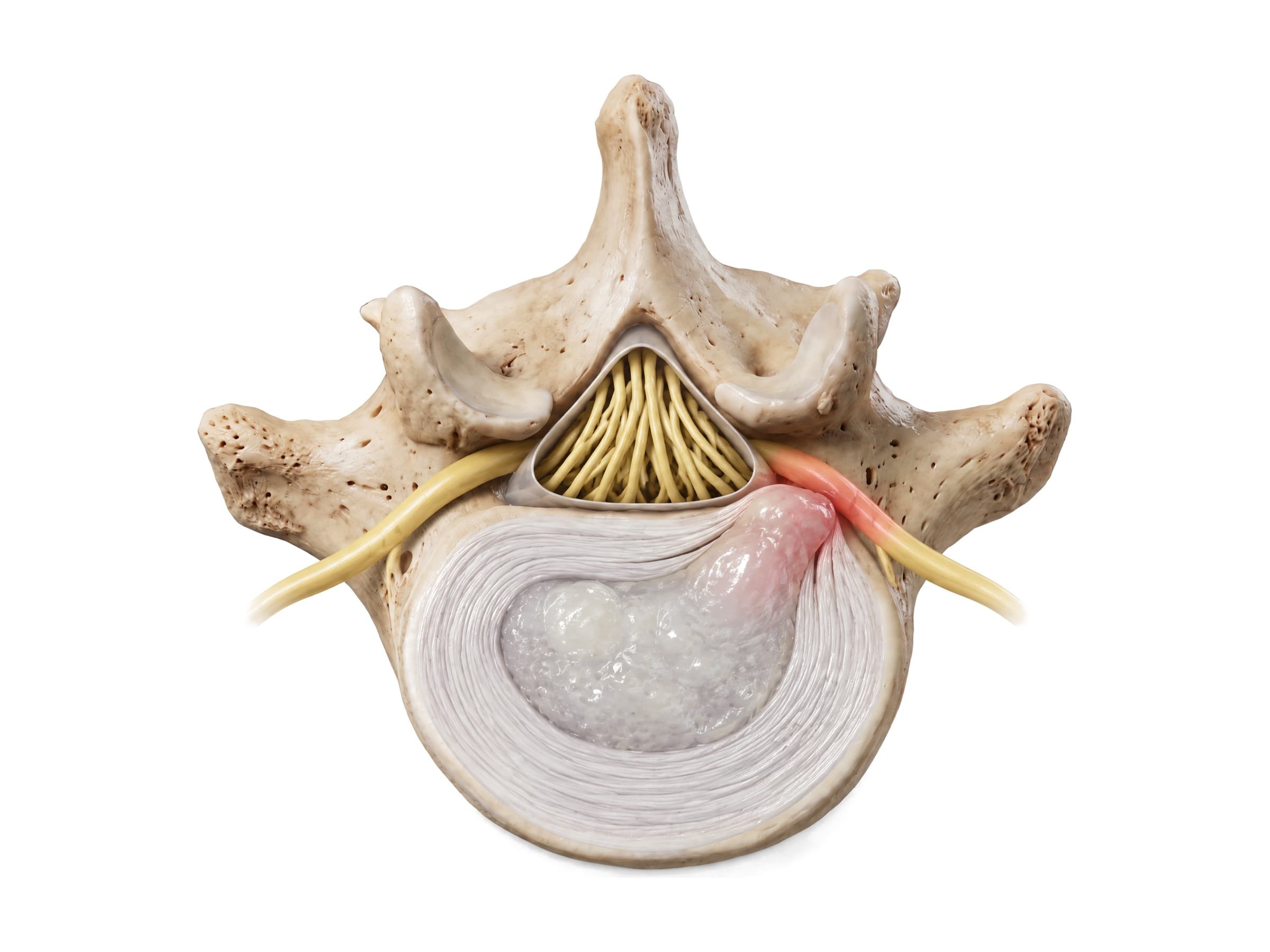
- Lumbar disc herniation: the nucleus pulposus pushes through a tear in the annulus, most often at L4-L5 or L5-S1, and presses on a nerve root, causing sciatica down the leg in that root's pattern. MRI shows the disc material and the compressed root. Persistent cases with weakness may be treated with microdiscectomy. A large central herniation can compress the whole cauda equina.
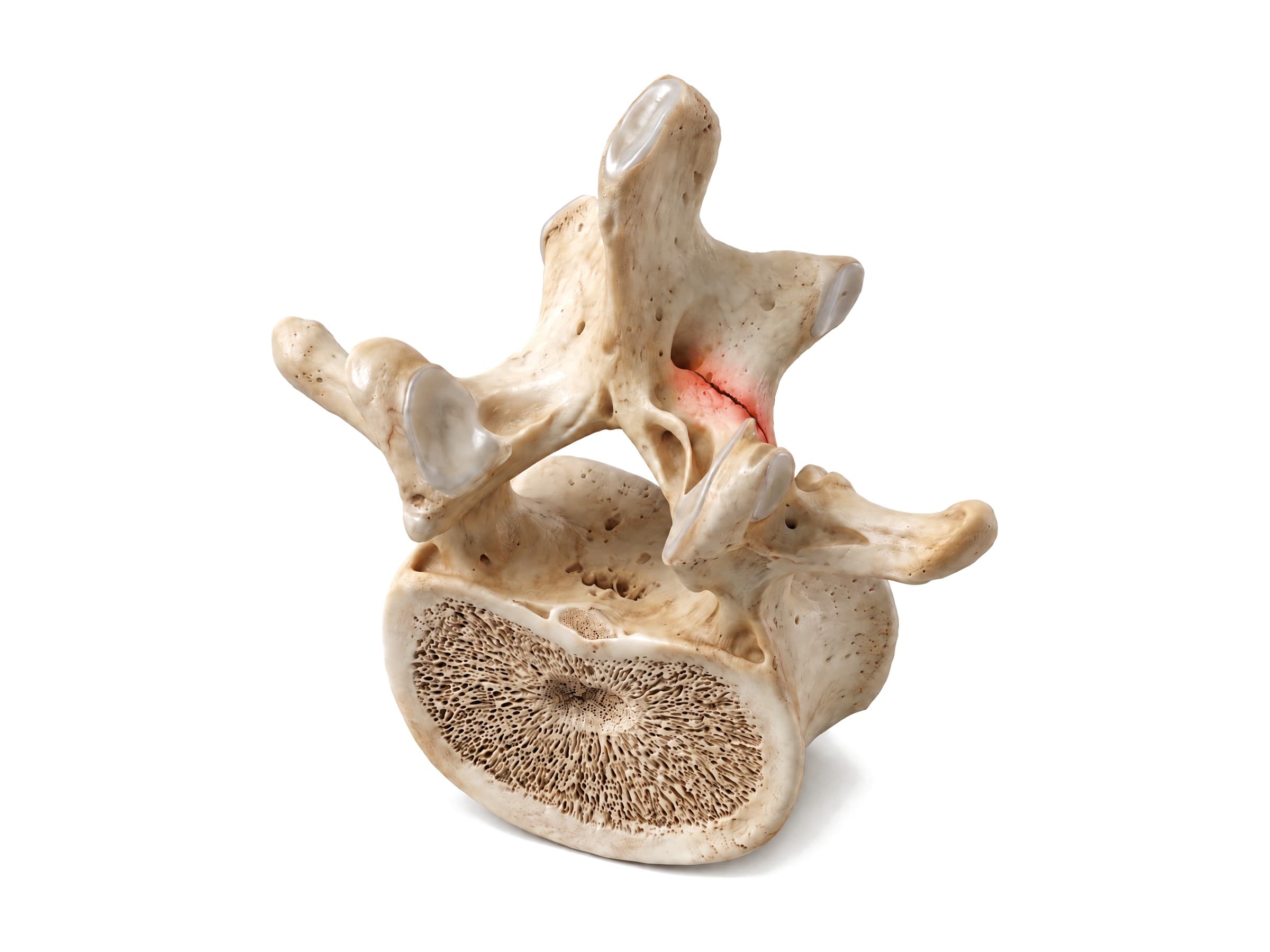
- Spondylolysis: a stress fracture of the pars interarticularis, most often at L5, from repeated arching of the back in sports such as gymnastics, football and cricket, or from acute hyperextension. On an angled (oblique) X-ray the defect looks like a collar on the "Scotty dog" outline; CT shows the break and MRI shows early stress reaction.
- Spondylolisthesis (anterolisthesis): a forward slip of one vertebra on the one below, graded I to V by how far it has slipped (grade I is less than a quarter of the vertebral body). Isthmic slips follow spondylolysis on both sides, usually L5 on S1; degenerative slips come from facet arthritis, usually L4 on L5 in adults over 50. The slip is measured on a standing side X-ray, and bending views show whether it moves.
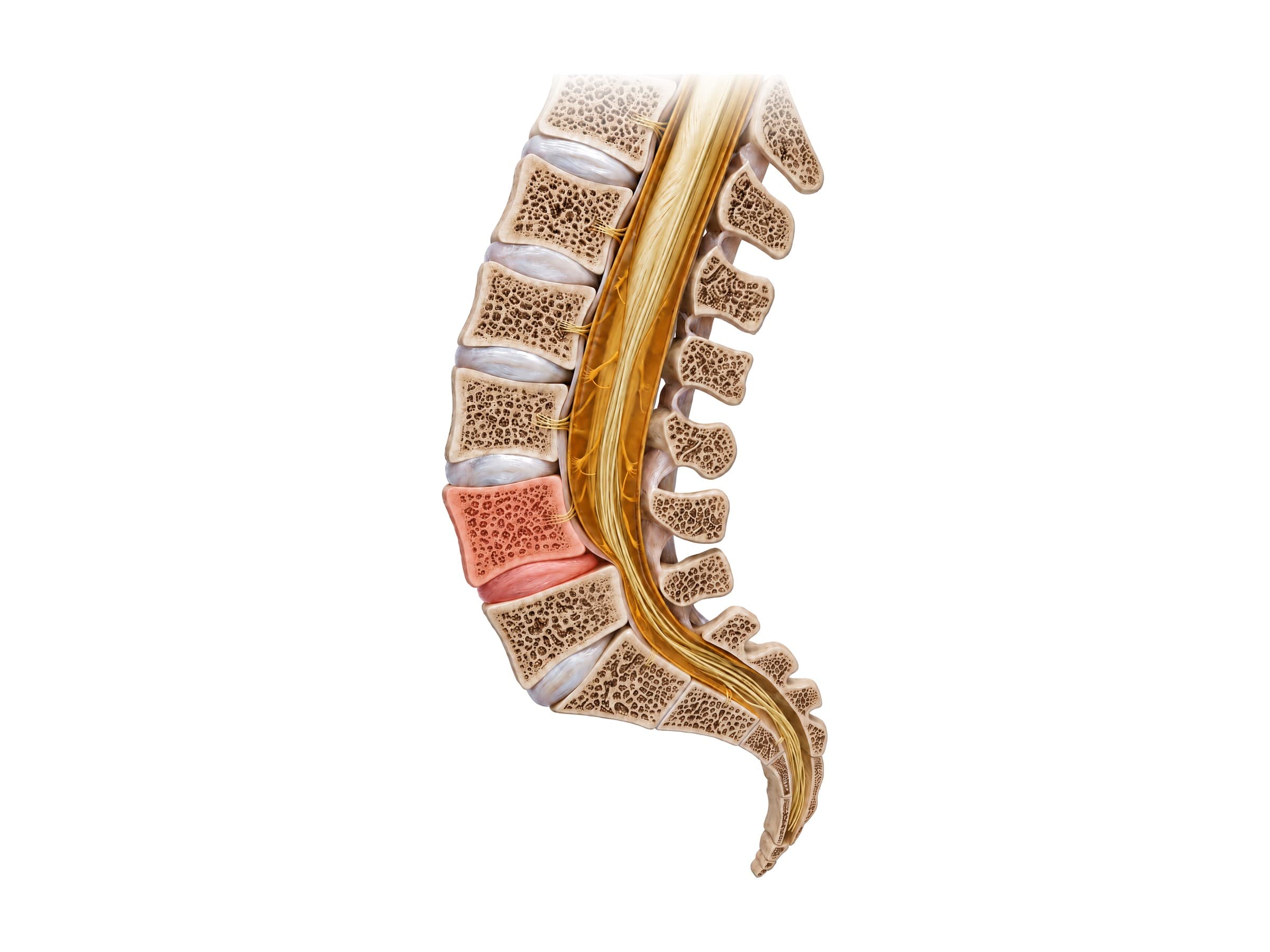
- Compression and burst fractures: the thoracolumbar junction (T11 to L2) is the most common site of spine fractures in falls and car crashes. In a compression fracture the front of the body collapses into a wedge while the back wall holds; in a burst fracture the back wall also breaks and bone fragments can push into the canal. In older adults with osteoporosis, compression fractures follow minor falls or no clear injury and may be treated with kyphoplasty.
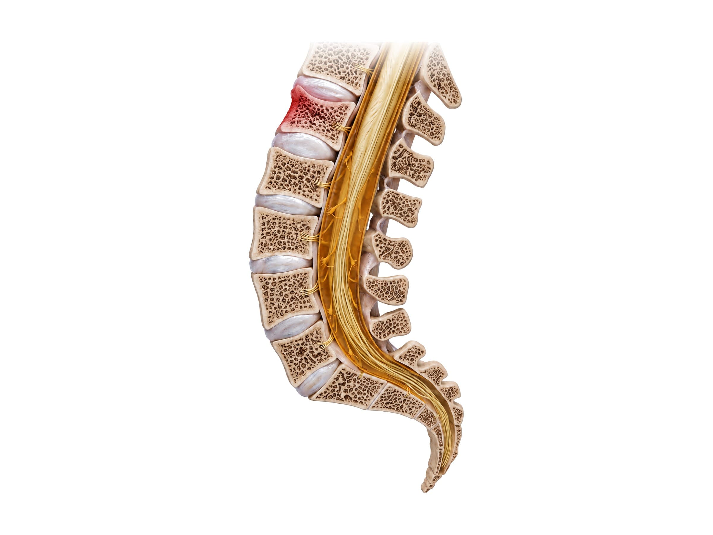
- Chance (seatbelt) fracture: a flexion-distraction injury in a car crash where a lap belt acts as a hinge, splitting the vertebra or its ligaments horizontally, usually at L1 to L3, often with abdominal injuries.
- Transverse process fractures: breaks of the thin side projections from a direct blow or violent muscle pull; usually stable but a marker of high-energy trauma.
- Lumbar spinal stenosis: narrowing of the canal or foramina from disc bulges, facet arthritis and a thickened ligamentum flavum, causing leg pain and heaviness on walking that eases on sitting or bending forward (neurogenic claudication).
- Cauda equina syndrome: compression of the whole bundle of nerve roots, usually by a large central disc herniation, causing saddle numbness, bladder or bowel dysfunction and leg weakness. It is a surgical emergency, and delays in diagnosis are a frequent subject of medical malpractice claims.
How the lumbar spine is shown in a personal injury case
Lower back injuries are the most common claims after car crashes, slip-and-fall accidents and workplace injuries, and among the most disputed. The defense often argues that a herniated disc or a slip was pre-existing degeneration, that the MRI findings are normal for the plaintiff's age, or that the leg symptoms do not match the scan. Each lumbar injury follows a recognizable mechanism of injury: a disc herniation from a lifting injury or a rear-end collision, a compression fracture from a fall onto the buttocks or a high-energy crash, a Chance fracture from a lap belt. A labeled illustration of the normal lumbar spine is the baseline exhibit. It lets an expert show the jury where the L4-L5 and L5-S1 discs sit, which nerve roots they touch, and why the plaintiff's numbness runs to the big toe or the outer foot, before showing the injury itself: the herniated disc pressing on the nerve, the collapsed vertebra, or the screws and rods of a lumbar fusion. Illustrations drawn from the plaintiff's own MRI or CT tie the scan, the anatomy and the symptoms together. In cauda equina malpractice cases, an illustration of the compressed nerve roots explains why hours mattered.
Natomy creates custom medical legal illustrations of lumbar spine injuries for demand packages and trial exhibits, and the rules for using these visuals at trial are covered in demonstrative evidence. For the sections above, see thoracic spine anatomy and cervical spine anatomy. Related atlas pages: leg nerves and dermatomes, back muscles anatomy, hip anatomy, knee anatomy, ankle anatomy and foot bones.
FAQ
How many vertebrae are in the lumbar spine?+
Most people have five lumbar vertebrae, numbered L1 to L5 from the top. They are the largest movable vertebrae in the spine because they carry the weight of the upper body. A few people have a variation at the lumbosacral junction: L5 can be partly fused to the sacrum (sacralization), or the top of the sacrum can be separate and act like a sixth lumbar vertebra (lumbarization). These variants are usually harmless but matter when counting levels on an X-ray or before surgery.
What is meant by L1, L2, L3, L4, L5?+
L1 to L5 are the names of the five lumbar vertebrae, counted from the top of the lower back down. L1 sits just below the last rib-bearing vertebra (T12), and L5 sits on top of the sacrum. A line between the tops of the hip bones (the iliac crests) usually crosses at the L4 vertebra or the L4-L5 disc, which is how clinicians find the levels by touch. Discs are named for the two vertebrae they sit between, such as L4-L5 or L5-S1.
What are the first signs of L4-L5 compression?+
A disc herniation or stenosis at L4-L5 most often presses on the L5 nerve root. Early signs are pain that runs from the buttock down the outside of the leg to the top of the foot and big toe, numbness or tingling in the same area, and weakness lifting the big toe or the front of the foot. Severe L5 weakness causes foot drop, where the toes catch while walking. A herniation far out to the side at L4-L5 can instead pinch the L4 root, causing pain at the front of the thigh and shin.
What nerve is affected at L5-S1?+
A herniated disc at L5-S1 usually compresses the S1 nerve root. That causes sciatica down the back of the thigh and calf into the sole and outer edge of the foot, numbness along the little toe, weakness pushing off or standing on tiptoe, and a reduced or absent ankle reflex. Together, L4-L5 and L5-S1 account for the large majority of lumbar disc herniations because they carry the most load and movement.
What are the red flags for lower back problems?+
Most lower back pain is mechanical and improves within weeks. Red flags that need urgent evaluation are signs of cauda equina syndrome (numbness in the groin and inner thighs, called saddle anesthesia; new trouble urinating or controlling the bladder or bowels; and rapidly worsening weakness in both legs), and signs of a serious underlying cause such as fever, a history of cancer, unexplained weight loss, significant trauma, or new severe pain in someone over 50 or on long-term steroids.
Where does the spinal cord end?+
In adults the spinal cord ends at about the level of the L1-L2 disc, where it tapers into the conus medullaris. Below that, the lumbar and sacral nerve roots continue down the canal as the cauda equina, a bundle that looks like a horse's tail, and the dural sac ends at about S2. Because the cord stops above L2, a lumbar puncture is done below it, at the L3-L4 or L4-L5 level, where a needle can enter the fluid without reaching the cord.
What is the lumbosacral joint?+
The lumbosacral joint is the junction between the fifth lumbar vertebra (L5) and the top of the sacrum (S1). It includes the L5-S1 disc in front and a pair of facet joints behind, and it is reinforced by the iliolumbar ligaments that tie L5 to the pelvis. Because the sacrum tilts forward, the L5-S1 disc is wedge shaped and the joint carries strong shear forces, which makes it a common site for disc herniation, degeneration and isthmic spondylolisthesis.
What is the difference between spondylolysis and spondylolisthesis?+
Spondylolysis is a stress fracture or defect in the pars interarticularis, the narrow bridge of bone in the back of a vertebra, most often at L5. It is common in young athletes who repeatedly arch the back, such as gymnasts and football linemen. Spondylolisthesis is the forward slip of one vertebra on the one below. It can follow spondylolysis on both sides (isthmic, usually L5 on S1) or develop from arthritis of the facet joints (degenerative, usually L4 on L5 in adults over 50). Forward slip is also called anterolisthesis.
Need a custom illustration of this joint?
Generate a labeled or exhibit-ready figure for a specific patient, injury, or case in minutes.
Try Natomy →