Back Muscles Anatomy: Upper & Lower Back Muscles Diagram
Labeled back muscles diagrams: trapezius, latissimus dorsi, rhomboids, erector spinae, quadratus lumborum and the lumbar fascia, plus common back injuries.
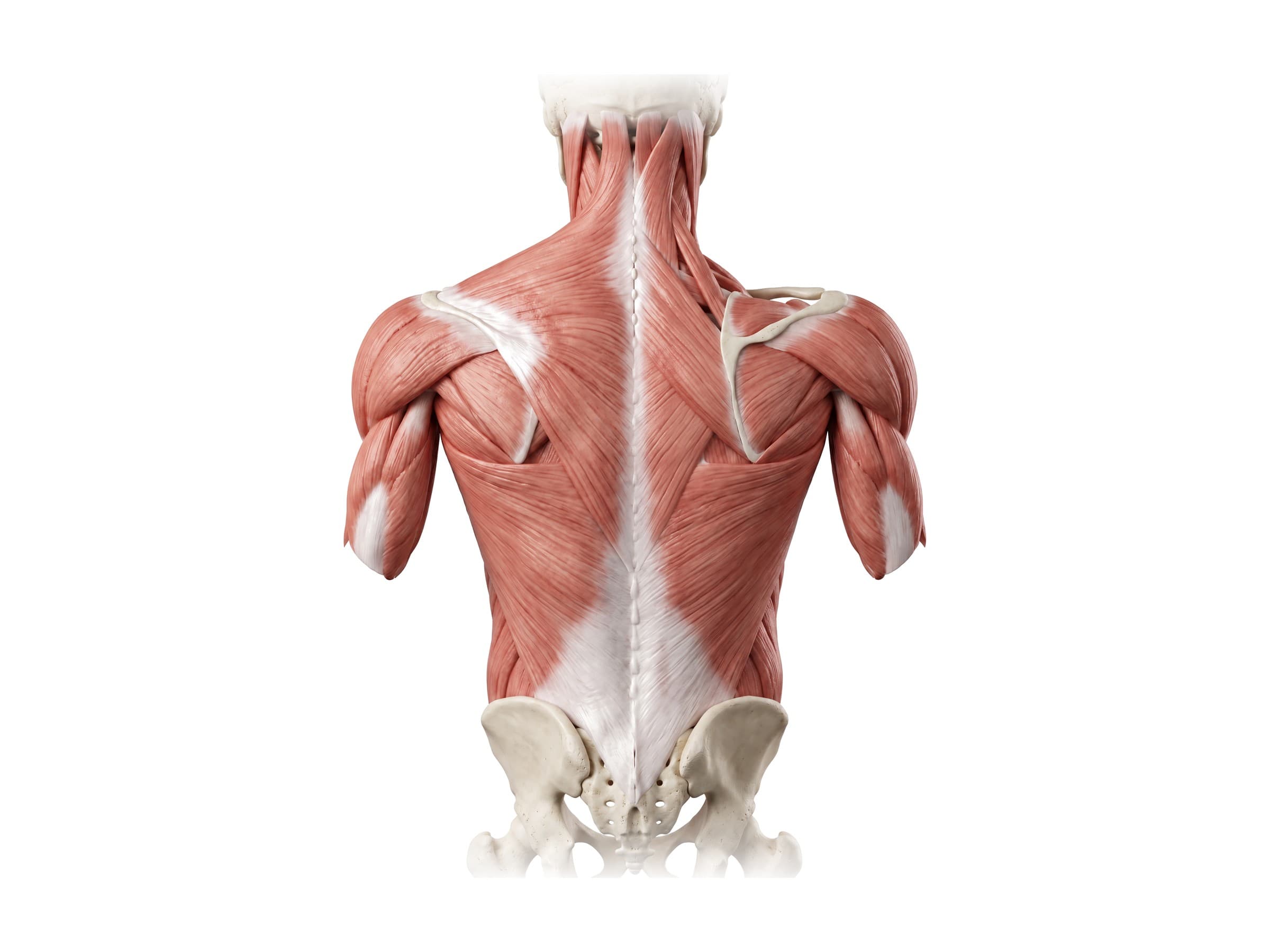
The back muscles are the layered muscles that run from the skull, spine and pelvis to the ribs, shoulder blades and arms. They fall into three layers: a superficial layer (the trapezius, latissimus dorsi, rhomboids and levator scapulae) that moves the shoulder and arm, a thin intermediate layer that attaches to the ribs, and a deep or intrinsic layer (the erector spinae, multifidus and their neighbors) that holds the spine upright and moves it. In the lower back, the quadratus lumborum and psoas major join them, and the thoracolumbar fascia wraps the whole lumbar group. Back muscle strains are the most common cause of acute low back pain, and back and neck soft-tissue injuries are among the most frequent claims after a motor vehicle collision or fall.
The three layers of back muscles
Anatomists divide the back muscles by depth and by what they move.
- Superficial (extrinsic) layer: trapezius, latissimus dorsi, rhomboid major and minor, and levator scapulae. These start on the spine and end on the scapula or humerus, so despite sitting on the back, they mainly move the shoulder and arm. They are supplied by nerves from the front of the spinal cord (the brachial plexus and the spinal accessory nerve), not by the nerves of the back.
- Intermediate layer: serratus posterior superior and serratus posterior inferior, two thin sheets that run from the spine to the ribs and assist breathing.
- Deep (intrinsic) layer: the "true" back muscles that move the spine itself. From superficial to deep: the splenius muscles in the neck, the erector spinae, the transversospinales (semispinalis, multifidus and rotatores), and the small interspinales and intertransversarii between neighboring vertebrae. All are supplied by the dorsal rami of the spinal nerves.
Superficial back muscles
From behind, the trapezius is the large diamond-shaped muscle covering the upper back and the back of the neck. It starts on the base of the skull and the spinous processes from C7 to T12, and converges on the clavicle, acromion and spine of the scapula. Below it, the latissimus dorsi is the broadest muscle in the body, fanning from the lower thoracic spine, the thoracolumbar fascia and the iliac crest up to a narrow tendon on the front of the humerus.
On the right side of the figure, the trapezius has been cut back to show the layer beneath it. The rhomboid major and rhomboid minor run downward and outward from the spinous processes to the medial border of the scapula, and the levator scapulae runs from the upper cervical vertebrae down to the top corner of the scapula. The infraspinatus and teres major visible on the scapula belong to the shoulder group.
- Trapezius: elevates, retracts and rotates the scapula, and extends the neck. Its upper part is the muscle most often sore after a whiplash injury. Supplied by the spinal accessory nerve (cranial nerve XI).
- Latissimus dorsi: extends, adducts and internally rotates the arm, the main muscle for pull-ups, rowing and swimming. Supplied by the thoracodorsal nerve.
- Rhomboid major and minor: pull the scapula toward the spine and hold it against the rib cage. Supplied by the dorsal scapular nerve.
- Levator scapulae: lifts the scapula and bends the neck to the side. A common site of neck and shoulder-blade pain. Supplied by the dorsal scapular nerve and branches of C3 and C4.
Intermediate back muscles
The serratus posterior superior runs from the spinous processes of C7 to T3 down to ribs 2 to 5, and the serratus posterior inferior runs from T11 to L2 up to the lowest four ribs. Both are thin and lie just under the rhomboids and latissimus dorsi. They were traditionally described as breathing muscles (the superior one raising the ribs, the inferior one lowering them), though their contribution is small. They are rarely the focus of imaging or injury claims but explain the "layer cake" a surgeon sees when operating on the thoracic spine.
Deep back muscles: the erector spinae
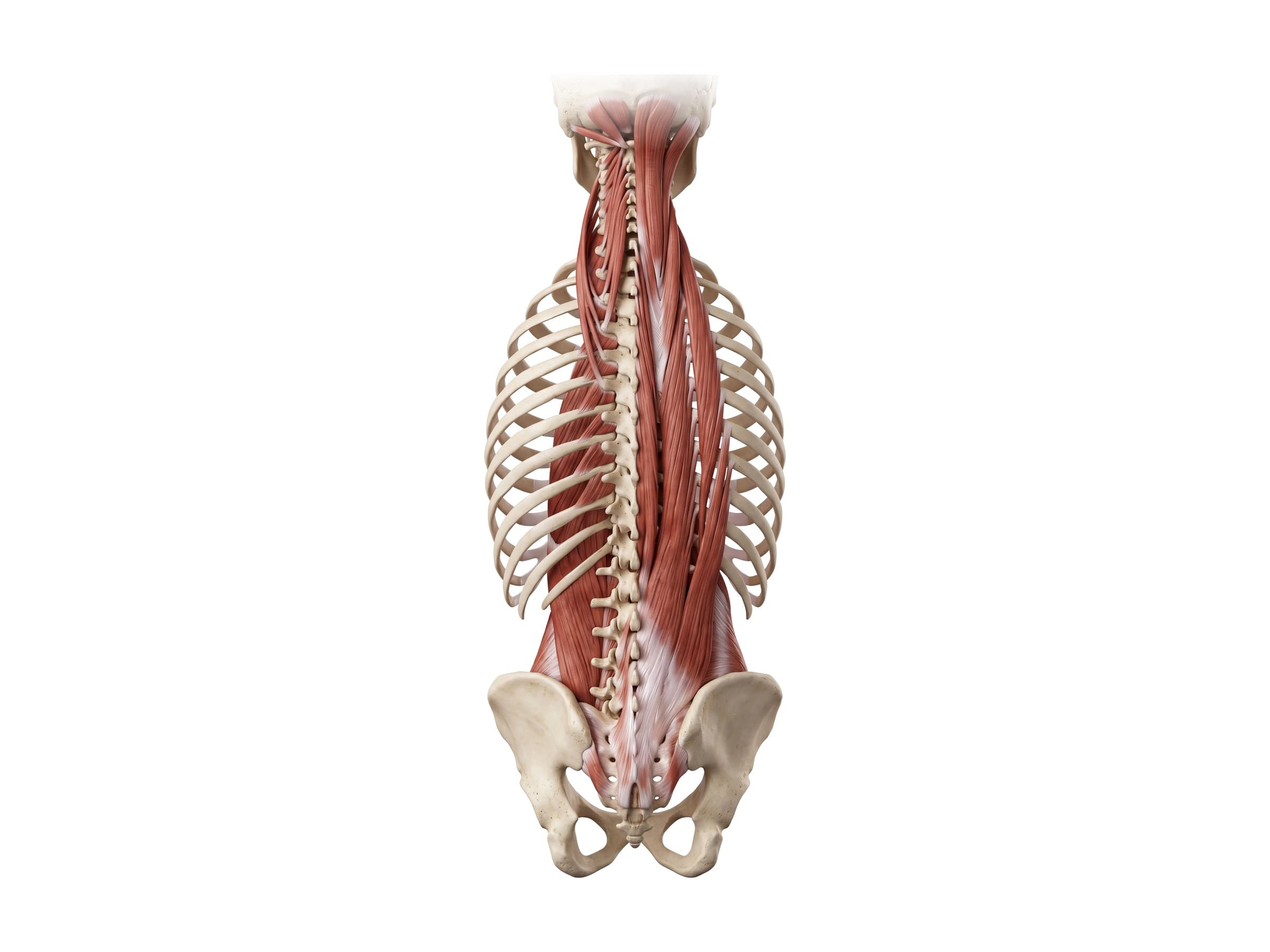
With the superficial and intermediate layers removed, the erector spinae fills the groove on each side of the spine from the sacrum to the skull. It starts as a thick tendon and muscle mass over the sacrum and lumbar spine and splits upward into three parallel columns:
- Iliocostalis: the outermost column, running from the iliac crest and sacrum to the angles of the ribs and up to the cervical transverse processes.
- Longissimus: the middle and longest column, running to the transverse processes of the vertebrae and, in the neck, all the way to the mastoid process of the skull.
- Spinalis: the innermost and smallest column, running between spinous processes, mainly in the thoracic region.
Acting on both sides, the erector spinae straightens the back from a bent-forward position and controls bending forward against gravity; acting on one side, it bends the spine to that side. In the neck, the splenius capitis wraps over the upper end of the erector spinae and turns the head.
Multifidus and the transversospinales
Beneath the erector spinae, filling the angle between the spinous and transverse processes, is the transversospinales group. The semispinalis (capitis, cervicis and thoracis) spans four to six vertebrae and is largest in the neck, where the semispinalis capitis forms most of the muscle bulk at the back of the neck. The multifidus spans two to four vertebrae and is thickest in the lumbar spine, where it is the muscle closest to the spine and the main segmental stabilizer of each lumbar level. The tiny rotatores span one or two vertebrae and are best developed in the thoracic spine.
The multifidus matters clinically: it shrinks and is replaced by fat on the painful side within weeks of a lumbar disc injury, a change visible on MRI, and it is the main target of spine-stabilization rehabilitation.
Quadratus lumborum and the lumbar muscles
The quadratus lumborum is a flat, four-sided muscle in the lower back, lying deeper than the erector spinae and to each side of the lumbar spine. It runs from the back of the iliac crest up to the 12th rib and the tips of the transverse processes of L1 to L4. In front of it, beside the vertebral bodies, the psoas major runs down from the lumbar spine to join the iliacus and insert on the femur as the iliopsoas, the main hip flexor.
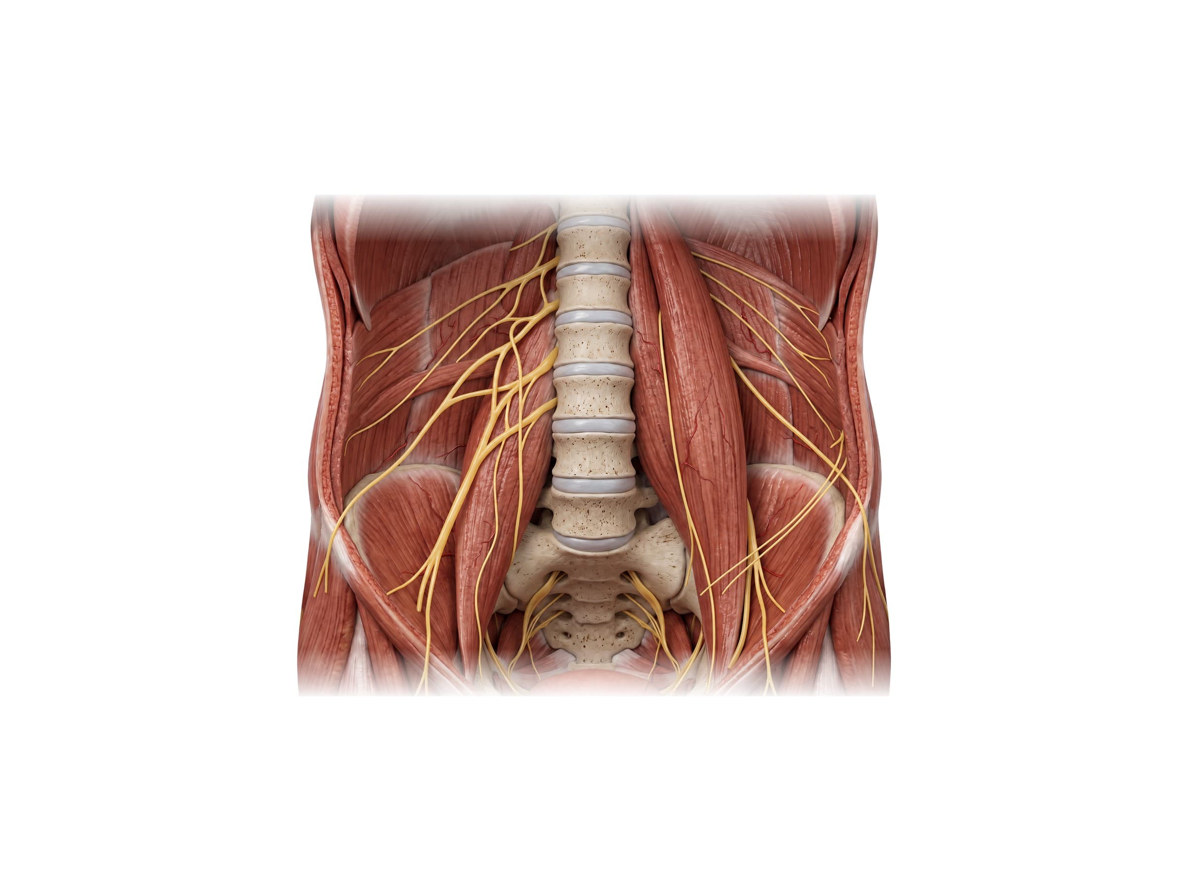
- Quadratus lumborum: bends the trunk to the side and hikes the hip; both sides together stabilize and extend the lumbar spine and fix the 12th rib during breathing. Supplied by branches of T12 to L4. A frequent source of deep, one-sided low back pain.
- Psoas major: flexes the hip and helps stabilize the lumbar spine. Supplied by the lumbar plexus (L1 to L3).
- Iliacus: fills the inside of the pelvic bone and joins the psoas to flex the hip. Supplied by the femoral nerve.
Thoracolumbar fascia
A cross-section through the lower back shows how the lumbar muscles are packed into compartments. The thoracolumbar fascia splits into three layers around them: a posterior layer over the back of the erector spinae, a middle layer between the erector spinae and the quadratus lumborum, attached to the tips of the transverse processes, and an anterior layer in front of the quadratus lumborum. The layers fuse at the outer edge of the erector spinae, where the abdominal wall muscles (the transversus abdominis and internal oblique) attach.
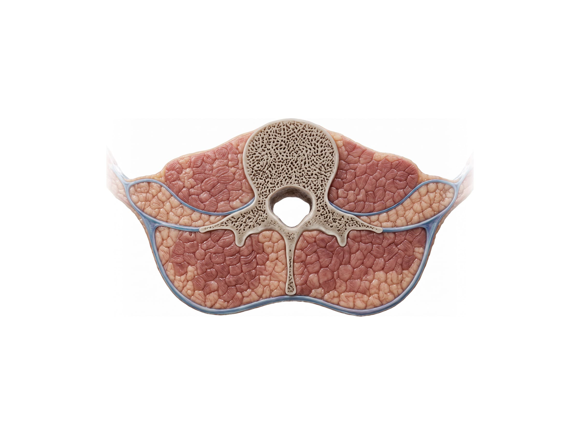
The posterior layer is also where the latissimus dorsi and gluteus maximus attach, which is why the fascia is described as a bridge that transfers load between the arms, trunk and legs. In the cross-section, the erector spinae and multifidus fill the large compartment behind the transverse processes, on either side of the spinous process, while the quadratus lumborum and psoas major sit in front of them, beside the vertebral body.
Nerves of the back muscles
The deep back muscles are supplied by the dorsal (posterior) rami of the spinal nerves. Each dorsal ramus leaves the spinal nerve just outside the intervertebral foramen, turns backward, and splits into medial and lateral branches that supply the muscles, facet joints and skin at that level. The medial branches are the nerves targeted in a medial branch block or radiofrequency ablation for facet joint pain.
The superficial back muscles are supplied from the front: the trapezius by the spinal accessory nerve, the latissimus dorsi by the thoracodorsal nerve, and the rhomboids and levator scapulae by the dorsal scapular nerve, all from the neck and brachial plexus. In the lower back, the lumbar plexus (L1 to L4) forms inside the psoas major and gives off the iliohypogastric and ilioinguinal nerves, which cross the front of the quadratus lumborum, the genitofemoral nerve on the front of the psoas, and the femoral nerve to the front of the thigh.
Common back muscle injuries and how they appear on imaging
- Lumbar muscle strain: an overstretching or partial tear of the erector spinae or multifidus, usually from lifting, twisting, or a sudden load such as a fall or collision. It causes pain, spasm and stiffness in the lower back without leg symptoms. A strain is diagnosed by examination; X-rays are normal, and MRI, when done, may show edema (bright signal on fluid-sensitive images) in the injured muscle.
- Cervical and upper back strain (whiplash): strain of the trapezius, levator scapulae, splenius and deep neck extensors from the rapid flexion and extension of a rear-end collision. Imaging is usually normal; the diagnosis rests on the history and examination.
- Muscle spasm and loss of lordosis: protective spasm of the paraspinal muscles can straighten the normal curve of the lumbar or cervical spine, sometimes noted on X-ray as "straightening of the lordosis." It is a nonspecific sign and can be positional.
- Multifidus atrophy: fatty replacement of the multifidus on MRI, often on the side of a disc herniation, is a sign of a longer-standing problem, which can help date an injury.
- Muscle hematoma or contusion: a direct blow can bleed into the paraspinal muscles or the psoas, visible on MRI or CT as a collection within the muscle.
- Herniated disc: not a muscle injury, but the main condition a back strain is distinguished from. Pain radiating down the leg, numbness, or weakness points to a disc pressing on a nerve root, which is seen on MRI.
How the back muscles are shown in a personal injury case
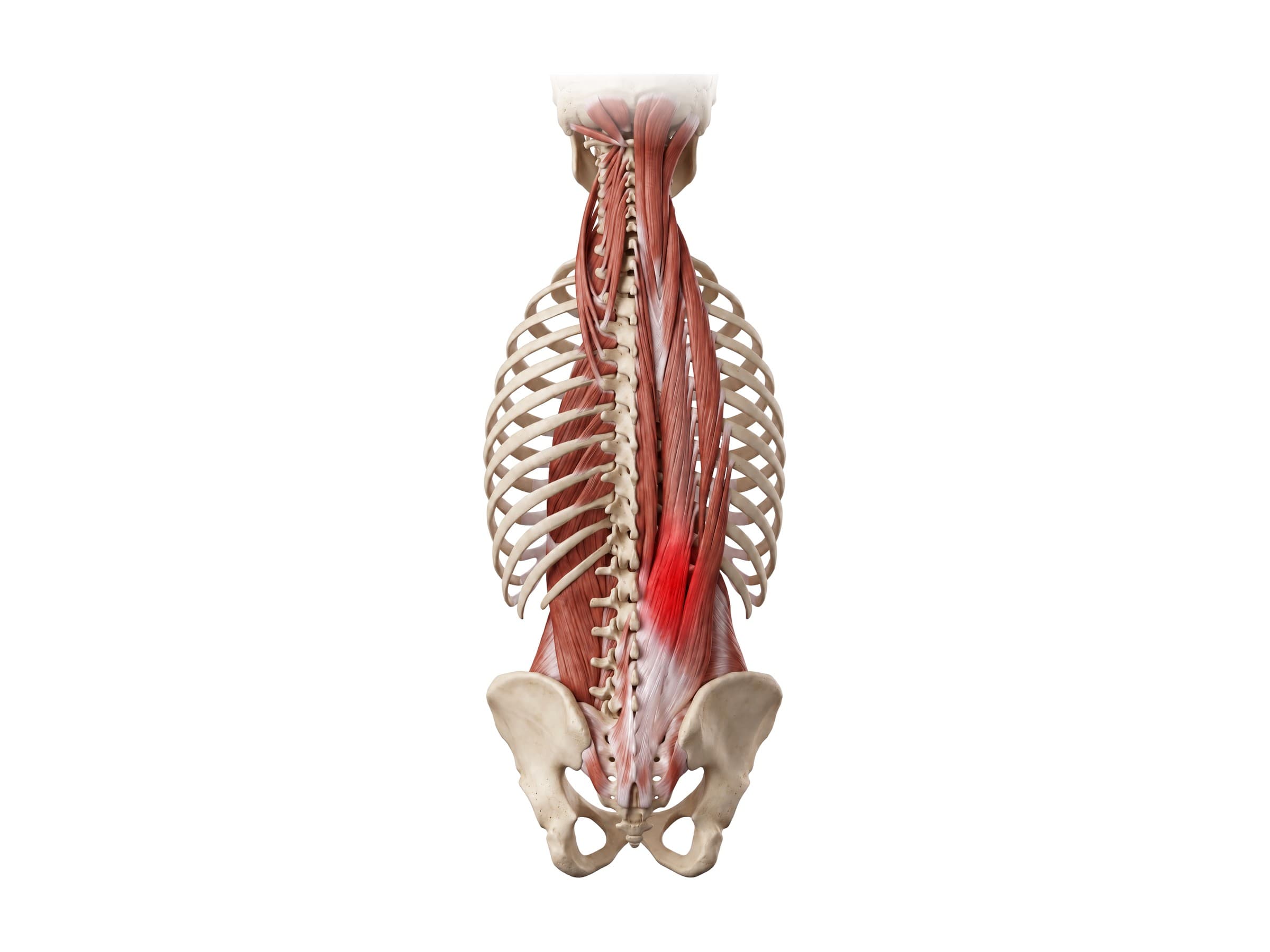
A labeled illustration of normal back muscle anatomy is the baseline exhibit in many soft-tissue injury claims after a motor vehicle collision, a slip and fall, or a lifting injury at work. Because a muscle strain does not show on X-ray, the illustration often carries more weight than the imaging: it shows a jury where the erector spinae, multifidus and quadratus lumborum sit, how they attach to the spine and pelvis, and how the mechanism of injury, the twisting or the sudden flexion and extension, loaded them. A paired exhibit then highlights the injured muscle in red, as in the strain figure above, or overlays the patient's own MRI findings such as edema or multifidus atrophy.
Natomy creates custom medical legal illustrations of back and spine injuries for demand packages and trial exhibits. For more on how normal anatomy is used in court, see demonstrative evidence. Related atlas pages: skull anatomy, shoulder anatomy, hand and wrist anatomy, elbow anatomy, hip anatomy, knee anatomy and ankle anatomy.
FAQ
What are the main muscles of the back?+
The largest and most visible back muscles are the trapezius across the upper back and neck, the latissimus dorsi across the mid and lower back, and the rhomboids and levator scapulae beneath the trapezius. Deeper, running the length of the spine, is the erector spinae (iliocostalis, longissimus and spinalis), and under it the multifidus and other small transversospinales muscles. In the lower back, the quadratus lumborum sits on either side of the lumbar spine between the lowest rib and the pelvis.
What are the three layers of back muscles?+
Back muscles are grouped into three layers. The superficial layer (trapezius, latissimus dorsi, rhomboids and levator scapulae) connects the spine to the shoulder blade and arm and moves the upper limb. The intermediate layer (serratus posterior superior and inferior) is thin and attaches to the ribs, helping with breathing. The deep or intrinsic layer (splenius, erector spinae, transversospinales and the small segmental muscles) moves and stabilizes the spine itself and holds the body upright.
What does the quadratus lumborum do?+
The quadratus lumborum is a flat, roughly rectangular muscle on each side of the lumbar spine, running from the iliac crest of the pelvis up to the 12th rib and the transverse processes of L1 to L4. Working on one side, it bends the trunk to that side and hikes the hip up; working on both sides, it extends and stabilizes the lumbar spine and anchors the 12th rib during breathing. It is a common source of deep, one-sided low back pain, often felt just above the back of the pelvis.
What muscles are in the lower back?+
The lower back (lumbar region) contains the lumbar part of the erector spinae, the multifidus closest to the spine, and the quadratus lumborum deeper and to each side. In front of the lumbar spine, the psoas major runs from the vertebrae down to the thigh. All of these are wrapped by the thoracolumbar fascia, which also anchors the lower edge of the latissimus dorsi and the abdominal wall muscles.
What is the erector spinae?+
The erector spinae is the large group of muscles that runs in two columns on either side of the spine from the sacrum and pelvis up to the skull. Each side has three parallel columns: iliocostalis (outermost, attaching to the ribs), longissimus (middle, the longest), and spinalis (innermost, next to the spinous processes). The erector spinae straightens the back from a bent position, bends it backward and to the side, and works constantly to keep the trunk upright. It is the muscle injured in most lower back strains.
How can you tell a back muscle strain from a disc problem?+
A back muscle strain usually causes pain and spasm that stays in the back, is tender to the touch over the muscle, and is worse with bending or twisting. A herniated disc more often causes pain that travels down the leg (sciatica), numbness or tingling in a specific pattern, or weakness, because the disc presses on a nerve root. A strain is a clinical diagnosis and does not show on X-ray; a herniated disc is seen on MRI. Any loss of bladder or bowel control, numbness in the groin, or progressive leg weakness needs urgent medical care.
What is the thoracolumbar fascia?+
The thoracolumbar fascia is a thick sheet of connective tissue that encloses the deep back muscles in the lower back. In the lumbar region it has three layers: a posterior layer over the back of the erector spinae, a middle layer between the erector spinae and the quadratus lumborum, and an anterior layer in front of the quadratus lumborum. The latissimus dorsi, the gluteus maximus, and the abdominal wall muscles all attach to it, so it transfers load between the trunk, pelvis and limbs.
What nerves supply the back muscles?+
The deep (intrinsic) back muscles, including the erector spinae and multifidus, are supplied by the dorsal (posterior) rami of the spinal nerves, segment by segment along the spine. The superficial back muscles that move the arm are supplied differently: the trapezius by the spinal accessory nerve (cranial nerve XI), the latissimus dorsi by the thoracodorsal nerve, and the rhomboids and levator scapulae by the dorsal scapular nerve. The quadratus lumborum is supplied by branches of T12 to L4, and the psoas major by the lumbar plexus.
Need a custom illustration of this joint?
Generate a labeled or exhibit-ready figure for a specific patient, injury, or case in minutes.
Try Natomy →