Thoracic Spine Anatomy: T1 to T12 Vertebrae, Ribs and T12
Thoracic spine anatomy labeled: the T1 to T12 vertebrae, rib joints, ligaments and the T12 thoracolumbar junction, plus compression and burst fractures.
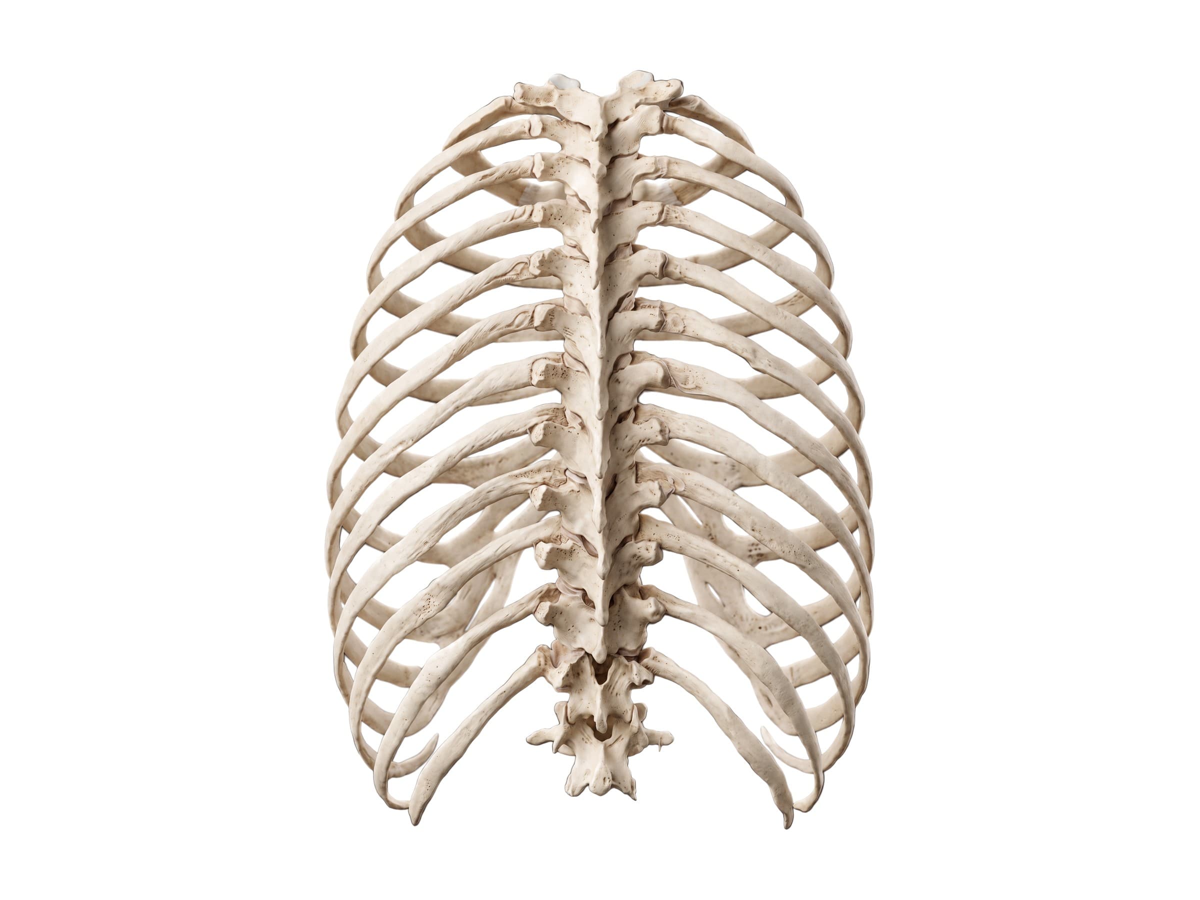
The thoracic spine is the middle section of the spine: twelve vertebrae, T1 to T12, between the neck and the lower back. It is the longest part of the spine and the only part joined to the ribs, which together with the sternum form the rib cage that protects the heart and lungs. That connection makes the thoracic spine far stiffer than the neck or lower back. Thoracic spine anatomy covers the vertebrae, the costovertebral and costotransverse joints where the ribs attach, the discs and ligaments, the thoracic spinal cord and nerves, and the thoracolumbar junction at T12, where the stiff thoracic spine meets the mobile lumbar spine and where most spine fractures happen. Knowing these parts is the first step to reading a thoracic MRI report, explaining a compression fracture to a patient, or illustrating a back injury claim.
The twelve thoracic vertebrae
The labeled view above shows the thoracic spine from behind, with a pair of ribs at every level. From the side, the thoracic spine curves gently backward, a curve called the thoracic kyphosis, normally about 20 to 45 degrees. An increased curve (hyperkyphosis, the rounded upper back) can come from poor posture, Scheuermann's disease in teenagers, or multiple compression fractures in older adults.
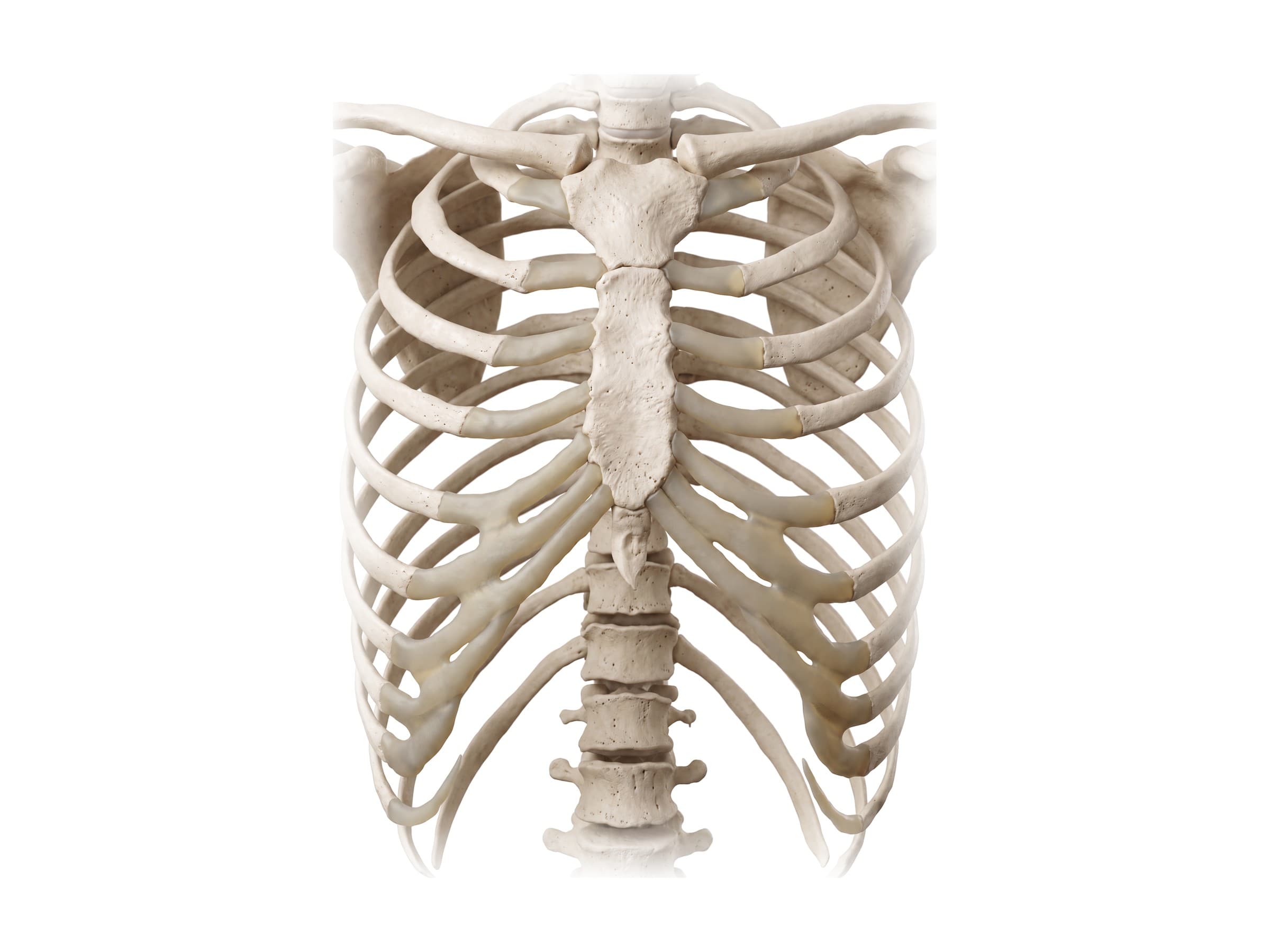
From the front, the thoracic vertebrae form the back of the rib cage. Ribs 1 to 7 (the true ribs) join the sternum directly through their own costal cartilages. Ribs 8 to 10 (false ribs) join the cartilage of the rib above. Ribs 11 and 12 (floating ribs) end free in the muscles of the flank. The thoracic vertebrae are often grouped as:
- Upper (T1 to T4): small, with T1 similar to a cervical vertebra. T1 carries the first rib and contributes to the nerves of the hand.
- Middle (T5 to T8): the typical thoracic vertebrae, closest to the heart and the descending aorta.
- Lower (T9 to T12): larger, increasingly like lumbar vertebrae, with T11 and T12 carrying the floating ribs.
Anatomy of a thoracic vertebra
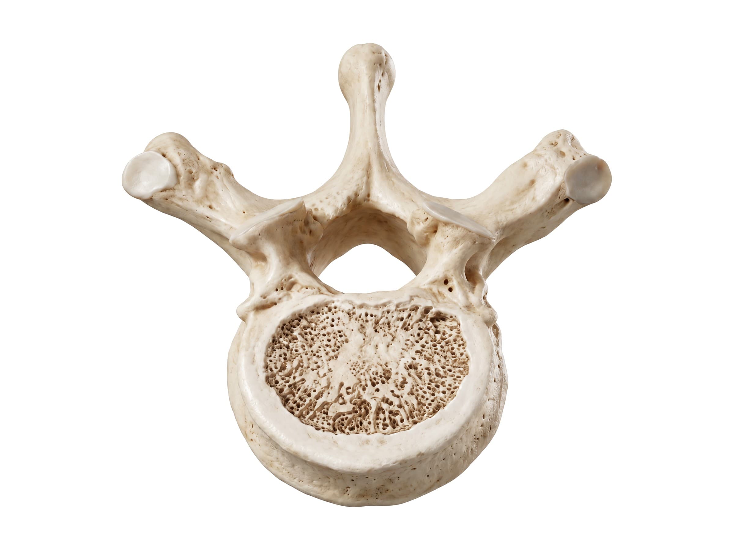
A typical thoracic vertebra, seen here from above with the front at the bottom, has:
- Vertebral body: heart-shaped, medium-sized, between the small cervical bodies and the large lumbar bodies.
- Vertebral canal: small and round. The thoracic canal is the narrowest part of the spinal canal relative to the cord, which leaves little room if a fracture fragment or disc pushes into it.
- Pedicles and laminae: the arch around the canal. The laminae overlap like roof tiles.
- Transverse processes: long and strong, pointing back and out. Each carries a costal facet where the tubercle of a rib joins it (T1 to T10).
- Spinous process: long and slender, sloping steeply downward so it overlaps the vertebra below. The tip you feel in the middle of the back usually sits at the level of the vertebral body below.
- Superior and inferior articular processes: the facet joints. Thoracic facets face mostly backward and forward, which allows rotation of the trunk while the ribs limit bending.
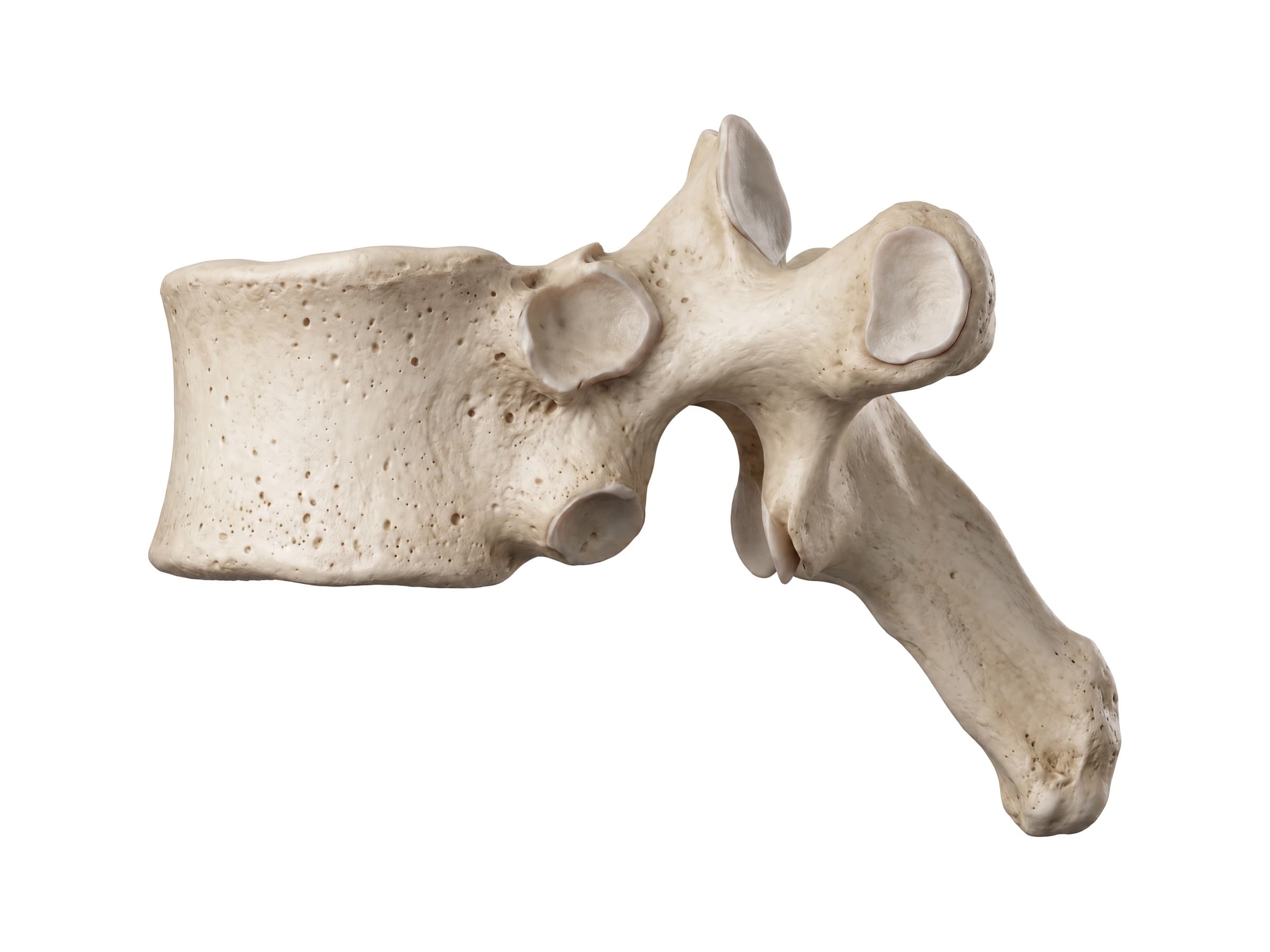
Seen from the side, the feature that defines a thoracic vertebra is its set of costal facets: a superior and an inferior costal facet (half-facets, or demifacets) on the side of the body where the head of a rib meets two neighboring vertebrae, and a transverse costal facet on the transverse process for the tubercle of the rib. T1, T10, T11 and T12 are atypical: their rib heads meet a single full facet on one vertebra.
The rib joints: costovertebral and costotransverse
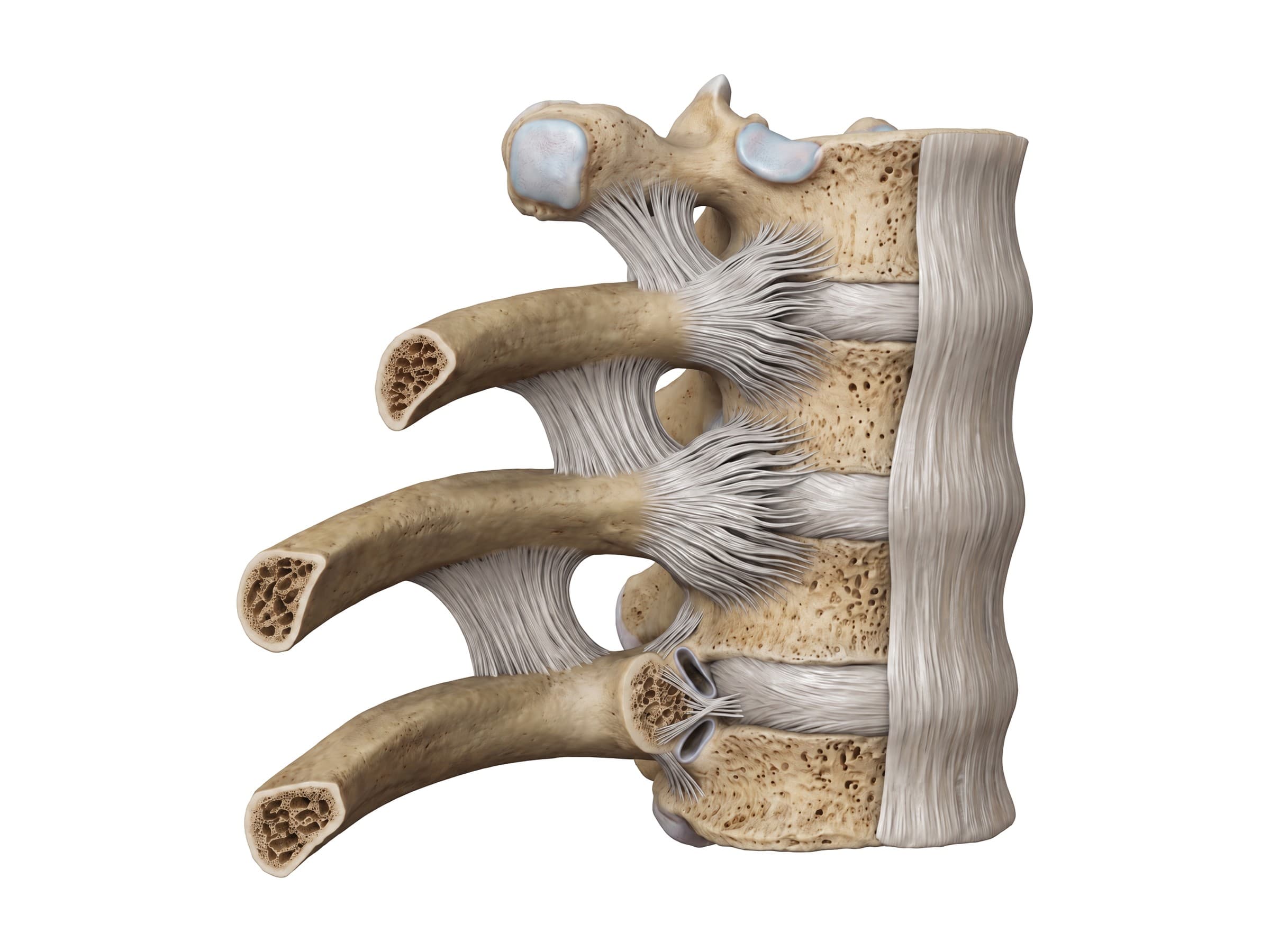
Each rib joins the spine at two joints:
- Costovertebral joint: the head of the rib sits in the hollow formed by the costal facets of two vertebral bodies and the disc between them (for ribs 2 to 9). A fan-shaped radiate ligament holds it to the front of the bodies, and an interarticular ligament ties the rib head to the disc.
- Costotransverse joint: the tubercle of the rib sits against the costal facet of the transverse process, held by the costotransverse ligaments.
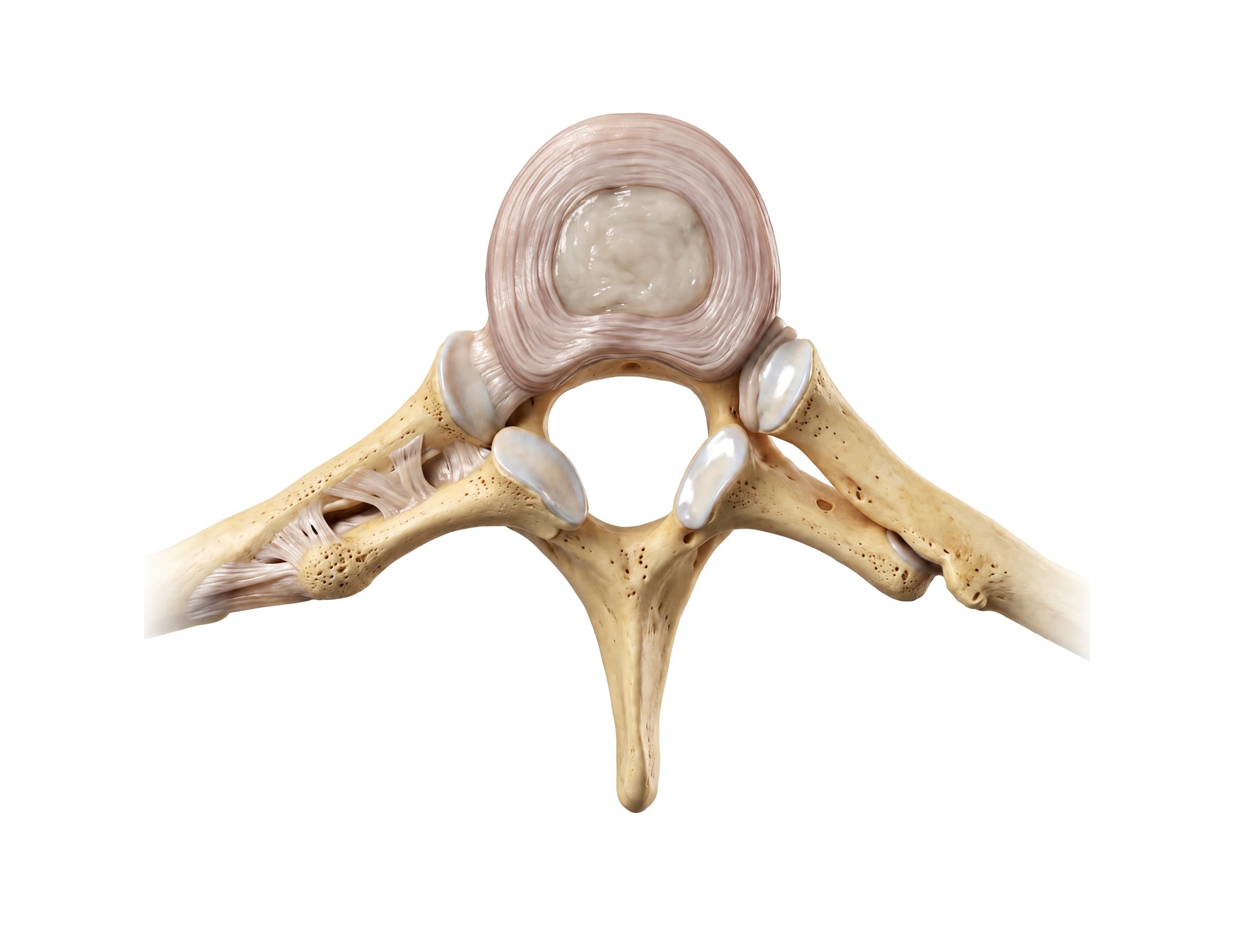
Seen from above, the two joints make each rib a lever that pivots as you breathe: the upper ribs lift the sternum forward (the pump-handle movement) and the lower ribs swing outward (the bucket-handle movement). Sprains of these joints are a common cause of sharp, one-sided mid back pain that wraps around the chest and worsens with a deep breath.
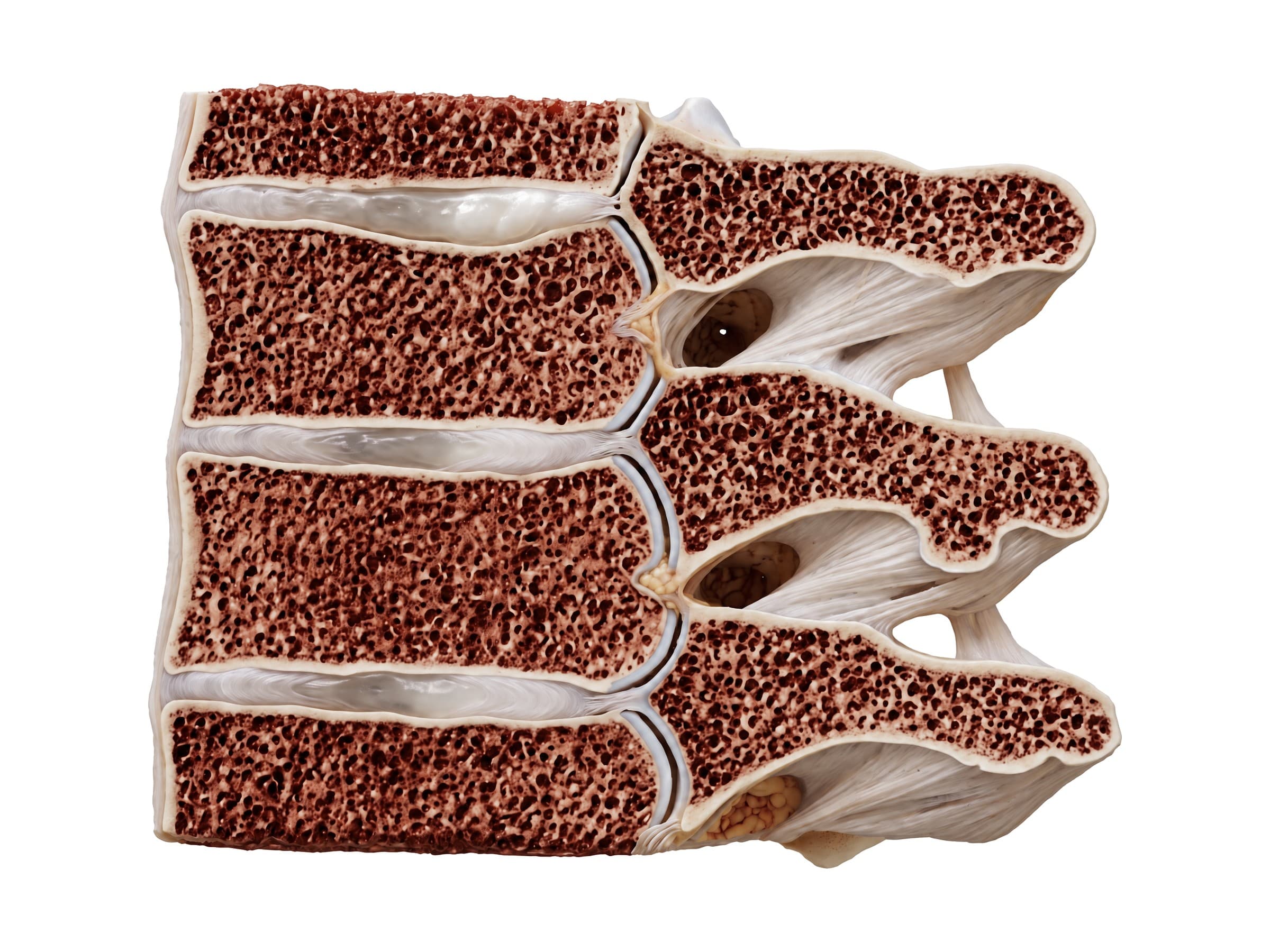
Cut through the level of the rib heads, the section shows the vertebral bodies and discs in front, the intervertebral foramina where the thoracic nerves leave, and the heads, necks and tubercles of the ribs running back to the transverse processes, tied in place by the costotransverse ligaments.
Discs and ligaments
The thoracic intervertebral discs are thinner than cervical and lumbar discs, and the rib cage protects them, so thoracic disc herniations are uncommon (under 1 percent of all disc herniations). When they do happen, usually in the lower thoracic spine, they can press on the spinal cord itself rather than on a single nerve root.
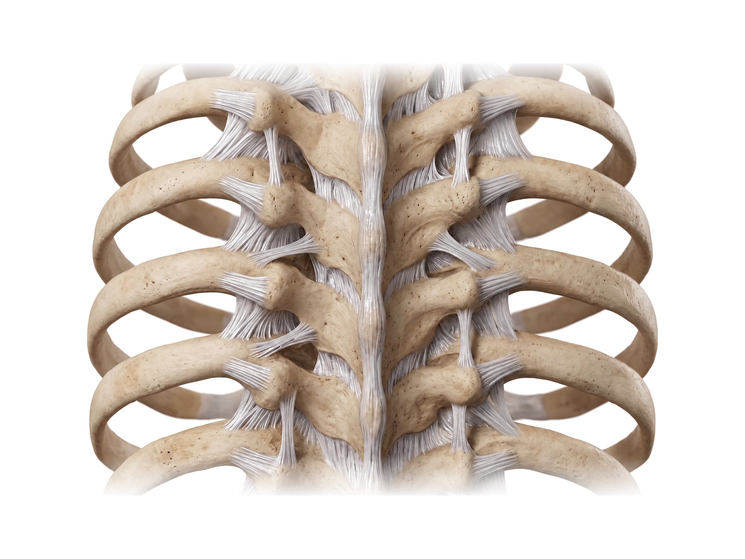
The ligaments are the same as in the rest of the spine: the anterior longitudinal ligament down the front of the bodies, the posterior longitudinal ligament inside the canal, the ligamentum flavum between the laminae, and the interspinous and supraspinous ligaments between the spinous processes. In addition, the costotransverse ligaments tie each rib to the transverse processes, and the rib cage and sternum act as a fourth column of support, which is why the thoracic spine is so stable.
T12 and the thoracolumbar junction
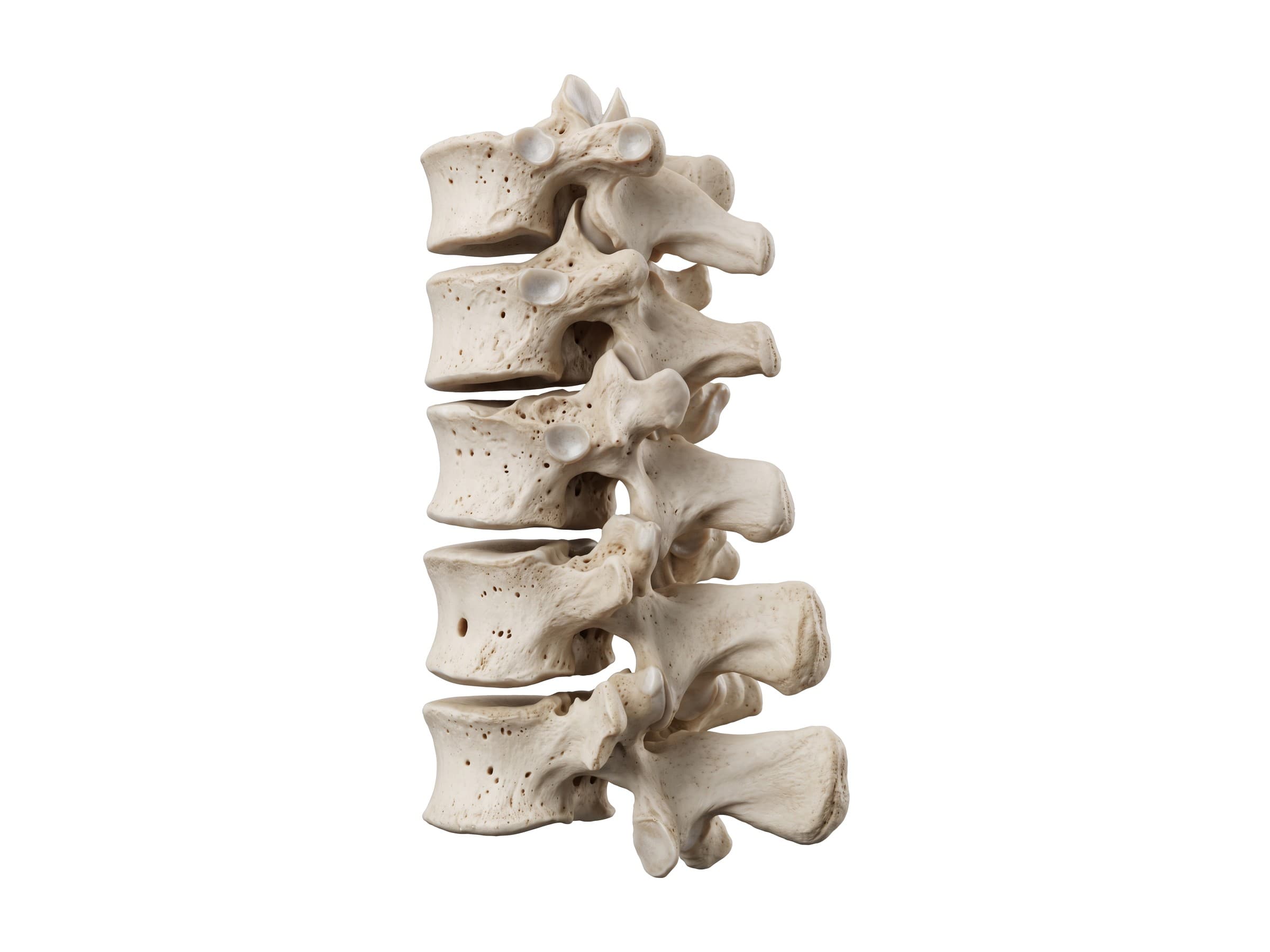
T12 is the last thoracic vertebra. It carries the twelfth rib on a single facet, has no costal facet on its transverse process, and is a transition vertebra: its upper facet joints face like thoracic joints, but its lower facet joints face inward like lumbar joints. Together with T11, L1 and L2 it forms the thoracolumbar junction, where the stiff, rib-supported thoracic spine meets the flexible lumbar spine.
That sudden change in stiffness concentrates force. In falls from a height, car crashes and osteoporosis, T12 and L1 are the most commonly fractured vertebrae in the spine. The spinal cord also ends near this level (around L1-L2), so injuries at T12 can damage the end of the cord (the conus medullaris) and the nerve roots of the cauda equina at the same time, affecting the legs, bladder and bowel.
Thoracic spinal cord and nerves
The thoracic spinal cord runs the length of the thoracic spine inside the narrow canal. It carries the signals to and from the legs and controls the trunk muscles. Between T1 and L2 it also contains the sympathetic nerve cells that control blood pressure, heart rate and sweating, which is why high thoracic cord injuries can cause dangerous swings in blood pressure.
There are twelve pairs of thoracic nerves, each leaving below the vertebra with the same number:
- T1: joins the brachial plexus and supplies the small muscles of the hand.
- T2 to T11 (intercostal nerves): run in a groove along the underside of each rib, between the intercostal muscles, and supply the chest wall. The lower intercostal nerves continue forward to supply the abdominal wall muscles and skin.
- T12 (subcostal nerve): runs below the twelfth rib to the lower abdominal wall.
Their skin bands, the thoracic dermatomes, wrap around the trunk in horizontal stripes. Useful landmarks are T4 at the nipple, T6 at the bottom of the sternum (xiphoid), T10 at the belly button and T12 just above the groin. Shingles (herpes zoster) in a thoracic nerve causes a painful rash in exactly one of these bands on one side. For the bands of the legs, see lower limb nerves and dermatomes.
Muscles of the thoracic spine
The erector spinae and multifidus run along the back of the thoracic spine and hold it upright; the rhomboids, trapezius and latissimus dorsi cover them and move the shoulder blades and arms; and the intercostal muscles fill the spaces between the ribs and move them for breathing. All of these are covered in back muscles anatomy.
Range of motion of the thoracic spine
The ribs limit bending, so the thoracic spine moves less than the neck and lower back, except in rotation. Typical values are about 20 to 45 degrees of flexion, 15 to 25 degrees of extension, 20 to 25 degrees of side bending and 30 to 45 degrees of rotation to each side, with most rotation in the middle and lower thoracic spine.
Common thoracic spine injuries and how they appear on imaging
X-rays show alignment and the height of the vertebral bodies. CT shows fractures in detail and is the standard after major trauma. MRI shows the spinal cord, discs and ligaments, and whether a fracture is new (bone marrow swelling) or old.
- Compression fracture: the front of the vertebral body collapses into a wedge while the back wall holds, from a fall onto the buttocks, a crash, or, in people with osteoporosis, a minor fall or no clear injury at all. On a side X-ray the front of the body is shorter than the back; the percentage of height lost guides treatment. Painful osteoporotic fractures may be treated with kyphoplasty or vertebroplasty.
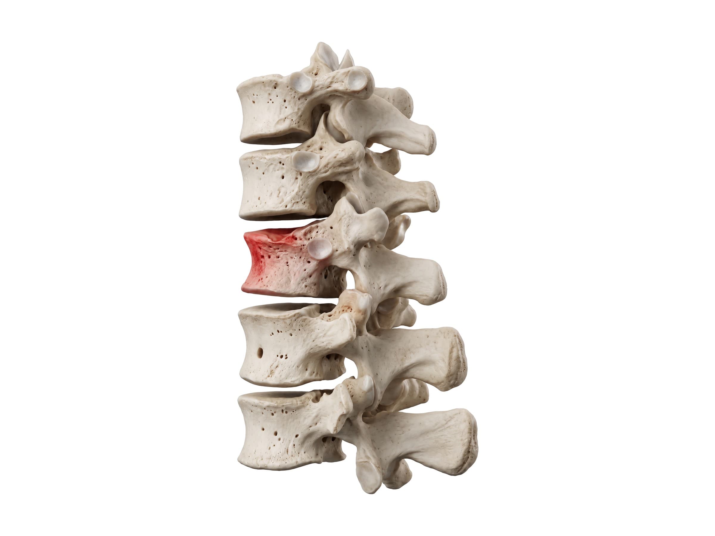
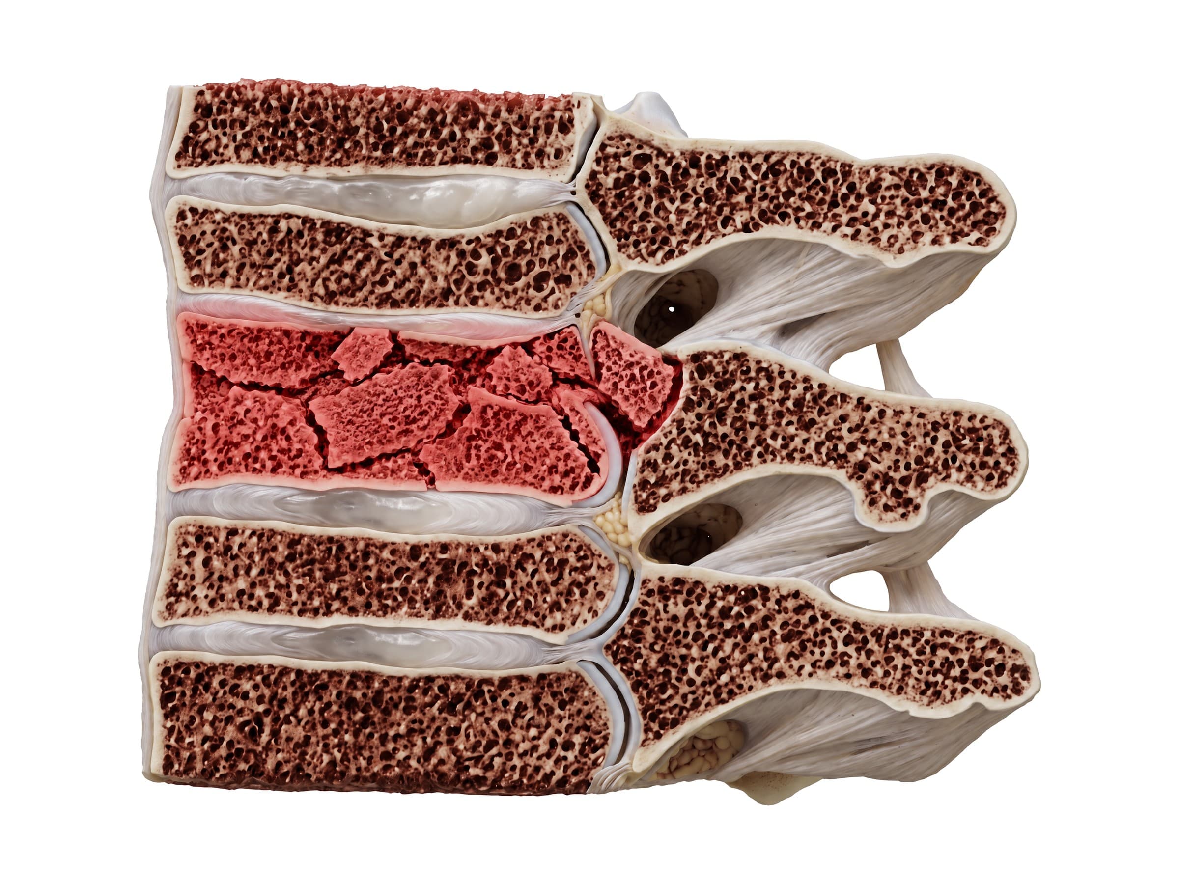
- Burst fracture: a high-energy axial load, such as a fall from a height onto the feet or a high-speed crash, crushes the entire vertebral body. Unlike a compression fracture, the back wall also breaks, and fragments can be pushed backward (retropulsed) into the spinal canal. CT shows the fragments and how much of the canal is narrowed; MRI shows any cord injury. Unstable burst fractures are fixed with screws and rods.
- Chance (seatbelt) fracture: in a head-on crash with only a lap belt, the body jackknifes over the belt and the vertebra splits horizontally from back to front, usually at the thoracolumbar junction (T12 to L2). It is often accompanied by injuries to the bowel and other abdominal organs.
- Fracture-dislocation: the most severe thoracic injury, where the vertebra shifts on the one below with failure of all the supporting structures. Because the thoracic canal is narrow, these injuries often cause complete paraplegia.
- Rib fractures: the most common chest injury after blunt trauma, often next to the spine at the angle of the rib. Several adjacent ribs broken in two places can create a flail segment that moves the wrong way with breathing.
- Thoracic disc herniation: uncommon, usually at T8 to T12; can cause band-like pain around the chest or, if it presses on the cord, leg weakness and balance problems.
How the thoracic spine is shown in a personal injury case
Thoracic fractures, especially at T12 and L1, are among the most common serious injuries in falls, motor vehicle collisions and workplace accidents, and they often lead to lasting pain, a permanent loss of vertebral height and, in severe cases, paralysis. The defense often argues that a compression fracture was old or caused by osteoporosis rather than the incident. Each fracture follows a recognizable mechanism of injury: a compression fracture from flexion and axial load in a fall, a burst fracture from a high-energy vertical load, a Chance fracture from a lap belt in a head-on crash. A labeled illustration of the normal thoracic spine and the thoracolumbar junction is the baseline exhibit. It lets an expert show the jury where T12 sits, why that level breaks, and how close the spinal cord is, before showing the injury itself: the wedge-shaped vertebra, the fragment pushed into the canal, or the screws and rods of the fusion. Illustrations drawn from the plaintiff's own CT or MRI, including the bone marrow swelling that shows a fracture is new, tie the scan to the incident.
Natomy creates custom medical legal illustrations of thoracic spine injuries for demand packages and trial exhibits, and the rules for using these visuals at trial are covered in demonstrative evidence. For the sections above and below, see cervical spine anatomy and lumbar spine anatomy. Related atlas pages: back muscles anatomy, shoulder anatomy and hip anatomy.
FAQ
How many thoracic vertebrae are there?+
There are 12 thoracic vertebrae, numbered T1 to T12 from the base of the neck to the bottom of the rib cage. Each one joins a pair of ribs, which is what sets the thoracic vertebrae apart from the cervical vertebrae above and the lumbar vertebrae below. They get gradually larger from T1 to T12 as they carry more of the body's weight.
What is the T12 vertebra?+
T12 is the twelfth and lowest thoracic vertebra, at the bottom of the rib cage just above the lower back. It carries the twelfth (floating) rib and sits on top of L1. Its upper joints face like a thoracic vertebra's and its lower joints face like a lumbar vertebra's, so it is the transition point between the stiff, rib-supported thoracic spine and the mobile lumbar spine.
Why is T12 commonly fractured?+
The thoracolumbar junction (T11 to L2) is where the stiff thoracic spine, braced by the rib cage, meets the mobile lumbar spine. Forces from a fall or crash concentrate at that change in stiffness, so T12 and L1 are the most commonly fractured vertebrae in the spine. In older adults with osteoporosis, T12 compression fractures can happen after a minor fall or even a cough or lift.
What happens if the thoracic vertebrae are damaged?+
Most thoracic fractures are stable compression fractures that cause sharp mid back pain and heal over about 8 to 12 weeks. Because the thoracic spinal canal is narrow and the cord there has a limited blood supply, unstable fractures, burst fractures with fragments in the canal, or dislocations can injure the spinal cord and cause weakness or paralysis of the legs (paraplegia), loss of feeling below the level of injury, and loss of bladder and bowel control.
What are the worst vertebrae to fracture?+
Fractures of the upper cervical spine (C1 and C2) and fracture-dislocations anywhere in the spine are the most dangerous because they can injure the spinal cord. High neck injuries can affect breathing. In the thoracic spine, the cord fills more of the narrow canal, so unstable thoracic fractures carry a high risk of complete paraplegia. Isolated compression fractures, by contrast, are usually stable and heal without surgery.
What are the red flags for the thoracic spine?+
Mid back pain is less often mechanical than neck or low back pain, so red flags matter more. Warning signs are pain after significant trauma, unexplained weight loss, fever, a history of cancer (the thoracic spine is the most common site of spinal metastases), pain that is constant and worse at night, new weakness or numbness in the legs, trouble walking or balance problems, and loss of bladder or bowel control. Pain that wraps around the chest can also come from the heart, lungs or gallbladder.
What nerves come out of the thoracic spine?+
Twelve pairs of thoracic spinal nerves leave the thoracic spine, each below the vertebra with the same number. T1 contributes to the brachial plexus and the hand. T2 to T11 become the intercostal nerves, which run along the underside of each rib to supply the chest wall muscles and skin, and the lower ones continue to the abdominal wall. T12 is the subcostal nerve below the last rib. Their skin bands (dermatomes) wrap around the trunk: T4 at the nipple, T10 at the belly button.
What is the difference between thoracic and lumbar vertebrae?+
Thoracic vertebrae have costal facets where the ribs attach to the body and transverse processes, a heart-shaped body, a small round canal, and long spinous processes that slope steeply down. Lumbar vertebrae have no rib facets, much larger kidney-shaped bodies, a triangular canal, and short, square spinous processes pointing straight back. Thoracic facet joints face mostly backward and allow rotation; lumbar facet joints face inward and limit it.
Need a custom illustration of this joint?
Generate a labeled or exhibit-ready figure for a specific patient, injury, or case in minutes.
Try Natomy →