Shoulder Anatomy: Bones, Muscles & Rotator Cuff
Labeled illustrations of the shoulder's bones, rotator cuff, muscles, ligaments, labrum and nerves, plus common shoulder injuries.
The shoulder is the most mobile joint in the body, and the trade-off for that range of motion is stability: the shoulder dislocates more often than any other major joint, and rotator cuff and labral tears are among the most common findings on shoulder MRI after a fall, sports injury, or motor vehicle collision. Shoulder anatomy comes down to three bones (the clavicle, scapula, and humerus), four joints, the rotator cuff and the larger muscles around it, a ring of cartilage called the labrum, and the nerves of the brachial plexus. Knowing how they fit together is the first step to reading a shoulder MRI report, explaining a diagnosis to a patient, or illustrating a personal injury claim.
Bones of the shoulder
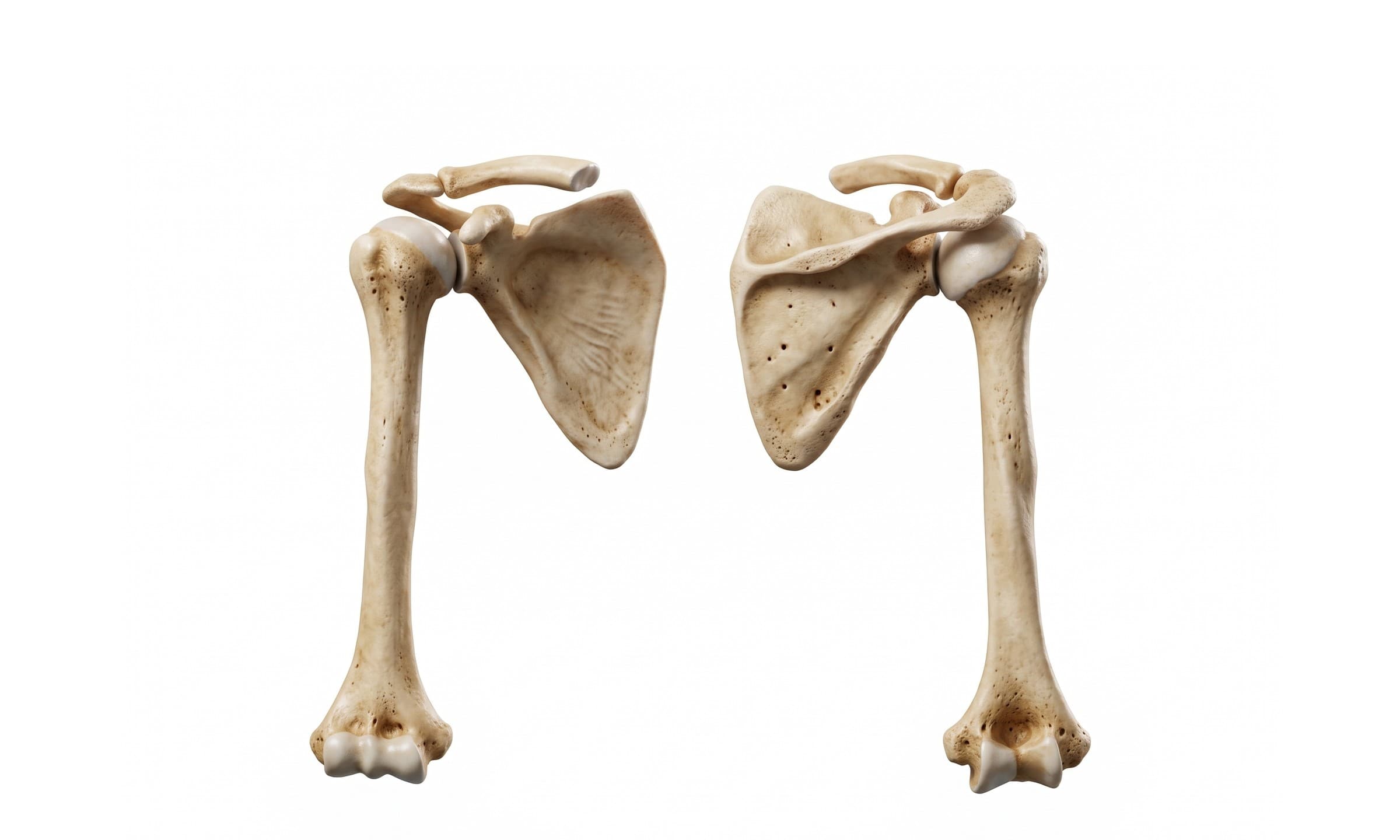
Three bones meet at the shoulder. In the anterior view above (left), the clavicle runs horizontally from the sternum out to the top of the shoulder, where it meets the acromion, the flat projection of the scapula that forms the roof of the joint. Just below and in front of that junction, the hook-shaped coracoid process curves forward from the scapula. The rounded humeral head sits against the glenoid fossa, a shallow, pear-shaped socket on the outer edge of the scapula. Just below the head, the greater tubercle (on the outside) and lesser tubercle (in front) are where the rotator cuff tendons attach, with the intertubercular (bicipital) groove between them carrying the long head of the biceps tendon.
- Clavicle: the collarbone, the only bony connection between the arm and the trunk. It acts as a strut that holds the shoulder out to the side, and it is one of the most commonly fractured bones in the body.
- Scapula: the shoulder blade, a flat triangular bone that glides over the back of the rib cage. It gives rise to the acromion, coracoid process, and glenoid fossa, and anchors 17 muscles.
- Humerus: the upper arm bone. Its proximal end has the humeral head, the anatomical neck just around the head's edge, the greater and lesser tubercles, and the surgical neck below them, the narrowing where proximal humerus fractures most often occur.
In the posterior view (right), the spine of the scapula runs across the back of the shoulder blade and ends laterally as the acromion. It divides the back of the scapula into the supraspinous fossa above and the infraspinous fossa below, the two hollows filled by the supraspinatus and infraspinatus muscles. The scapula's medial border runs parallel to the spine, and its inferior angle is the lowest point, the part that sticks out in scapular winging. On the front of the scapula, the matching hollow is the subscapular fossa, which holds the subscapularis.
Joints of the shoulder
What most people call "the shoulder joint" is one of four joints that move together every time the arm is raised.
- Glenohumeral joint: the main shoulder joint, a ball-and-socket synovial joint between the humeral head and the glenoid fossa. The socket covers only about a quarter to a third of the ball, which gives the shoulder its range of motion and makes it the most commonly dislocated major joint.
- Acromioclavicular (AC) joint: the small joint at the top of the shoulder where the clavicle meets the acromion. It is the joint injured in an AC separation (separated shoulder).
- Sternoclavicular (SC) joint: where the inner end of the clavicle meets the sternum, the only true joint connecting the arm to the skeleton of the trunk.
- Scapulothoracic joint: not a true joint but a gliding surface where the scapula slides over the back of the rib cage. Roughly one degree of scapular rotation accompanies every two degrees of glenohumeral motion when the arm is raised, a pattern called scapulohumeral rhythm.
Ligaments, joint capsule, and labrum
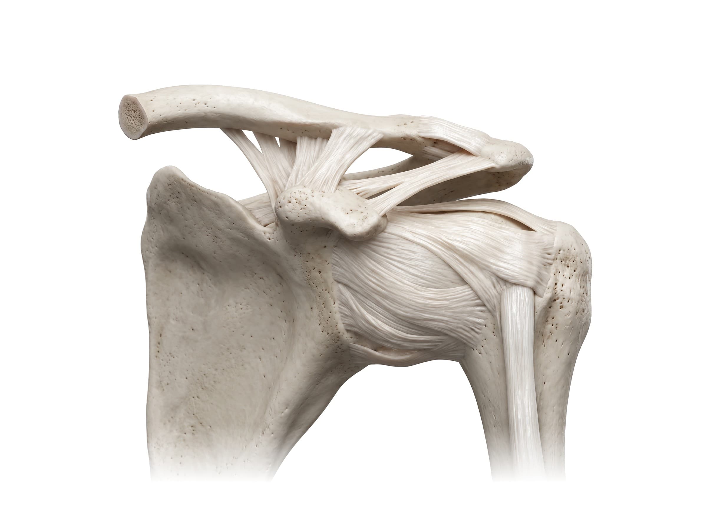
The glenohumeral joint is wrapped in a loose joint capsule, thickened at the front into three bands: the superior, middle, and inferior glenohumeral ligaments. The inferior glenohumeral ligament is the most important of the three, acting as a sling under the humeral head when the arm is raised and turned outward, the position in which most anterior dislocations happen. Above the joint, the coracohumeral ligament runs from the coracoid process to the tubercles of the humerus, and the coracoacromial ligament bridges the coracoid process to the acromion, forming, together with those two bones, the coracoacromial arch that the supraspinatus tendon slides beneath.
The AC joint is held together by the acromioclavicular ligament across the joint itself and, more strongly, by the coracoclavicular ligament, which runs up from the coracoid process to the underside of the clavicle in two parts: the cone-shaped conoid medially and the flatter trapezoid laterally. The coracoclavicular ligament is what keeps the clavicle from riding up in an AC separation; when it tears, the end of the clavicle becomes visibly prominent.
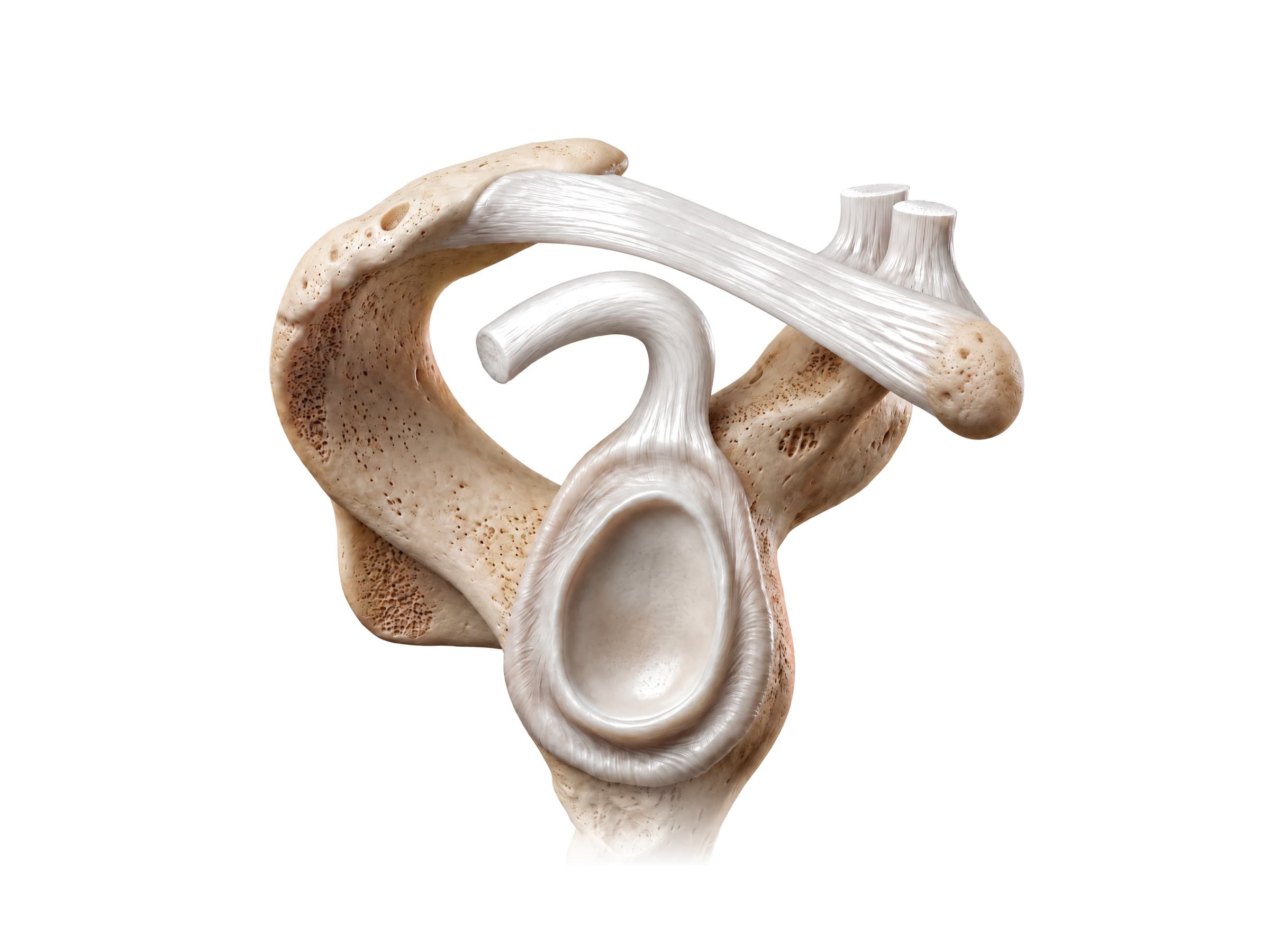
Looking straight into the socket with the humerus removed, the glenoid labrum is visible as a ring of fibrocartilage around the rim of the glenoid fossa. It deepens the shallow socket and acts as an attachment point for the glenohumeral ligaments. At the very top of the glenoid, the long head of the biceps tendon anchors into the labrum at the supraglenoid tubercle, the biceps anchor. This is the region injured in a SLAP tear. The front and lower part of the labrum is where it tears in a Bankart lesion after an anterior dislocation.
Shoulder joint in cross-section
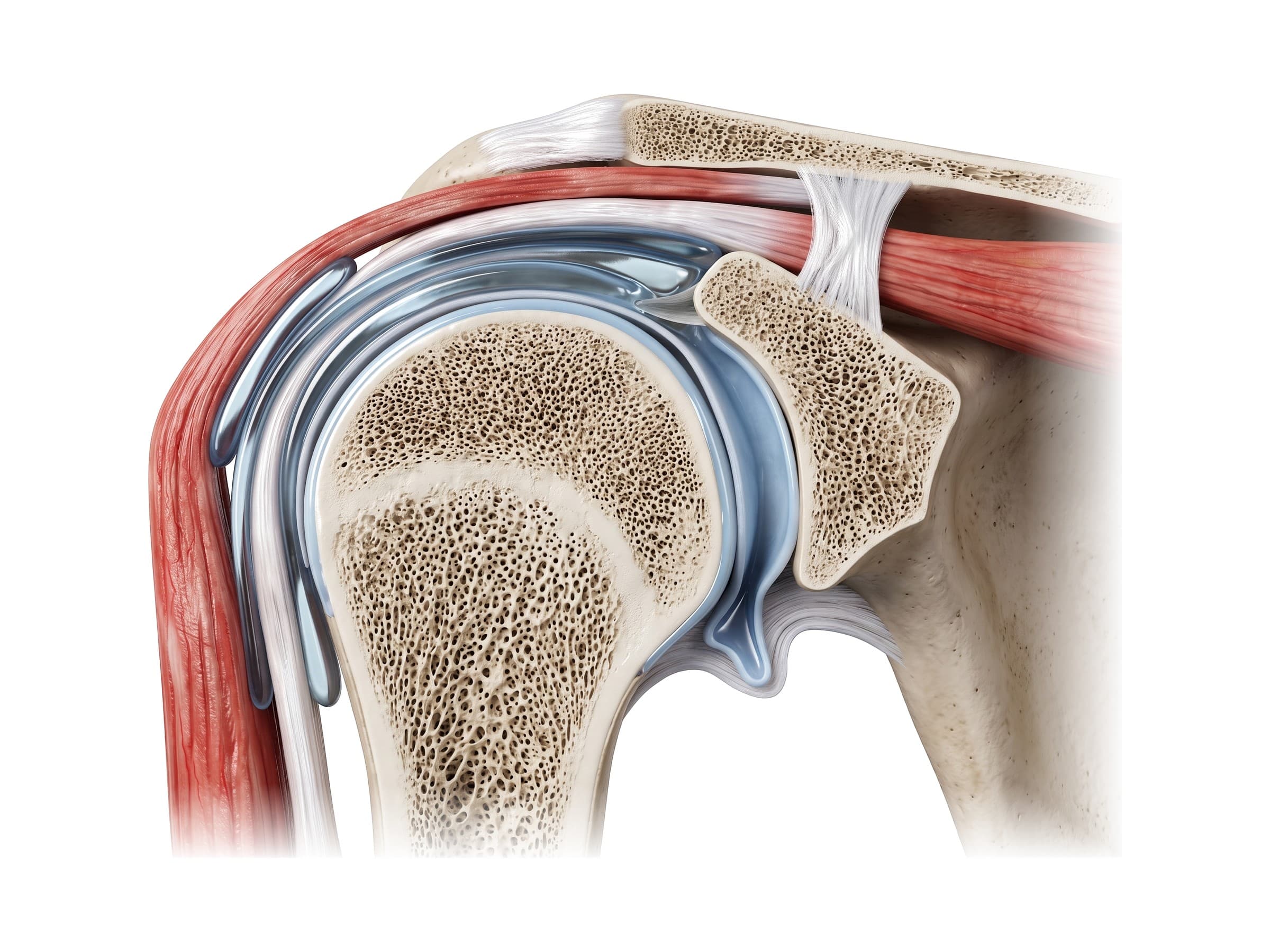
A coronal cut through the shoulder, the same plane a shoulder MRI is most often read in, shows how the layers stack from the skin down to the joint. On the outside is the deltoid. Beneath it, the subacromial-subdeltoid bursa is a thin fluid-filled sac that lets the rotator cuff glide under the acromion and deltoid. Below the bursa, the supraspinatus tendon passes over the top of the humeral head to insert on the greater tubercle. Deepest of all, the humeral head and the glenoid are each capped with articular cartilage, with the labrum visible as a small triangle at the upper and lower rims of the glenoid and the joint capsule enclosing the joint cavity. Above it all sit the acromion and the AC joint.
This view explains impingement: the supraspinatus tendon and bursa pass through a narrow space between the humeral head below and the acromion and coracoacromial ligament above, and anything that narrows that space (a bone spur, a thickened bursa, swelling) pinches them when the arm is raised.
Muscles of the shoulder
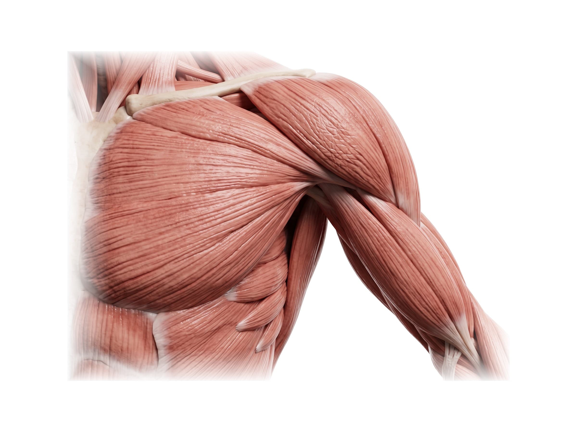
From the front, the deltoid caps the shoulder in three parts: the anterior part from the outer third of the clavicle, the middle part from the acromion, and the posterior part from the spine of the scapula. All three converge on the deltoid tuberosity halfway down the humerus. The deltoid is the main muscle that lifts the arm away from the body (abduction) once the supraspinatus has started the movement. Across the top of the shoulder, the trapezius pulls the scapula up and back. In front, the pectoralis major fans from the clavicle and sternum to the front of the humerus, and below the coracoid process, the coracobrachialis and the short head of the biceps brachii run down the front of the arm.
- Deltoid: abducts the arm; its anterior and posterior parts also flex and extend it. Supplied by the axillary nerve.
- Trapezius: elevates, retracts, and rotates the scapula. Supplied by the spinal accessory nerve (cranial nerve XI).
- Pectoralis major: adducts and internally rotates the arm and pulls it forward. Supplied by the medial and lateral pectoral nerves.
- Latissimus dorsi: the broad back muscle that extends, adducts, and internally rotates the arm. Supplied by the thoracodorsal nerve.
- Serratus anterior: holds the scapula flat against the rib cage and rotates it upward when the arm is raised overhead. Supplied by the long thoracic nerve, whose injury causes scapular winging.
- Biceps brachii and triceps brachii: the long heads of both cross the shoulder joint, the biceps from the top of the glenoid and the triceps from just below it.
Rotator cuff muscles
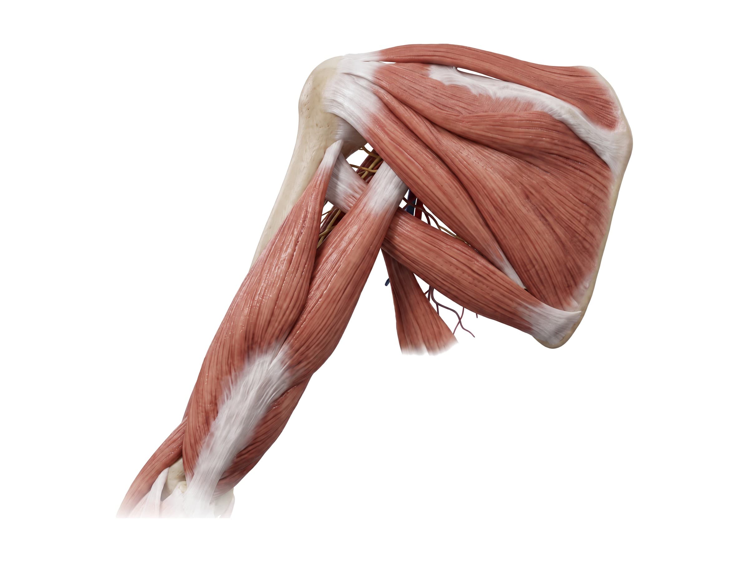
Under the deltoid, four smaller muscles form the rotator cuff, remembered as SITS: supraspinatus, infraspinatus, teres minor, and subscapularis. Each starts on the scapula and ends on the top of the humerus, and their tendons blend into a single cuff that wraps the humeral head on the top, back, and front. Their main job is not to move the arm but to hold the humeral head centered in the shallow glenoid while the larger muscles move it.
Seen from behind with the deltoid removed, the supraspinatus fills the supraspinous fossa above the spine of the scapula and passes under the acromion to the top of the greater tubercle. Below the spine, the infraspinatus fills the infraspinous fossa, and the teres minor runs along the scapula's lateral border; both insert on the back of the greater tubercle. Below them, teres major runs to the front of the humerus instead, and the long head of the triceps passes between teres minor and teres major.
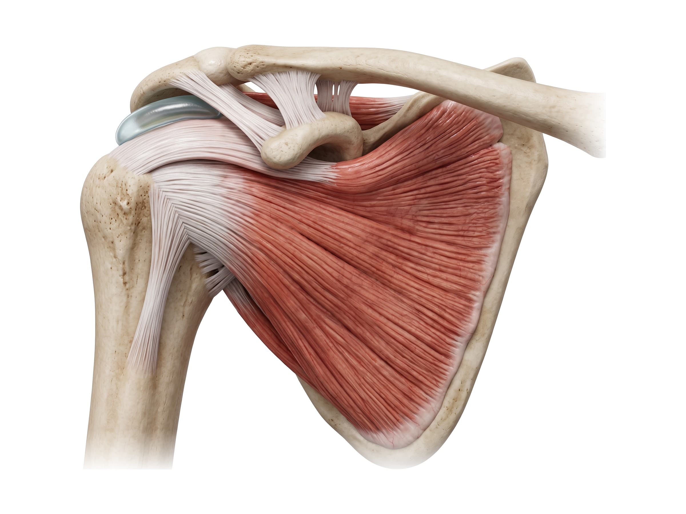
The fourth cuff muscle, the subscapularis, is the only one on the front of the scapula. It fills the subscapular fossa on the surface that faces the ribs and converges into a broad tendon that crosses the front of the shoulder joint to insert on the lesser tubercle. It is the largest and strongest of the four, and the main internal rotator of the arm.
- Supraspinatus: from the supraspinous fossa to the top of the greater tubercle. Starts abduction (the first 15 degrees or so) and holds the humeral head down against the glenoid. Supplied by the suprascapular nerve. The most commonly torn rotator cuff tendon.
- Infraspinatus: from the infraspinous fossa to the back of the greater tubercle. The main external rotator of the arm. Supplied by the suprascapular nerve.
- Teres minor: from the upper lateral border of the scapula to the lowest facet of the greater tubercle. Assists external rotation. Supplied by the axillary nerve.
- Subscapularis: from the subscapular fossa to the lesser tubercle. Internally rotates the arm and stabilizes the front of the joint. Supplied by the upper and lower subscapular nerves.
- Teres major (not part of the rotator cuff): from the inferior angle of the scapula to the front of the humerus. Internally rotates, adducts, and extends the arm alongside the latissimus dorsi. Supplied by the lower subscapular nerve.
Nerves of the shoulder
Every nerve to the shoulder and arm, including the median, ulnar and radial nerves that supply the hand and wrist, comes from the brachial plexus, a network formed by the spinal nerve roots C5 through T1. The roots leave the neck between the anterior and middle scalene muscles and join into the upper, middle, and lower trunks. Each trunk splits into anterior and posterior divisions behind the clavicle, and the divisions re-form into the lateral, posterior, and medial cords around the axillary artery in the armpit. The cords end as the five main nerves of the arm: the musculocutaneous, axillary, radial, median, and ulnar nerves.
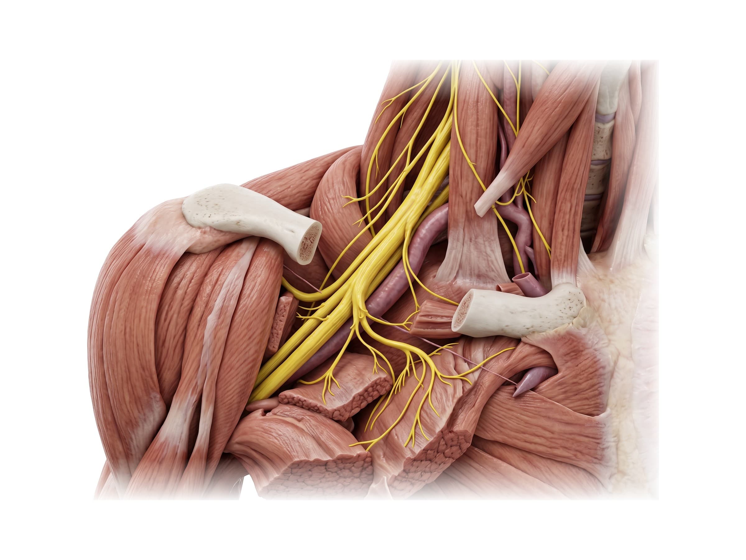
- Axillary nerve (C5, C6): from the posterior cord, through the quadrangular space below the joint, and around the surgical neck of the humerus to the deltoid and teres minor. The nerve most often injured in anterior dislocations and surgical neck fractures.
- Suprascapular nerve (C5, C6): from the upper trunk, through the suprascapular notch at the top of the scapula, to the supraspinatus and infraspinatus. Can be compressed at the notch or by a ganglion cyst from a labral tear.
- Long thoracic nerve (C5 to C7): runs down the side of the chest wall to the serratus anterior; injury causes winging of the scapula.
- Musculocutaneous nerve (C5 to C7): from the lateral cord, through the coracobrachialis, to the biceps and brachialis.
- Upper trunk (C5, C6): the part of the plexus injured when the head and shoulder are forced apart, as in a motorcycle crash or a difficult delivery (Erb's palsy), leaving the arm hanging at the side and turned inward.
Common shoulder injuries and how they appear on imaging
- Rotator cuff tear: usually the supraspinatus tendon, either partial-thickness or full-thickness. On MRI, a full-thickness tear shows fluid signal running through the tendon from the bursa to the joint, often with retraction of the torn edge. Ultrasound is also accurate in experienced hands.
- Anterior shoulder dislocation: more than 95% of shoulder dislocations go forward. X-rays show the humeral head out of the socket, and after reduction the classic paired injuries are a Bankart lesion (a tear of the front-lower labrum, sometimes with a fragment of glenoid bone) and a Hill-Sachs lesion (a dent in the back of the humeral head where it struck the glenoid rim).
- SLAP tear: a tear of the superior labrum at the biceps anchor, from a fall on an outstretched arm, traction, or repetitive overhead throwing. Best seen on MR arthrography, where contrast tracks into the tear.
- AC separation: from a fall directly onto the point of the shoulder. Graded on X-ray by how far the clavicle rides up relative to the acromion (Rockwood types I to VI), which reflects whether the AC and coracoclavicular ligaments are torn.
- Clavicle fracture: about 80% occur in the middle third of the bone, typically from a fall onto the shoulder or seatbelt loading in a motor vehicle collision. Visible on a standard X-ray.
- Proximal humerus fracture: most often at the surgical neck, especially in older adults after a fall. Classified by the number of displaced parts (head, greater tubercle, lesser tubercle, shaft) using the Neer classification, usually on X-ray with CT for surgical planning.
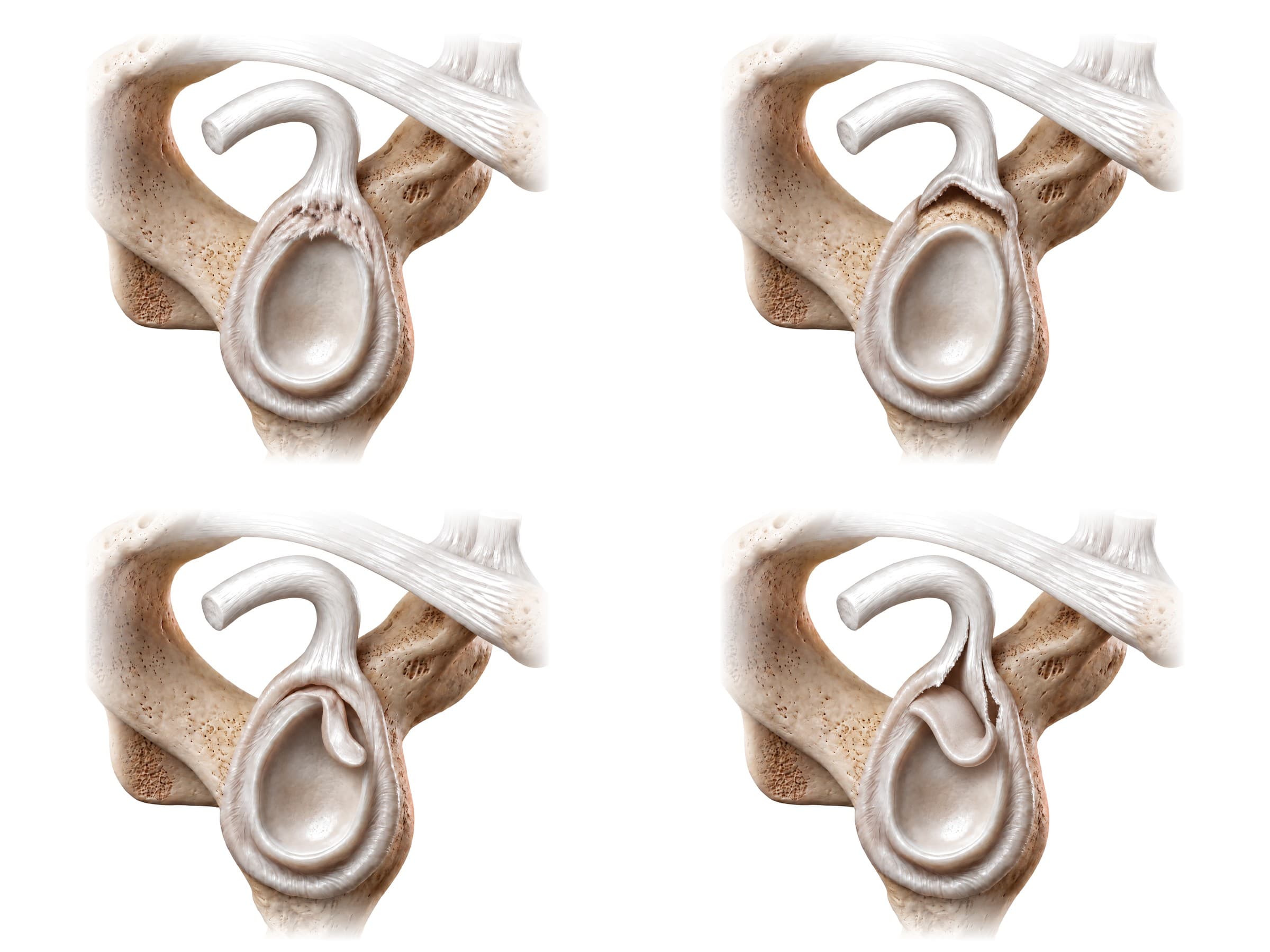
SLAP tears are graded using the Snyder classification, which describes four patterns of injury to the superior labrum and the biceps anchor. Type I is fraying and degeneration of the superior labrum with the biceps anchor still firmly attached. Type II, the most common, is detachment of the superior labrum and the biceps anchor from the glenoid. Type III is a bucket-handle tear of the superior labrum, where a flap of torn labrum can displace into the joint, but the biceps anchor itself is intact. Type IV is a bucket-handle tear that extends up into the biceps tendon. Later classifications added further types for tears that extend around the labrum, but Types I to IV are the ones most reports use.
How the shoulder is shown in a personal injury case
A labeled illustration of normal shoulder anatomy is the baseline exhibit in most shoulder-injury claims. It shows a jury where the rotator cuff, labrum, and AC joint sit in an uninjured shoulder before the injury-specific exhibit (the torn supraspinatus tendon, the detached labrum, the surgical anchors and sutures) is introduced. Shoulder cases often turn on whether a rotator cuff tear is acute or degenerative, since partial tears become common with age, so a side-by-side exhibit that pairs the normal-anatomy figure with the patient's own MRI findings, and points to signs of an acute injury such as bone bruising, fluid around the tear, or a fresh, sharply retracted tendon edge, is typically clearer to a lay jury than either image alone.
Natomy creates custom medical legal illustrations of shoulder injuries for demand packages and trial exhibits. For more on how normal anatomy is used in court, see demonstrative evidence. Related atlas pages: skull anatomy, back muscles anatomy, elbow anatomy, hand and wrist anatomy, hip anatomy, knee anatomy and ankle anatomy.
FAQ
What are the bones of the shoulder?+
Three bones make up the shoulder: the clavicle (collarbone), the scapula (shoulder blade), and the humerus (upper arm bone). The rounded head of the humerus sits in the glenoid fossa, a shallow socket on the outer edge of the scapula. The clavicle connects the shoulder to the rest of the skeleton at the sternum and meets the acromion of the scapula at the acromioclavicular (AC) joint.
What are the four rotator cuff muscles?+
The rotator cuff is made of four muscles, often remembered as SITS: supraspinatus, infraspinatus, teres minor, and subscapularis. All four start on the scapula and end on the top of the humerus, where their tendons blend into a single cuff around the humeral head. Supraspinatus helps lift the arm out to the side, infraspinatus and teres minor rotate it outward, and subscapularis rotates it inward. Together they hold the ball of the humerus centered in its shallow socket.
Is teres major part of the rotator cuff?+
No. Teres major sits just below teres minor on the back of the scapula and the two are easy to confuse, but teres major inserts on the front of the humerus (the medial lip of the intertubercular groove), not on the humeral head, and it does not help hold the joint together. It works with the latissimus dorsi to rotate the arm inward and pull it down and back. Teres minor is the rotator cuff muscle.
What is the glenohumeral joint?+
The glenohumeral joint is the main shoulder joint: a ball-and-socket synovial joint where the head of the humerus (the ball) meets the glenoid fossa of the scapula (the socket). The socket is only about a quarter to a third the size of the ball, which is why the shoulder has the largest range of motion of any joint in the body, and why it dislocates more often than any other major joint. The labrum, joint capsule, glenohumeral ligaments, and rotator cuff make up for the shallow socket.
What muscles are in the shoulder?+
The muscles that move the shoulder fall into three groups. The superficial muscles are the deltoid, which gives the shoulder its rounded shape and lifts the arm, and the trapezius across the top of the shoulder and upper back. The rotator cuff (supraspinatus, infraspinatus, teres minor, and subscapularis) sits deeper and stabilizes the joint. A third group moves the arm and scapula against the trunk: pectoralis major and minor in front, latissimus dorsi and teres major below, and the serratus anterior, rhomboids, and levator scapulae, which control the scapula. The biceps and triceps also cross the shoulder through their long heads.
What nerve supplies the deltoid?+
The axillary nerve (C5 and C6) supplies the deltoid and teres minor. It leaves the posterior cord of the brachial plexus, passes through the quadrangular space below the shoulder joint, and wraps around the surgical neck of the humerus. That path is why an anterior shoulder dislocation or a fracture of the surgical neck can injure it, causing weakness lifting the arm and a patch of numbness over the outer shoulder.
What is a SLAP tear?+
A SLAP tear (superior labrum anterior to posterior) is a tear of the top of the glenoid labrum, where the long head of the biceps tendon anchors. It is graded with the Snyder classification: Type I is fraying with the anchor intact, Type II (the most common) is detachment of the labrum and biceps anchor from the glenoid, Type III is a bucket-handle tear of the labrum, and Type IV is a bucket-handle tear that extends into the biceps tendon. It is best seen on MR arthrography.
What is the AC joint?+
The acromioclavicular (AC) joint is where the outer end of the clavicle meets the acromion of the scapula, at the top of the shoulder. It is held together by the acromioclavicular ligament and, more importantly, by the coracoclavicular ligament (its conoid and trapezoid parts) running from the coracoid process up to the clavicle. A fall directly onto the point of the shoulder can tear these ligaments, an injury called an AC separation or separated shoulder.
Need a custom illustration of this joint?
Generate a labeled or exhibit-ready figure for a specific patient, injury, or case in minutes.
Try Natomy →