Spine Diagram: Vertebral Column Labeled, Vertebrae and Discs
Labeled spine diagram of the whole vertebral column: the 33 vertebrae by region, spinal curves, discs, ligaments, spinal cord and common spine injuries.
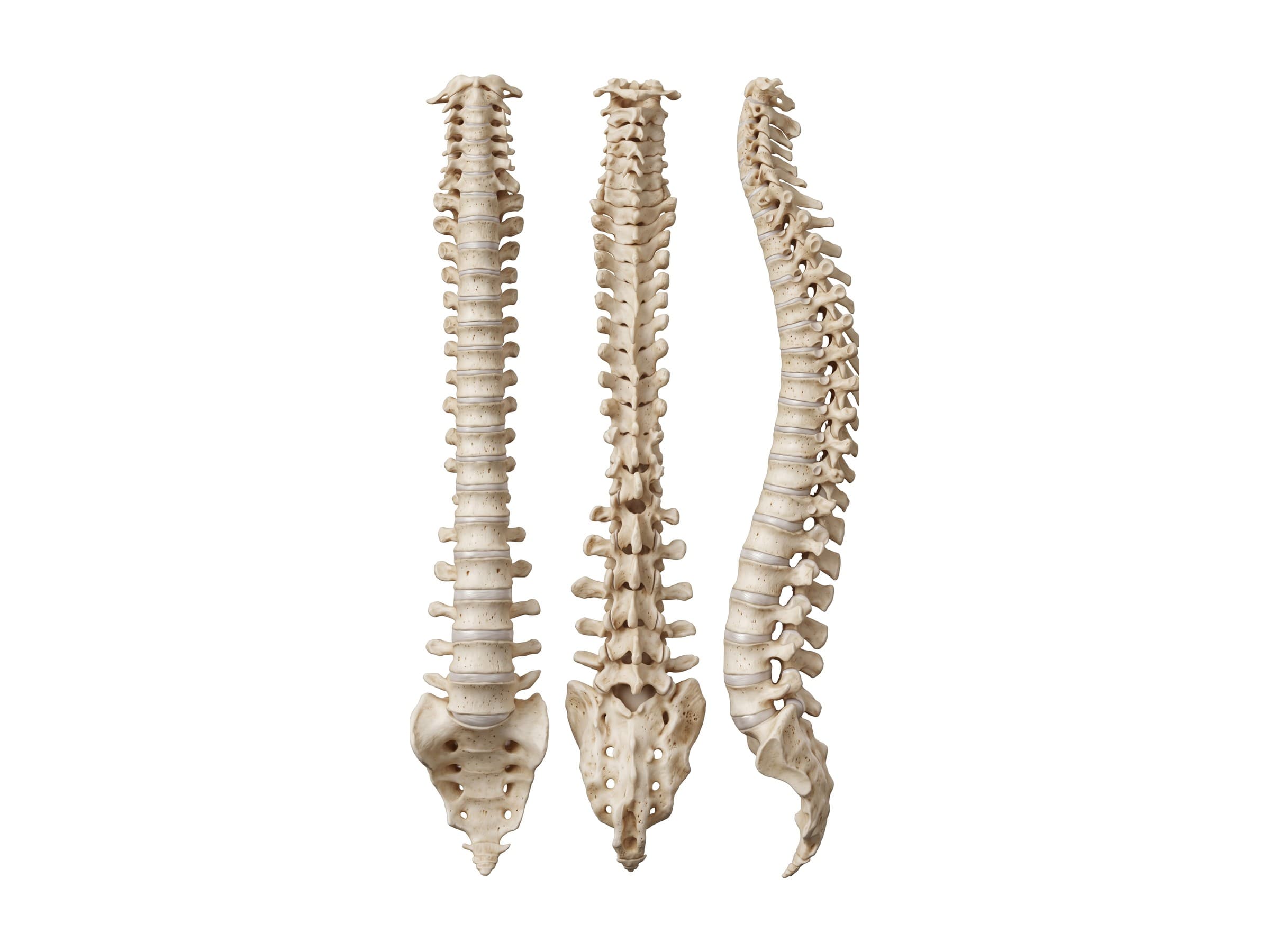
The spine, also called the vertebral column, spinal column or backbone, is the stack of 33 vertebrae that runs from the base of the skull to the pelvis. It has five regions: the cervical spine (7 vertebrae in the neck), the thoracic spine (12 in the chest), the lumbar spine (5 in the lower back), the sacrum (5 fused) and the coccyx (the tailbone). Between the movable vertebrae sit 23 intervertebral discs, and the column is held together by facet joints and long ligaments. The spine has three jobs: it holds the body upright and carries its weight, it lets the trunk and neck bend and twist, and it protects the spinal cord and the nerve roots that leave it at every level. This page is the overview of the whole column; each region has its own detailed atlas page linked below.
Spine diagram: the vertebral column from the front, back and side
The labeled spine diagram above shows the same vertebral column three ways. From the front (left), the vertebral bodies stack into a column that widens from the neck to the lower back, then rests on the triangular sacrum. From the back (middle), you see the vertebral arches and the row of spinous processes, the bumps you can feel down the middle of your back. From the side (right), the column is not straight: it forms an S shape of four curves, and the openings between the vertebrae (intervertebral foramina) are where the spinal nerves leave.
Vertebrae labeled: how the spine is numbered
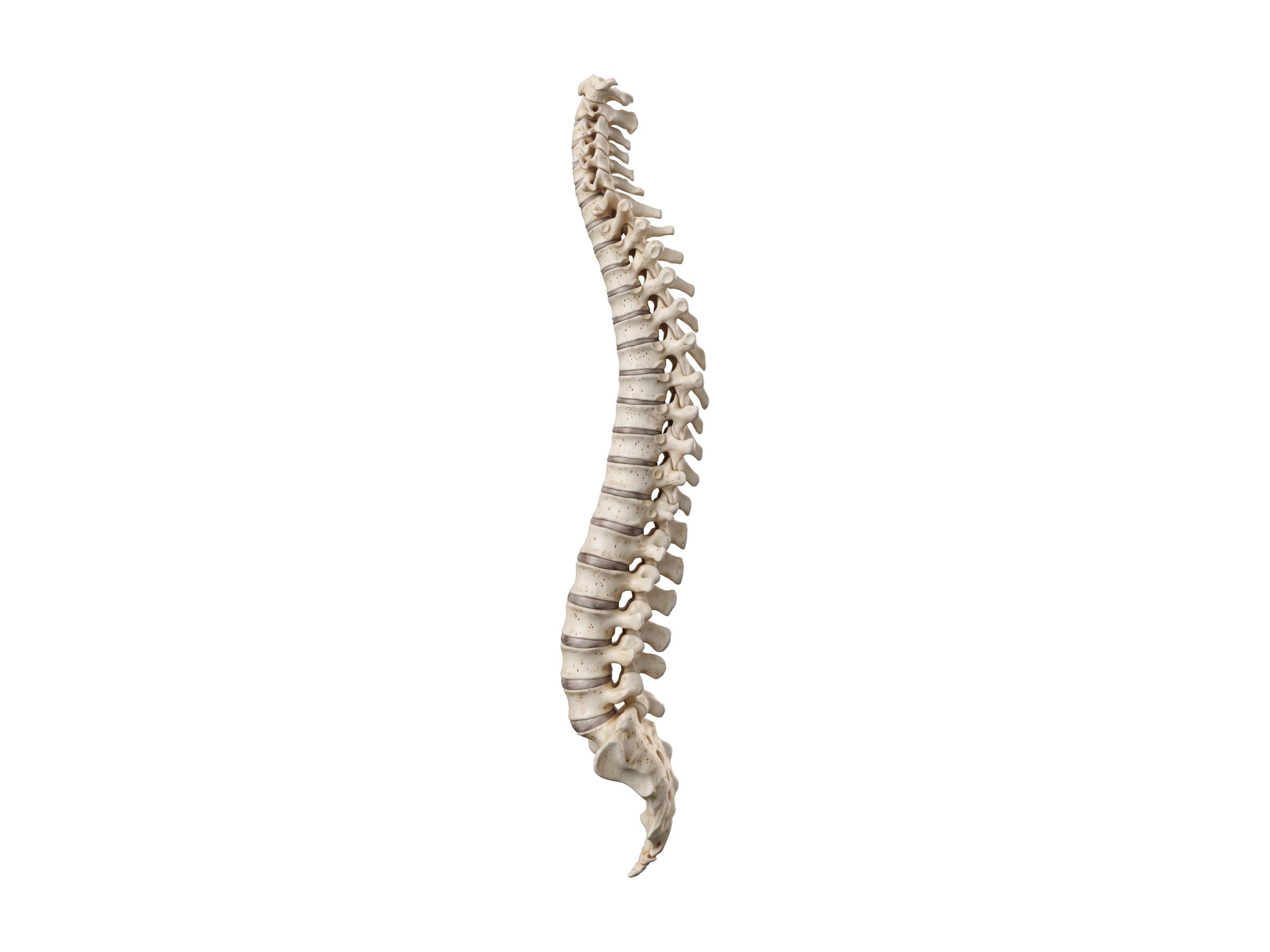
Each vertebra is named by its region and its position from the top: C for cervical, T for thoracic (sometimes written D, for dorsal), L for lumbar and S for sacral. So the top of the neck is C1, the first rib-bearing vertebra is T1, the first vertebra below the ribs is L1, and the top of the sacrum is S1. Discs and joints are named for the two vertebrae they join, such as the C5-C6 disc or the L5-S1 joint.
Clinicians find the levels by touch with a few landmarks: the C7 spinous process is the prominent bump at the base of the neck; a line between the lower tips of the shoulder blades crosses about T7; and a line between the tops of the hip bones (the iliac crests) crosses about L4 or the L4-L5 disc. On an X-ray or MRI, levels are counted down from C2 or up from the sacrum, and a transitional vertebra at the lumbosacral junction is a well-known cause of counting errors.
The five regions of the spine
| Region | Vertebrae | Number | Main feature |
|---|---|---|---|
| Cervical | C1 to C7 | 7 | Small bodies, holes for the vertebral arteries, the most mobile region |
| Thoracic | T1 to T12 | 12 | Joints for the ribs, long downward spinous processes, the stiffest region |
| Lumbar | L1 to L5 | 5 | The largest bodies, carry most of the body's weight |
| Sacral | S1 to S5 | 5 (fused) | One triangular bone, the back wall of the pelvis |
| Coccygeal | Co1 to Co4 | 3 to 5 (fused) | The tailbone |
Cervical spine (C1 to C7)
The neck holds up the head and gives it its wide range of motion. The top two vertebrae are specialized: the atlas (C1) is a ring with no body that carries the skull, and the axis (C2) has an upright peg, the dens, that the atlas turns around. The cervical vertebrae, the vertebral artery, the cervical nerves and neck injuries are covered in cervical spine anatomy.
Thoracic spine (T1 to T12)
The longest region, and the only one joined to the ribs. The rib cage makes it stiff, which protects the heart and lungs but concentrates stress where it ends at T12. The rib joints, the thoracolumbar junction and thoracic fractures are covered in thoracic spine anatomy.
Lumbar spine (L1 to L5)
Five large vertebrae in the lower back that carry most of the upper body's weight and allow bending forward and back. The L4-L5 and L5-S1 levels at the bottom take the most load, and most disc herniations and slips happen there. See lumbar spine anatomy for the lumbar vertebrae, the cauda equina and the lumbar nerves.
Sacrum and coccyx
Below L5, five sacral vertebrae fuse into the sacrum, which locks into the pelvis at the sacroiliac joints, and three to five tiny coccygeal vertebrae fuse into the coccyx. Both are described briefly further down this page.
The normal curves of the spine
Seen from the side, a healthy spine has four curves:
- Cervical lordosis: the neck curves forward, typically about 20 to 40 degrees.
- Thoracic kyphosis: the upper back curves backward, about 20 to 45 degrees.
- Lumbar lordosis: the lower back curves forward, about 40 to 60 degrees.
- Sacral kyphosis: the sacrum and coccyx curve backward and down.
The thoracic and sacral curves are primary curves, present in the womb. The cervical and lumbar curves are secondary curves that develop as a baby learns to hold up its head and then to stand and walk. Together they work like a spring, absorbing load and keeping the head balanced over the pelvis with little muscle effort.
Abnormal curves have their own names. Hyperkyphosis is a rounded upper back, from posture, Scheuermann's disease in teenagers or compression fractures in older adults. Hyperlordosis is an exaggerated sway of the lower back. A flattened lordosis, especially in the neck, is common on X-rays after an injury from muscle spasm. Scoliosis is a sideways curve of 10 degrees or more seen from the front or back, usually with some rotation of the vertebrae.
Anatomy of a vertebra: cervical vs thoracic vs lumbar
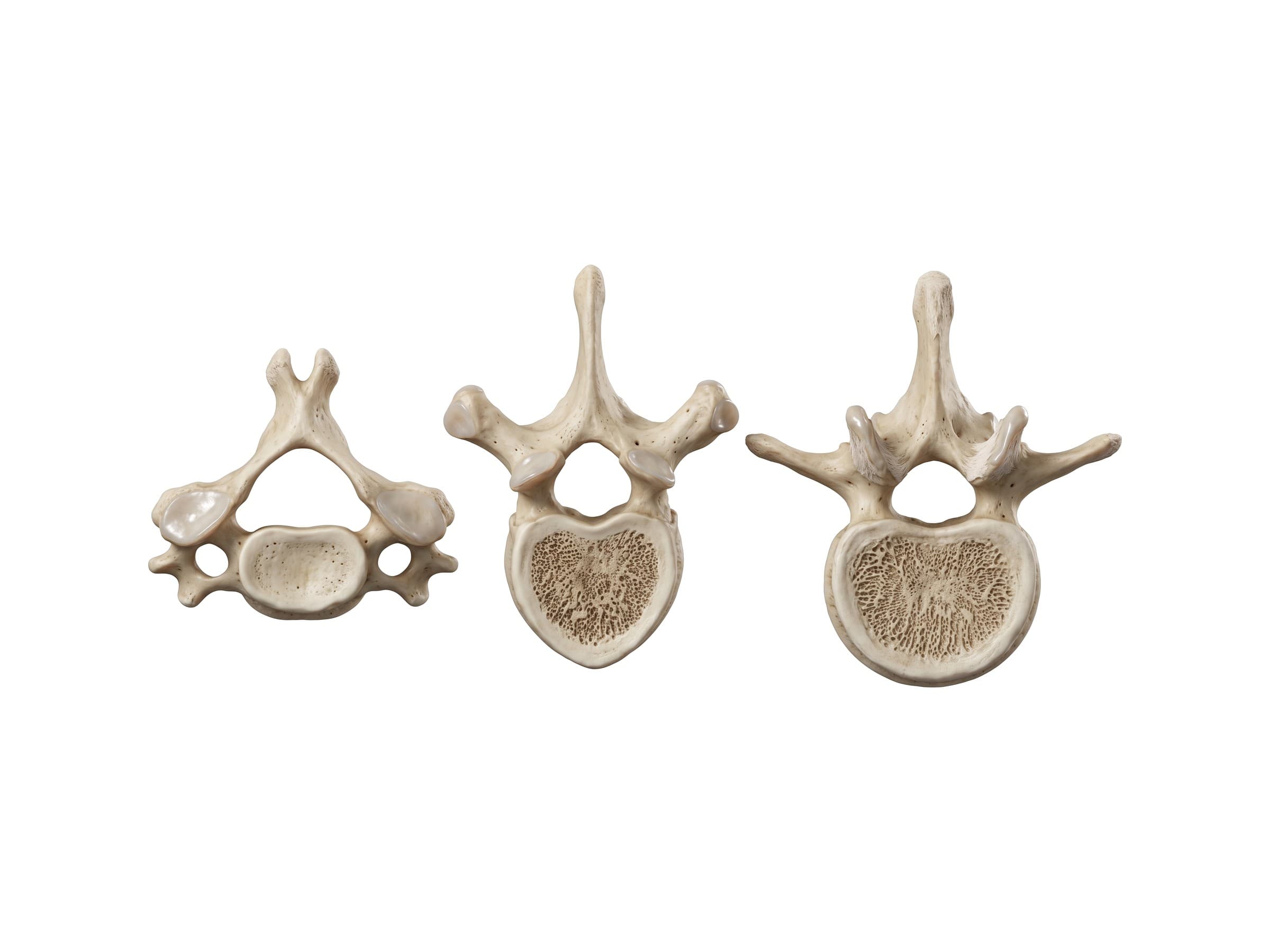
Every movable vertebra from C3 to L5 is built on the same plan:
- Vertebral body: the solid block in front that carries weight. The bodies get larger from the neck down.
- Vertebral arch: the ring of bone behind the body, made of two pedicles (joining the arch to the body) and two laminae (the roof). Body and arch together surround the vertebral foramen; stacked up, these form the spinal canal.
- Spinous process: the projection pointing back from the middle of the arch, which you can feel under the skin.
- Transverse processes: projections to each side that anchor muscles and ligaments.
- Superior and inferior articular processes: two pairs of small joint surfaces that form the facet joints with the vertebrae above and below.
The three regions differ in ways you can see at a glance:
| Feature | Cervical | Thoracic | Lumbar |
|---|---|---|---|
| Body | Small, wide and oval | Medium, heart-shaped | Large, kidney-shaped |
| Vertebral foramen | Large and triangular | Small and round | Triangular |
| Spinous process | Short and forked (bifid) | Long, pointing down | Short, thick and square |
| Transverse processes | Have a hole (transverse foramen) for the vertebral artery | Have facets for the ribs | Long and thin, no holes or rib facets |
| Facet joints | Flat and tilted, allow turning and tilting | Upright, allow rotation | Curved, limit twisting |
These differences are why a single loose vertebra can be identified by region: a hole in the transverse process means cervical, a rib facet means thoracic, and a large body with neither means lumbar.
Intervertebral discs
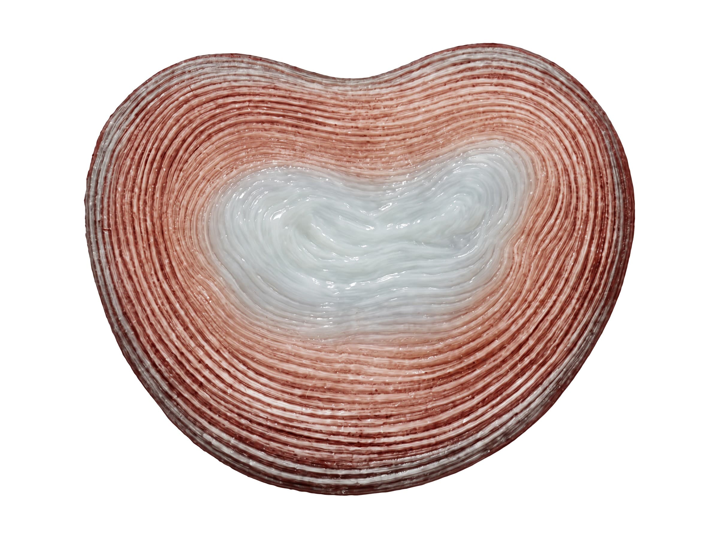
An intervertebral disc sits between each pair of vertebral bodies from C2-C3 down to L5-S1, 23 in all. Each disc has two parts:
- Annulus fibrosus: the tough outer ring, made of 15 to 25 layers of collagen fibers that cross at alternating angles like a radial tire. It holds the vertebrae together and resists twisting.
- Nucleus pulposus: the soft, gel-like center, mostly water. It spreads load evenly across the vertebral body like a water cushion.
Thin cartilage end plates cover the top and bottom of each disc where it meets the bone. Discs have almost no blood supply in adults and get their nutrients by diffusion through the end plates, which is why they heal poorly. They make up about a quarter of the height of the spine, and they slowly dry out and lose height with age, which is part of why people get shorter.
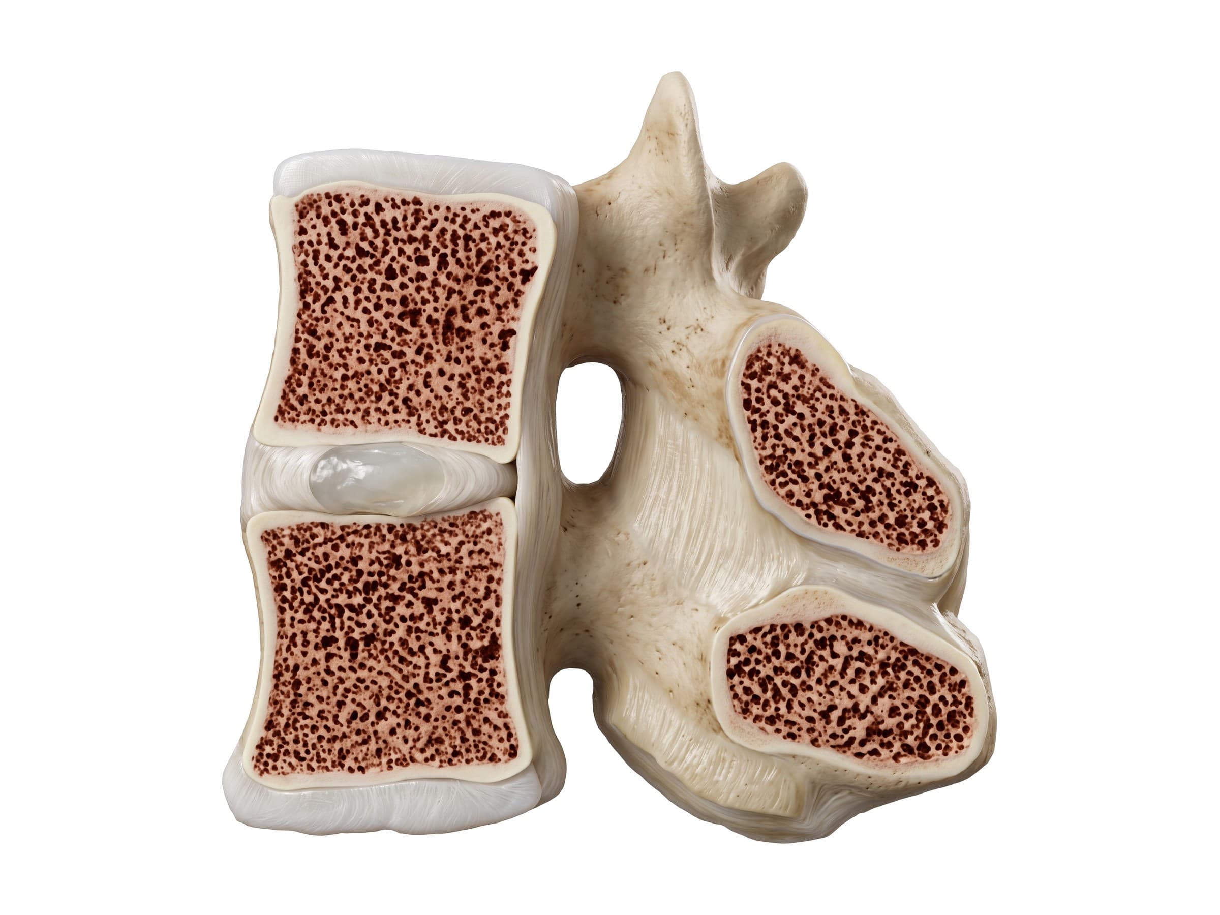
Cut down the middle, a spinal motion segment (two vertebrae and the disc between them) shows how the disc, the vertebral bodies and the arches behind fit together. The intervertebral foramen is the opening on each side, bounded by the disc in front, the pedicles above and below, and the facet joint behind, where the spinal nerve leaves the canal. A bulging disc, a bone spur or a thickened ligament can narrow it and pinch the nerve.
Joints and ligaments of the vertebral column
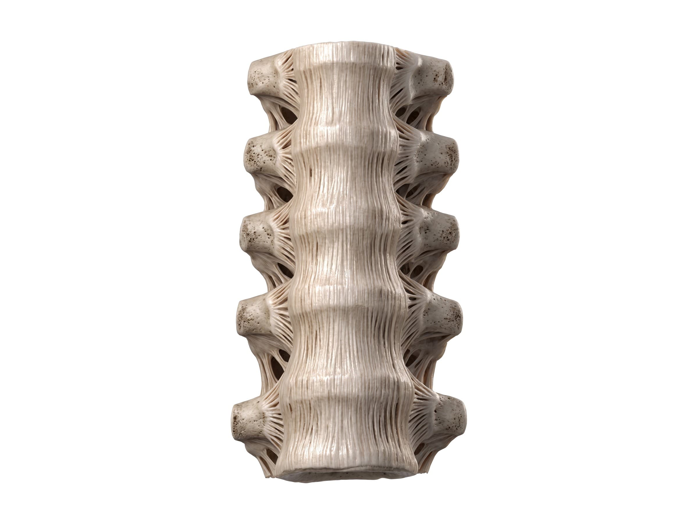
Each pair of vertebrae is joined in three places: the disc between the bodies in front, and the two facet joints (zygapophyseal joints) behind. Special joints sit at each end of the column: the atlanto-occipital joint between the skull and C1 (nodding), the atlantoaxial joint between C1 and C2 (turning the head), the small uncovertebral joints on the sides of the lower cervical bodies, the costovertebral joints where the ribs meet the thoracic spine, and the sacroiliac joints between the sacrum and the pelvis.
The main ligaments of the vertebral column run the whole length of the spine or join neighboring vertebrae:
- Anterior longitudinal ligament: a broad band down the front of the bodies and discs, from the skull to the sacrum. It limits bending backward and is the ligament stretched in a rear-end whiplash.
- Posterior longitudinal ligament: a narrower band down the back of the bodies, inside the spinal canal. Its narrow lumbar section is why most disc herniations go back and to one side.
- Ligamentum flavum: elastic yellow ligaments joining the laminae, forming part of the back wall of the canal. Thickening with age is a major cause of spinal stenosis.
- Interspinous and supraspinous ligaments: join the spinous processes and limit bending forward. In the neck the supraspinous ligament thickens into the ligamentum nuchae.
- Intertransverse ligaments: thin bands between the transverse processes.
- Facet joint capsules: thin capsules around each facet joint, a common source of neck and back pain.
Spinal canal, spinal cord and nerve roots
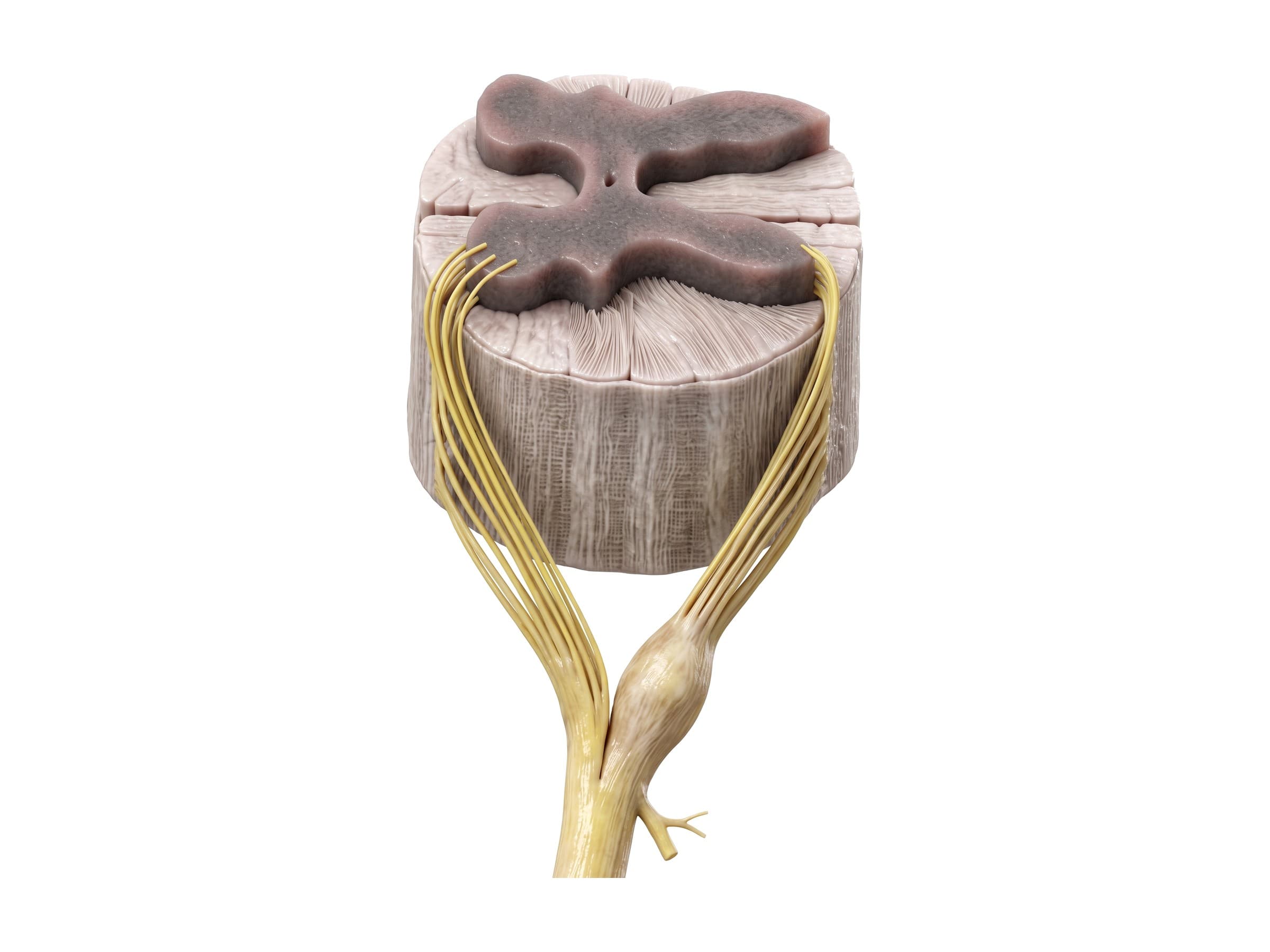
The vertebral foramina line up to form the spinal canal, which holds the spinal cord wrapped in its three membranes (the dura, arachnoid and pia mater) and bathed in cerebrospinal fluid. In adults the cord runs from the brainstem to about the L1-L2 level, where it tapers into the conus medullaris. Below that, the canal holds the cauda equina, a bundle of lumbar and sacral nerve roots.
At every level, two nerve roots leave the cord on each side. The anterior (ventral) root carries motor signals out to the muscles; the posterior (dorsal) root carries sensation in and has a swelling, the dorsal root ganglion, where the sensory nerve cells live. The two roots join into a spinal nerve that leaves through the intervertebral foramen and soon splits into a small dorsal ramus to the back and a larger ventral ramus to the front of the body and the limbs.
There are 31 pairs of spinal nerves: 8 cervical, 12 thoracic, 5 lumbar, 5 sacral and 1 coccygeal. The cervical nerves form the brachial plexus to the arm, the thoracic nerves run around the chest and abdomen, and the lumbar and sacral nerves form the plexuses to the leg, including the sciatic nerve. Each nerve supplies a band of skin, a dermatome, which is how a doctor can tell from the pattern of numbness which root is pinched. See leg nerves and dermatomes for the lower limb.
Sacrum and coccyx
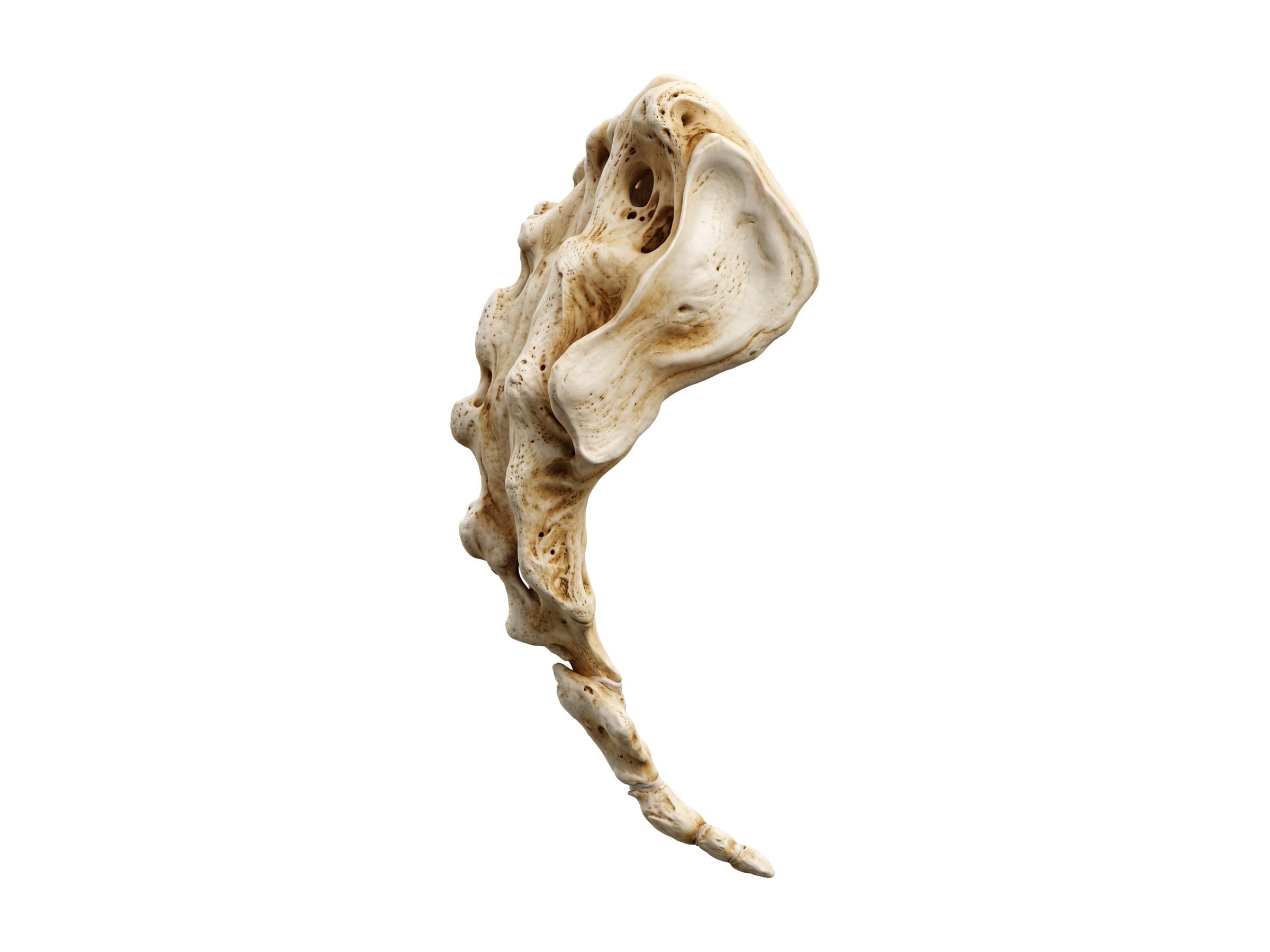
The sacrum is a large triangular bone made of five vertebrae (S1 to S5) that fuse between the late teens and about age 30. Its base carries L5 at the lumbosacral joint, its ear-shaped auricular surfaces form the sacroiliac joints with the hip bones, and the median sacral crest down its back is the remnant of the fused spinous processes. Four pairs of sacral foramina let the sacral nerves out to the pelvis and leg. The front, back and top of the sacrum are shown in detail in lumbar spine anatomy, and the sacroiliac joint in hip anatomy.
The coccyx is three to five small coccygeal vertebrae fused into a short, forward-curving tail, joined to the sacrum at the sacrococcygeal joint. It anchors the gluteus maximus and pelvic floor muscles, and it is often bruised or fractured in a fall onto the buttocks.
Muscles that support the spine
The spine is stabilized by layers of muscles. The erector spinae and multifidus run along the back of the column and hold it upright, the quadratus lumborum and psoas major brace the lumbar spine from the sides and front, and the abdominal wall muscles stiffen the trunk like a corset. All are shown in back muscles anatomy, and the neck muscles in cervical spine anatomy. Muscle strain is the most common cause of acute neck and back pain.
Common spine injuries and how they appear on imaging
X-rays show alignment, the curves, disc height and most fractures. CT shows fractures in detail and is the first test after major trauma. MRI shows the discs, ligaments, spinal cord and nerve roots, and is the test for nerve symptoms or suspected soft-tissue injury.
- Disc herniation: the nucleus pulposus pushes through a tear in the annulus fibrosus and presses on a nerve root, causing pain, numbness or weakness down the arm or leg in that root's pattern. Most occur at L4-L5 and L5-S1 in the lower back and C5-C6 and C6-C7 in the neck, and MRI shows the disc material and the compressed root. See lumbar disc herniation and cervical disc herniation.
- Compression fracture: the front of a vertebral body collapses into a wedge while the back wall holds. It is most common at the thoracolumbar junction, from a fall or crash, or from minor stress in osteoporosis, and it adds to the thoracic kyphosis. See thoracic spine anatomy.
- Burst fracture: a high-energy crush injury in which the back wall of the body also breaks and bone fragments can push into the spinal canal toward the cord. CT measures how much of the canal is blocked. See thoracic burst fractures.
- Spondylolisthesis (anterolisthesis): one vertebra slips forward on the one below, graded I to V by how far it has slipped. It is usually L5 on S1 after a pars fracture (spondylolysis) or L4 on L5 from facet arthritis, and is measured on a standing side X-ray. See lumbar spine anatomy.
- Whiplash: a sprain and strain of the neck from the rapid back-and-forth motion of a rear-end collision, stretching the anterior longitudinal ligament, facet capsules and neck muscles. X-rays are usually normal apart from a straightened cervical curve; MRI may show ligament or disc injury. See cervical spine anatomy.
- Spinal stenosis: narrowing of the spinal canal or the foramina by bulging discs, thickened ligamentum flavum and arthritic facet joints, which can pinch the cord in the neck or the nerve roots in the lower back.
- Spinal cord injury: damage to the cord from a fracture, dislocation or severe stenosis. The level of injury sets which functions are lost, which is why the neurological level is recorded by its spinal segment.
How the spine is shown in a personal injury case
Neck and back injuries are the most common claims after car crashes, slip-and-fall accidents and workplace injuries, and they are among the most contested. The defense often argues that a disc herniation, a slip or a compression fracture was pre-existing degeneration, or that the MRI findings are normal for the plaintiff's age. A labeled diagram of the whole spine is the starting exhibit: it shows the jury where the injured level sits in the column, how many vertebrae and discs there are, and why a problem at C6 causes symptoms in the hand while one at L5 causes symptoms in the foot. From there, an expert can zoom into the injured region and show the injury itself, following the mechanism of injury: a disc herniation from a rear-end collision, a compression fracture from a fall, a fusion with its screws and rods. Illustrations built from the plaintiff's own MRI or CT tie the scan, the anatomy and the symptoms together.
Natomy creates custom medical legal illustrations of spine injuries for demand packages and trial exhibits, and the rules for using these visuals at trial are covered in demonstrative evidence. For each region in detail, see cervical spine anatomy, thoracic spine anatomy and lumbar spine anatomy. Related atlas pages: back muscles anatomy, skull anatomy, brain anatomy, hip anatomy and leg nerves and dermatomes.
FAQ
How many vertebrae are in the human spine?+
The spine is made of 33 vertebrae: 7 cervical (neck), 12 thoracic (chest), 5 lumbar (lower back), 5 sacral that fuse into one sacrum, and usually 4 small coccygeal vertebrae that fuse into the coccyx. That leaves 24 separate, movable vertebrae in an adult, or 26 bones if the sacrum and coccyx are each counted as one. The numbers vary a little: some people have 11 or 13 thoracic vertebrae, a sixth lumbar vertebra, or 3 to 5 coccygeal segments, which matters when counting levels on an X-ray before surgery.
What are the five regions of the spine?+
From top to bottom: the cervical spine (C1 to C7) in the neck, the thoracic spine (T1 to T12) in the chest, where the ribs attach, the lumbar spine (L1 to L5) in the lower back, the sacrum (S1 to S5, fused) at the back of the pelvis, and the coccyx, or tailbone. The cervical and lumbar regions are the most mobile and the most often injured; the thoracic region is stiffened by the rib cage.
What are the normal curves of the spine?+
Seen from the side, a healthy spine has four curves that make a gentle S shape. The cervical and lumbar curves bend forward (lordosis), and the thoracic and sacral curves bend backward (kyphosis). Typical adult values are about 20 to 40 degrees of cervical lordosis, 20 to 45 degrees of thoracic kyphosis and 40 to 60 degrees of lumbar lordosis. From the front or back the spine should be straight; a sideways curve of 10 degrees or more is scoliosis.
What is the difference between the spine, the spinal column and the spinal cord?+
The spine, spinal column, vertebral column and backbone all mean the same thing: the stack of vertebrae, discs, joints and ligaments that runs from the skull to the pelvis. The spinal cord is the bundle of nerve tissue that runs inside it, through the spinal canal, from the brainstem down to about the L1-L2 level. The bones protect the cord; injuries to the bones and discs matter clinically when they press on the cord or its nerve roots.
How many intervertebral discs are in the spine?+
There are 23 intervertebral discs. The first sits between C2 and C3, and the last between L5 and S1. There is no disc between the skull and C1 or between C1 and C2, where the joints are built for nodding and turning the head, and the discs inside the sacrum and coccyx turn to bone or fibrous tissue as those vertebrae fuse. Each disc is named for the two vertebrae it sits between, such as C5-C6 or L4-L5.
How are the spinal levels and nerves numbered?+
Vertebrae are numbered from the top of each region: C1 to C7, T1 to T12, L1 to L5 and S1 to S5. There are 31 pairs of spinal nerves: 8 cervical, 12 thoracic, 5 lumbar, 5 sacral and 1 coccygeal. In the neck, each nerve exits above the vertebra with the same number (C6 exits between C5 and C6), and the extra C8 nerve exits below C7. From T1 down, each nerve exits below its vertebra (L4 exits between L4 and L5).
What part of the spine is most commonly injured?+
The two most mobile regions, the neck and the lower back, are injured most often. Neck sprains (whiplash) and disc herniations at C5-C6 and C6-C7 dominate after car crashes, and lumbar disc herniations at L4-L5 and L5-S1 dominate after lifting injuries and falls. Spine fractures cluster at the thoracolumbar junction (T11 to L2), where the stiff thoracic spine meets the mobile lumbar spine, and in the upper neck (C1 and C2) in high-energy trauma and falls in older adults.
Is the tailbone part of the spine?+
Yes. The coccyx, or tailbone, is the lowest part of the vertebral column: three to five small coccygeal vertebrae fused into a short triangle below the sacrum. It anchors the gluteus maximus, several pelvic floor muscles and ligaments, and it takes part of your weight when you lean back while sitting. A fall onto the buttocks can bruise, dislocate or fracture it, causing tailbone pain (coccydynia) that is worse when sitting.
Need a custom illustration of this joint?
Generate a labeled or exhibit-ready figure for a specific patient, injury, or case in minutes.
Try Natomy →