Elbow Anatomy: Bones, Ligaments & Nerves
Labeled illustrations of the elbow bones, olecranon, ulnar collateral and annular ligaments, cubital fossa, ulnar nerve and common elbow fractures.
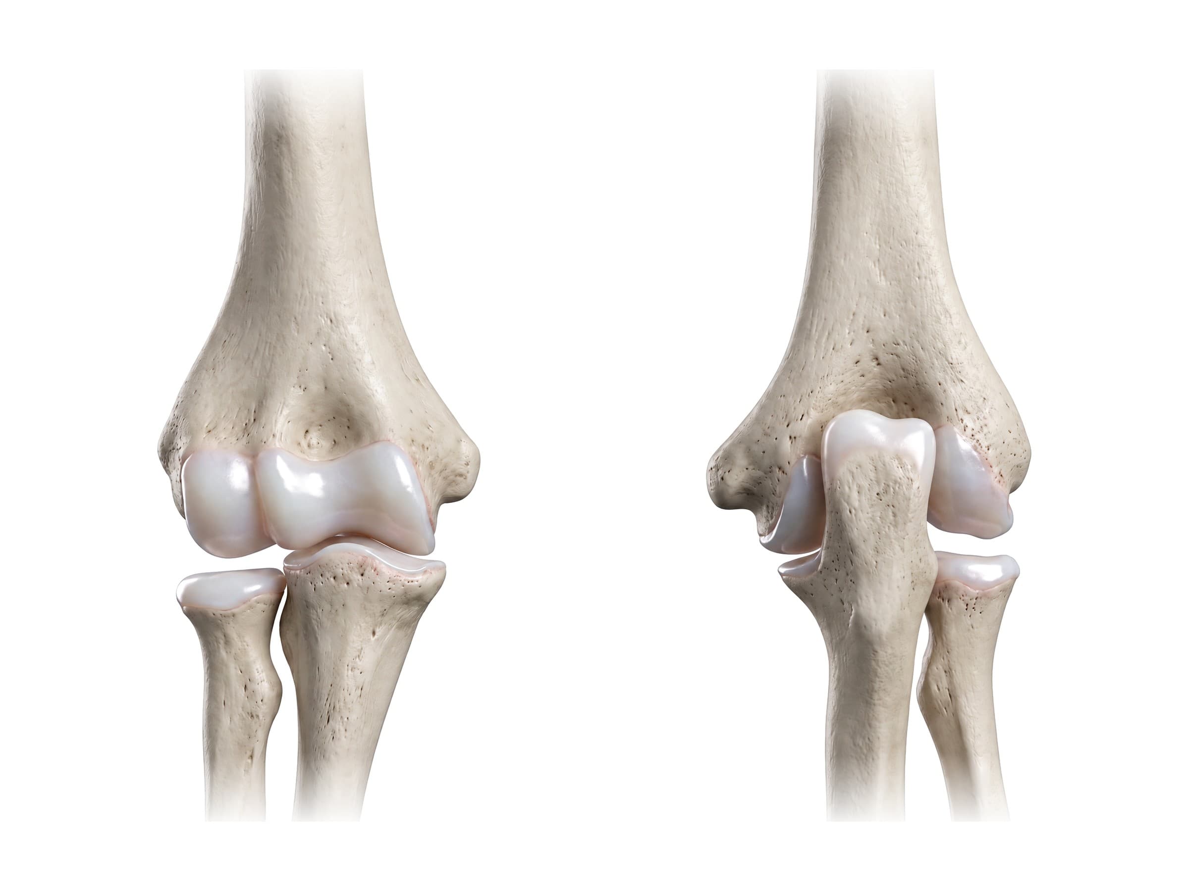
The elbow is the hinge joint between the upper arm and the forearm. Elbow anatomy comes down to three bones (the humerus, radius and ulna) meeting in three joints inside one capsule, the ligaments that hold them together on the inner and outer sides, the muscles that bend, straighten and rotate the forearm, and three nerves that pass close to the bone: the ulnar nerve behind the inner side (the funny bone), the median nerve in front, and the radial nerve on the outer side. Because so much is packed into a small space, a single fall can fracture a bone, tear a ligament and injure a nerve at once, which is why the anatomy matters for reading an elbow X-ray report, explaining a diagnosis to a patient, or illustrating a personal injury claim.
Bones of the elbow
The elbow joins three bones. In the front and back views of the elbow above:
- Humerus: the upper arm bone. Its lower end widens into the medial and lateral epicondyles and carries two joint surfaces: the capitulum and the trochlea.
- Radius: the forearm bone on the thumb side. Its disc-shaped head sits against the capitulum.
- Ulna: the forearm bone on the little finger side. Its upper end hooks around the trochlea.
The lower end of the humerus
- Capitulum: the rounded knob on the outer side that meets the cupped top of the radial head.
- Trochlea: the spool-shaped surface on the inner side that the ulna wraps around. Its shape is what makes the elbow a stable hinge.
- Medial epicondyle: the larger bump on the inner side, where the ulnar collateral ligament and the forearm flexor muscles start. The ulnar nerve runs in a groove just behind it.
- Lateral epicondyle: the smaller bump on the outer side, where the radial collateral ligament and the forearm extensor muscles start.
- Fossae: three hollows that make room for the forearm bones at the ends of movement: the coronoid fossa and radial fossa in front (filled when the elbow bends fully) and the deep olecranon fossa behind (filled when it straightens fully).
The upper ends of the ulna and radius
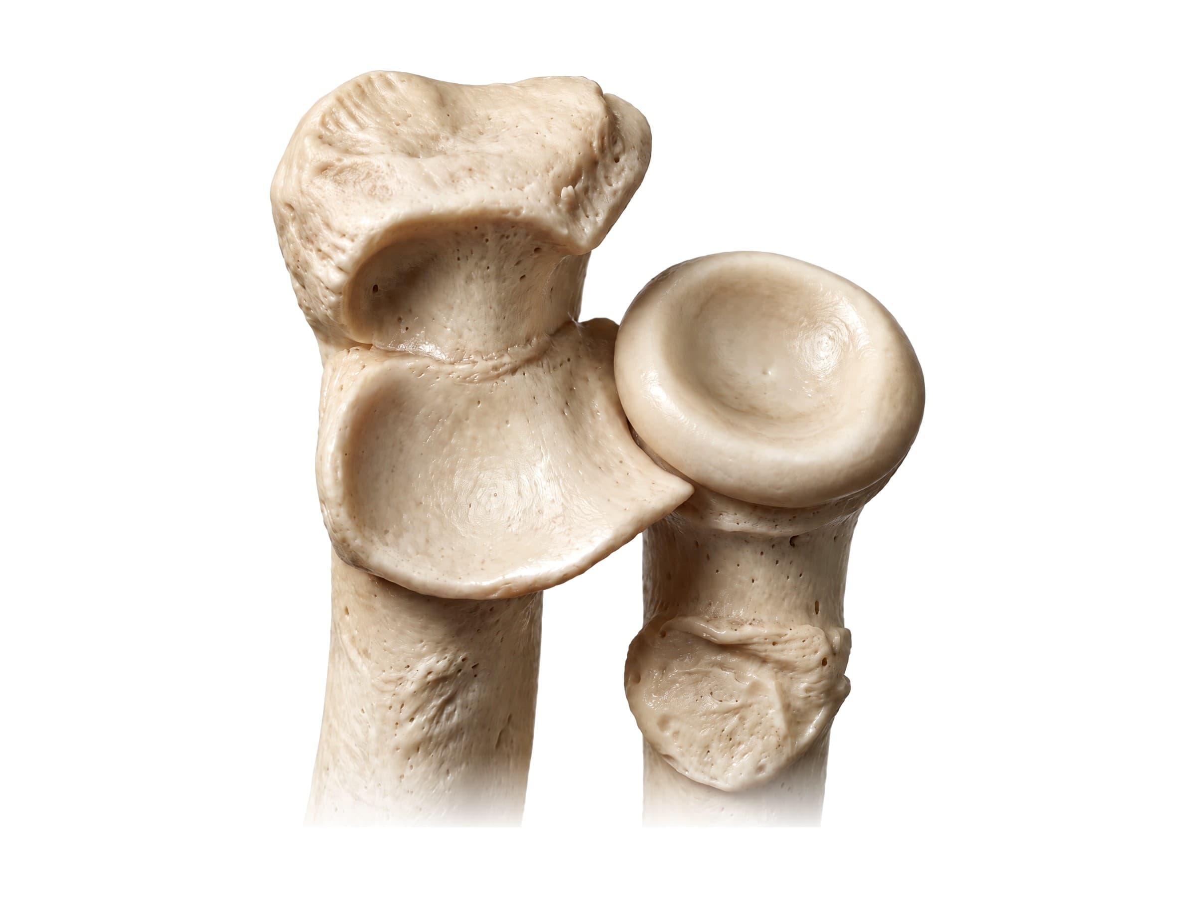
- Olecranon: the curved top of the ulna that forms the point of the elbow. The triceps attaches to it.
- Trochlear notch: the deep C-shaped notch between the olecranon and the coronoid process that grips the trochlea.
- Coronoid process: the front lip of the notch, which stops the ulna from sliding backward. The brachialis attaches just below it.
- Radial notch of the ulna: a small curved surface on the side of the ulna where the radial head spins.
- Radial head, neck and tuberosity: the radius ends in a flat disc (the head) on a narrow neck. Below the neck is the radial tuberosity, where the biceps tendon attaches.
Joints of the elbow
Three joints share a single joint capsule:
- Humeroulnar joint: between the trochlea and the trochlear notch of the ulna. A hinge that bends (flexion) and straightens (extension) the elbow, from straight to about 145 degrees.
- Humeroradial joint: between the capitulum and the radial head. It moves with bending and lets the radial head spin.
- Proximal radioulnar joint: between the radial head and the radial notch of the ulna, held by the annular ligament. A pivot that turns the palm down (pronation) and up (supination), about 75 to 85 degrees each way.
When the arm hangs straight with the palm forward, the forearm angles slightly away from the body. This carrying angle, about 5 to 15 degrees and usually larger in women, can change after a childhood elbow fracture.
Ligaments of the elbow
Ulnar (medial) collateral ligament
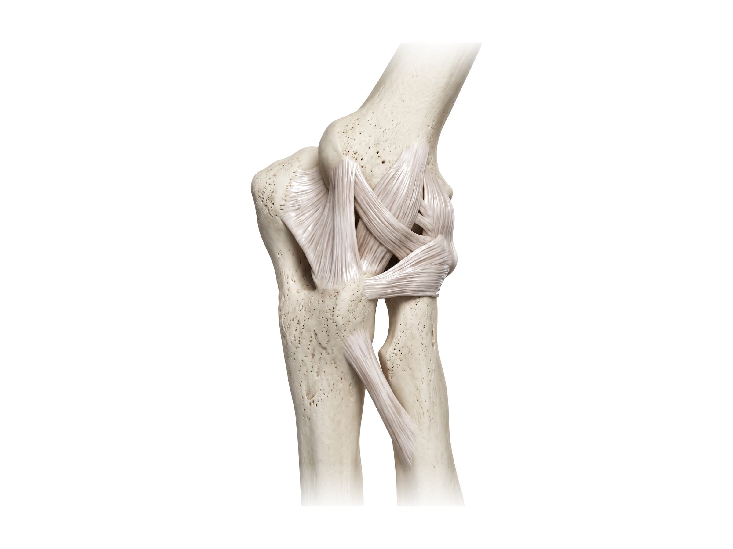
The ulnar collateral ligament (UCL), also called the medial collateral ligament, runs from the medial epicondyle to the ulna in three parts. The strong anterior bundle runs down to the coronoid process and is the main restraint against the elbow bending outward (valgus stress); the fan-shaped posterior bundle runs back to the olecranon and forms the floor of the cubital tunnel, where the ulnar nerve lies; and a small transverse bundle joins the olecranon to the coronoid process along the ulna. In front of it, the thinner anterior ligament of the joint capsule crosses the front of the joint, and below, the oblique cord runs from the coronoid process to the radius. Each pitch in baseball loads it close to its breaking point, and repeated throwing can stretch or tear it; reconstructing it with a tendon graft is known as Tommy John surgery.
Radial (lateral) collateral ligament complex
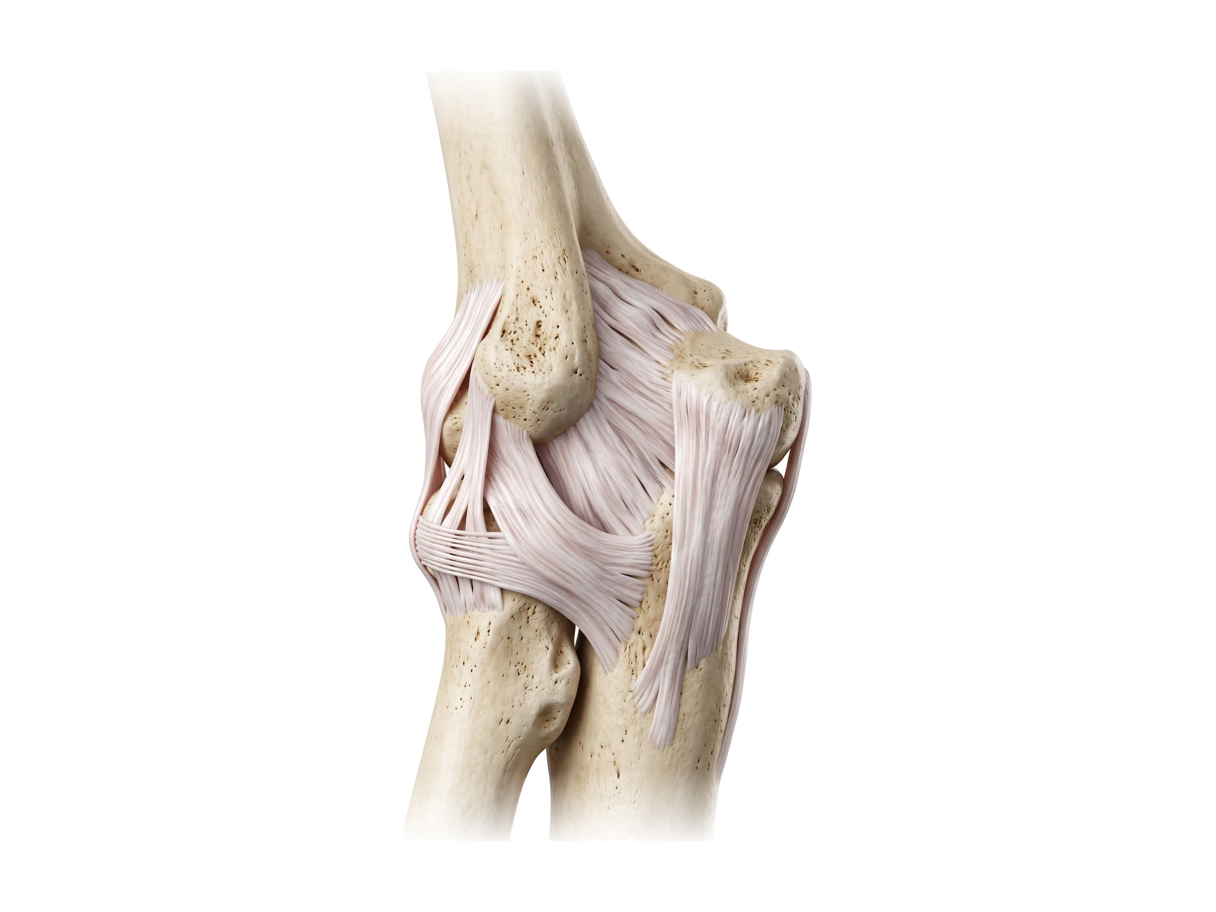
On the outer side, the lateral collateral ligament complex runs from the lateral epicondyle:
- Radial collateral ligament: fans from the lateral epicondyle into the annular ligament.
- Lateral ulnar collateral ligament (LUCL): runs behind the radial head to the ulna. It is the main lateral stabilizer, and it is usually the first ligament torn in an elbow dislocation. When it fails, the forearm can rotate out of joint (posterolateral rotatory instability).
Annular ligament
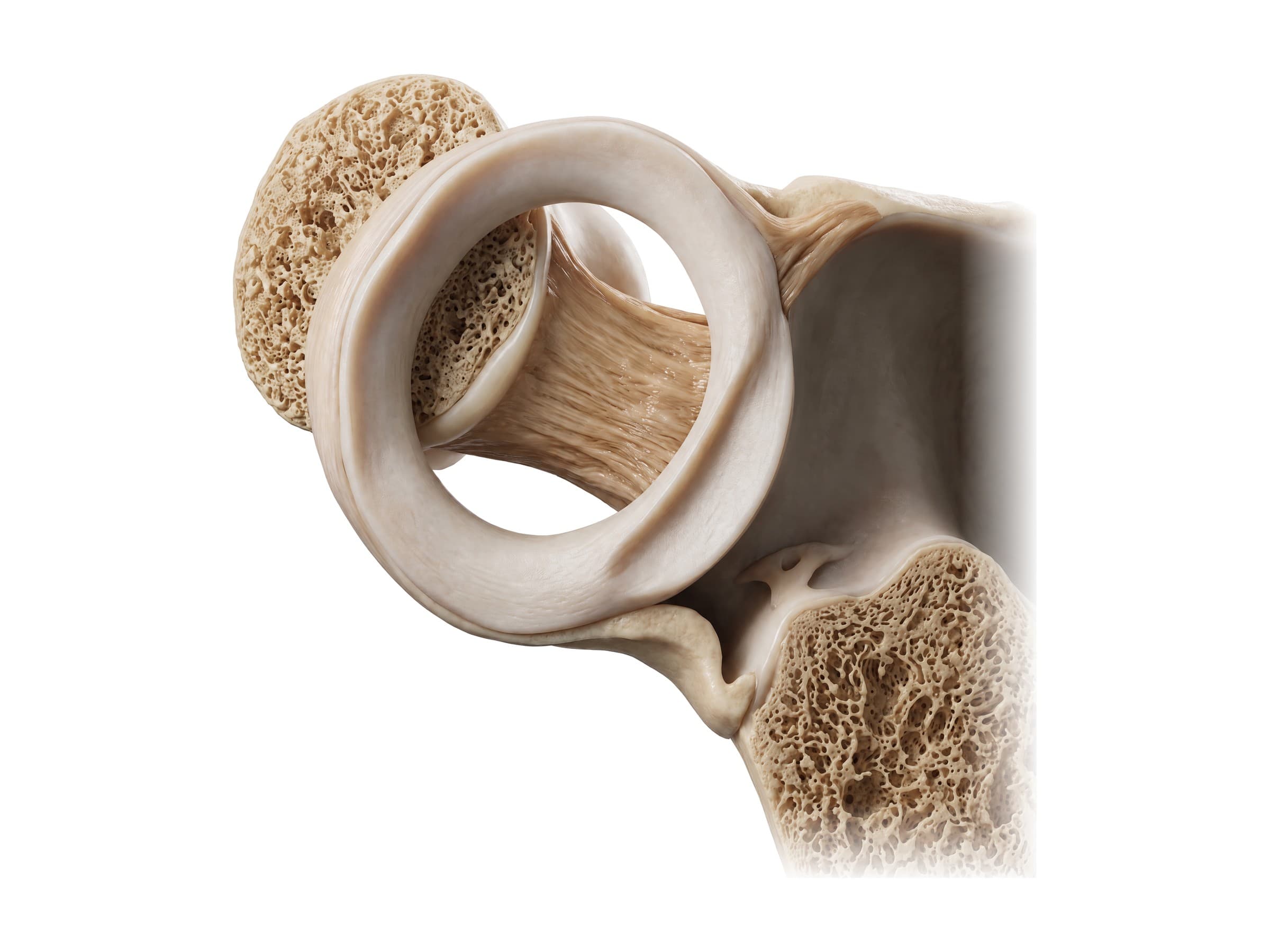
The annular ligament is a strong band that wraps around the radial head like a collar, attached at both ends to the edges of the radial notch of the ulna. It holds the radial head against the ulna while letting it spin, and the small quadrate ligament below it limits how far the radius can rotate. In young children the radial head is small and the ligament loose, so a sudden pull on the hand can slip the head partly out of the collar, which is nursemaid's elbow.
Muscles and tendons of the elbow
- Biceps brachii: the muscle on the front of the arm. Its tendon attaches to the radial tuberosity, so it both bends the elbow and turns the palm up; a flat sheet from the tendon, the bicipital aponeurosis, fans into the forearm. A distal biceps tendon rupture pulls the muscle up toward the shoulder.
- Brachialis: lies under the biceps and attaches to the ulna just below the coronoid process. It is the main elbow flexor.
- Brachioradialis: runs from the outer edge of the humerus to the wrist and bends the elbow, especially with the thumb pointing up.
- Triceps brachii: the muscle on the back of the arm, attaching to the olecranon. It is the main elbow extensor, helped by the small anconeus.
- Pronator teres and supinator: turn the palm down and up.
- Forearm flexors and extensors: the muscles that bend the wrist and fingers start from a common tendon on the medial epicondyle; those that straighten them start from a common tendon on the lateral epicondyle. Overuse of these tendons causes golfer's elbow and tennis elbow.
The cubital fossa
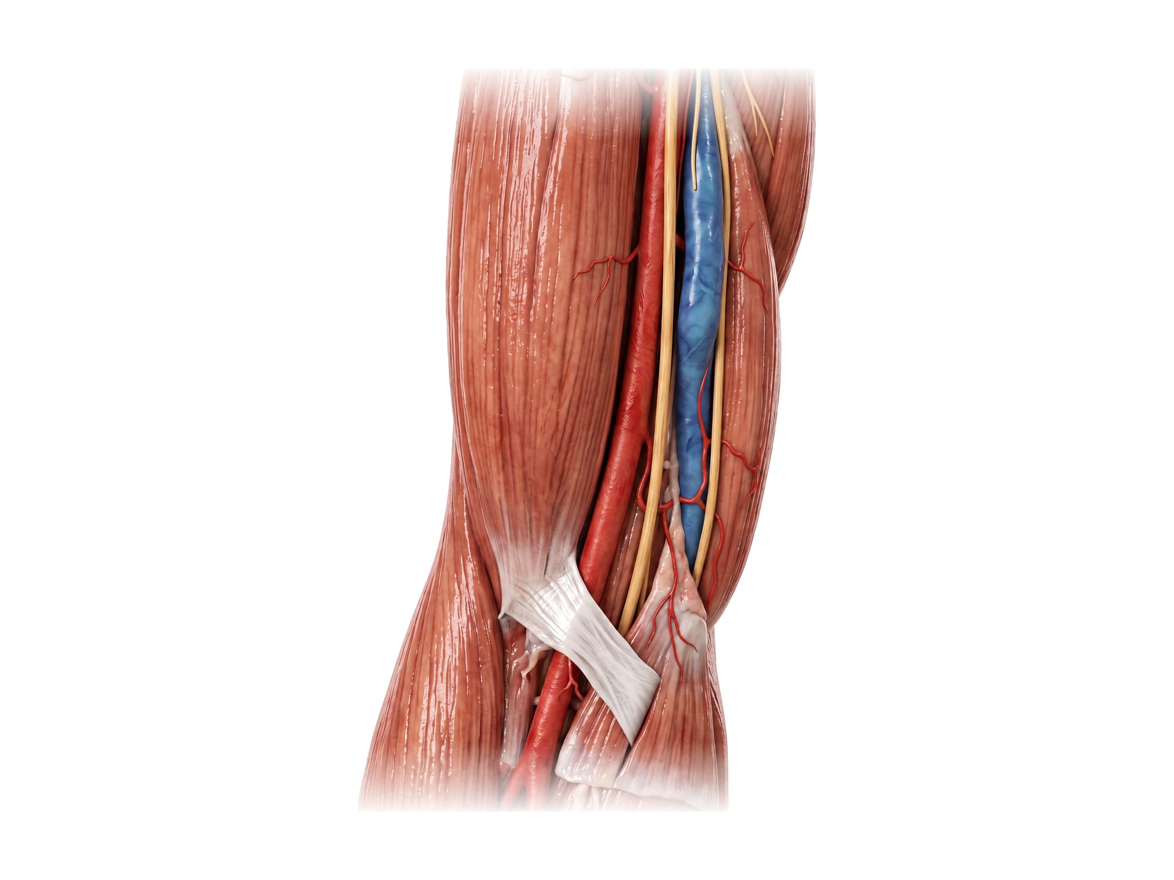
The cubital fossa is the triangular hollow at the front of the elbow, bordered by the brachioradialis on the outer side, the pronator teres on the inner side and a line between the epicondyles above. Its roof is the bicipital aponeurosis. From the outer side inward it contains the biceps tendon, the brachial artery and the median nerve (remembered as TAN: tendon, artery, nerve). The brachial artery divides here into the radial and ulnar arteries. Just under the skin, the median cubital vein crosses the fossa, the usual site for drawing blood.
Nerves of the elbow
- Ulnar nerve: passes behind the medial epicondyle through the cubital tunnel, close under the skin, which is why bumping the "funny bone" sends tingling into the ring and little fingers. Pressure or stretching here causes cubital tunnel syndrome, the second most common nerve compression in the arm after carpal tunnel syndrome. It continues into the hand to supply most of its small muscles.
- Median nerve: crosses the front of the elbow in the cubital fossa beside the brachial artery and passes between the two heads of the pronator teres. It is at risk in supracondylar fractures in children.
- Radial nerve: crosses in front of the lateral epicondyle and divides into a superficial sensory branch and the deep posterior interosseous nerve, which supplies the muscles that straighten the wrist and fingers. It can be injured in radial head fractures and dislocations.
All three are branches of the brachial plexus in the shoulder.
The olecranon bursa
A thin fluid-filled sac, the olecranon bursa, sits between the olecranon and the skin and lets the skin glide over the point of the elbow. A direct blow, leaning on the elbow for long periods, gout or infection can inflame it, causing olecranon bursitis: a soft, sometimes golf-ball-sized swelling at the point of the elbow. An infected bursa is red, hot and painful and needs drainage and antibiotics.
Common elbow injuries and how they appear on imaging
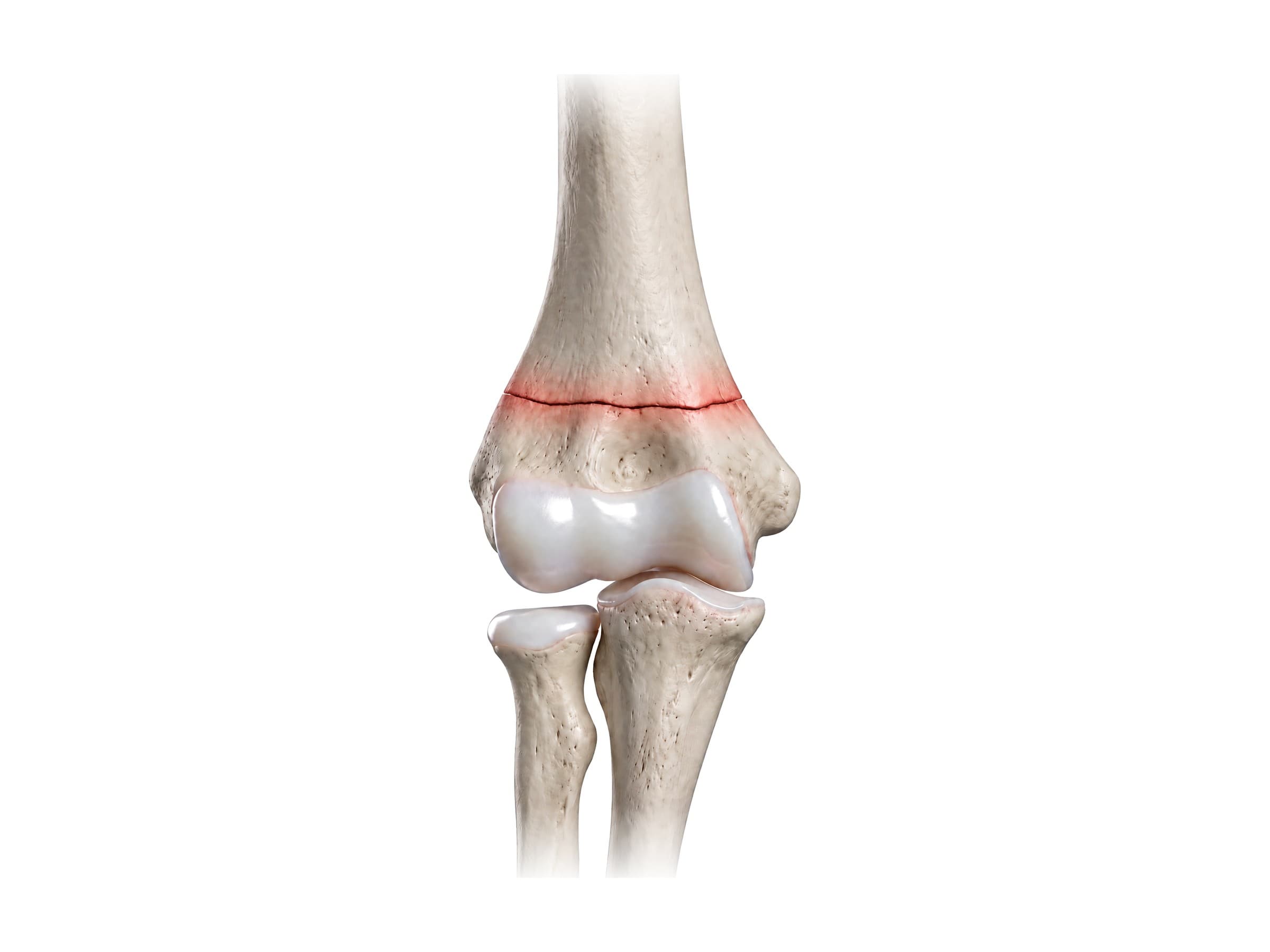
- Supracondylar fracture: a break across the lower humerus just above the joint, the most common elbow fracture in children, typically aged about 5 to 7, from a fall on an outstretched hand off playground equipment, a trampoline or a bed. Displaced fractures can injure the brachial artery and the median, radial or ulnar nerve, and a missed vascular injury can lead to compartment syndrome and Volkmann's ischemic contracture. Graded on X-ray by the Gartland classification; displaced fractures are usually pinned.
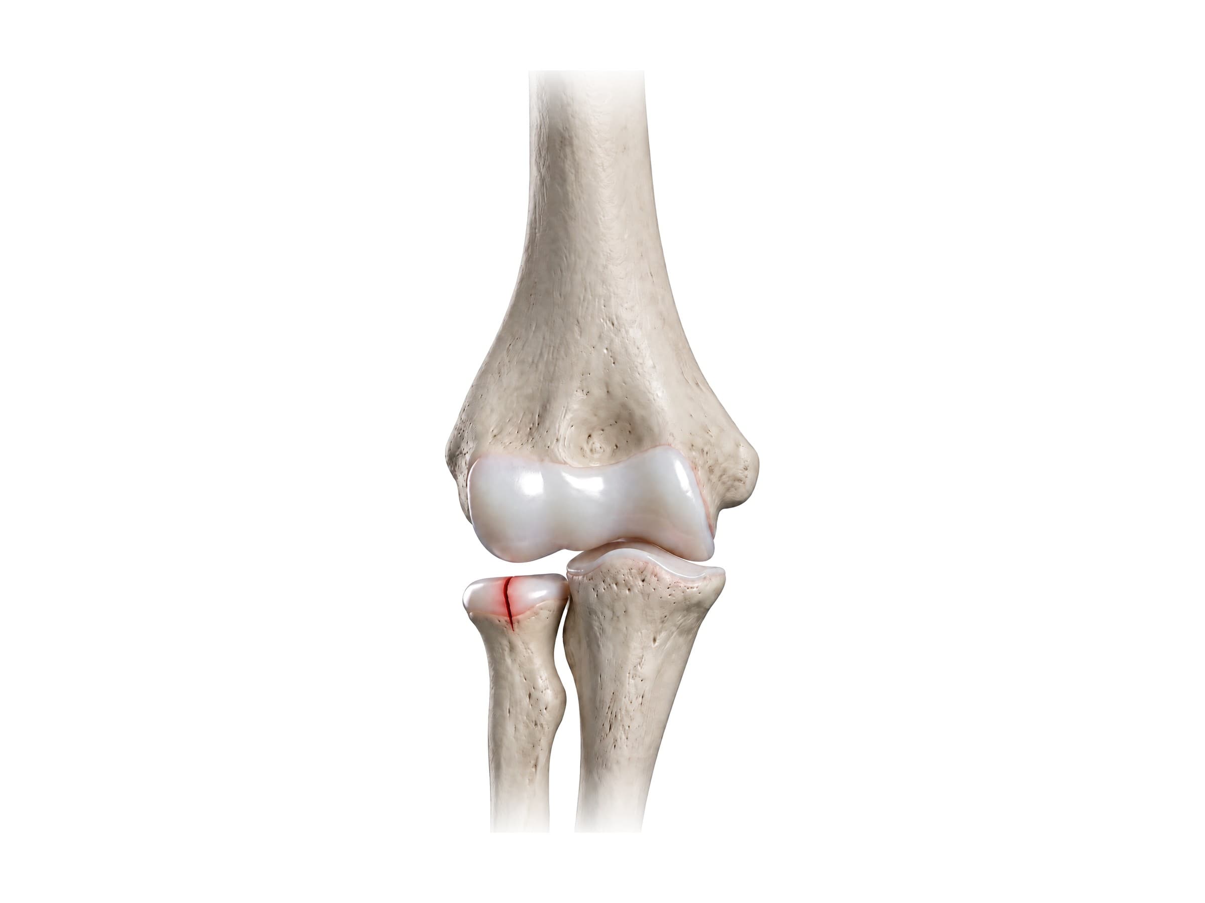
- Radial head fracture: the most common elbow fracture in adults, from a fall on an outstretched hand that drives the radial head into the capitulum. The fracture line can be hard to see, but fluid in the joint lifts the fat pads in front of and behind the humerus, visible on a lateral X-ray as the fat pad sign (a "sail sign" in front). Small fractures are treated with early motion; displaced ones are fixed or replaced.
-
Olecranon fracture: a break through the point of the elbow, usually from falling directly onto it. The triceps pulls the fragment upward and apart, so displaced fractures are usually fixed with a tension band wire or a plate.
-
Elbow dislocation: the forearm bones are forced out of joint, almost always backward (posterior), after a fall on an outstretched hand. It is the second most commonly dislocated major joint in adults after the shoulder. When a dislocation comes with a radial head fracture and a coronoid fracture, it is called the terrible triad because it is so unstable. X-rays confirm the dislocation and CT shows the fractures.
-
Ulnar collateral ligament tear: pain on the inner elbow in throwing athletes, sometimes after a single pop. Seen on MRI or MR arthrogram, with stress ultrasound to measure gapping of the joint.
-
Lateral epicondylitis (tennis elbow): degeneration of the tendon of extensor carpi radialis brevis where it attaches to the lateral epicondyle, causing pain on the outer elbow with gripping and lifting. Most cases are not from tennis. Ultrasound or MRI shows a thickened, torn tendon.
-
Medial epicondylitis (golfer's elbow): the same process in the flexor-pronator tendon on the medial epicondyle.
-
Distal biceps tendon rupture: a sudden pop at the front of the elbow when lifting a heavy load, most often in men aged 40 to 60. The muscle bunches up the arm and the tendon can't be felt in the cubital fossa (the hook test). Confirmed on MRI and usually repaired within a few weeks.
-
Cubital tunnel syndrome: numbness in the ring and little fingers and weakness of the hand from ulnar nerve compression, diagnosed with nerve conduction studies.
-
Nursemaid's elbow: radial head subluxation in a young child after a pull on the arm. X-rays are usually normal, and the radial head is slipped back into the annular ligament by turning the forearm.
-
Olecranon bursitis: swelling over the point of the elbow, seen on ultrasound as fluid in the bursa.
The mechanism of injury largely predicts which of these occurs: a fall on an outstretched hand loads the radial head, the lower humerus in children, or the ligaments in a dislocation; a direct blow breaks the olecranon; and repeated throwing or gripping wears out the UCL or the epicondyle tendons.
How the elbow is shown in a personal injury case
Elbow injuries appear in slip-and-fall and trip-and-fall claims, motor vehicle collisions (a driver's or passenger's elbow often strikes the door or console in a side impact), workplace accidents, and playground and premises cases involving children. A labeled illustration of the normal elbow, showing the three bones, the ligaments and the nerves that pass close to them, establishes the anatomy before the injury exhibit is introduced: the radial head fracture, the dislocated joint, the plate on the olecranon, or the injured ulnar nerve. Illustrations help explain why a supracondylar fracture in a child can threaten the blood supply to the whole forearm, why a "simple" radial head fracture left a patient unable to fully straighten the arm, and why numbness in the ring and little fingers after an elbow injury points to the ulnar nerve. In medical malpractice cases involving a missed vascular injury or compartment syndrome after a supracondylar fracture, an illustration of the brachial artery's path across the fracture site makes the timeline clear to a jury.
Natomy creates custom medical legal illustrations of elbow injuries for demand packages and trial exhibits. For more on how normal anatomy is used in court, see demonstrative evidence. Related atlas pages: shoulder anatomy, hand and wrist anatomy, hip anatomy, knee anatomy, ankle anatomy, back muscles anatomy and skull anatomy.
FAQ
What bones make up the elbow?+
The elbow is formed by three bones: the humerus (the upper arm bone) and the two forearm bones, the radius on the thumb side and the ulna on the little finger side. The lower end of the humerus has two joint surfaces: the rounded capitulum, which meets the head of the radius, and the spool-shaped trochlea, which fits into the trochlear notch of the ulna. The radius and ulna also meet each other just below the elbow.
What are the parts of your elbow called?+
The bony point at the back of the elbow is the olecranon, the top of the ulna. The bumps on either side are the medial epicondyle (inner side) and lateral epicondyle (outer side) of the humerus. The hollow at the front of the elbow is the cubital fossa. Inside the joint, the capitulum and trochlea of the humerus meet the radial head and the trochlear notch of the ulna, and the elbow is held together by the ulnar collateral ligament on the inner side, the radial collateral ligament on the outer side and the annular ligament around the radial head.
What is the most commonly damaged ligament in the elbow?+
It depends on how the injury happens. In throwing athletes such as baseball pitchers, the ulnar collateral ligament (UCL) on the inner side of the elbow is the most commonly injured, worn down by repeated stress until it tears; reconstruction is known as Tommy John surgery. In falls and elbow dislocations, the lateral ligaments on the outer side, especially the lateral ulnar collateral ligament, usually tear first.
What is the funny bone?+
The funny bone is not a bone at all but the ulnar nerve, which runs in a shallow groove behind the medial epicondyle on the inner side of the elbow, covered only by skin and a thin layer of tissue. Bumping it presses the nerve against the bone and sends a tingling or electric feeling down the forearm into the ring and little fingers. Long-term pressure on the nerve here causes cubital tunnel syndrome.
What is nursemaid's elbow?+
Nursemaid's elbow (radial head subluxation) is a common injury in children aged about one to four in which the head of the radius slips partly out of the annular ligament, usually when a child is lifted, swung or pulled by the hand or wrist. The child holds the arm still and refuses to use it. A clinician can usually slip the radial head back into place in seconds by turning the forearm, and X-rays are often not needed.
What is the cubital fossa?+
The cubital fossa is the triangular hollow at the front of the elbow, bordered by the brachioradialis on the outer side, the pronator teres on the inner side and an imaginary line between the two epicondyles above. From the outer side inward it contains the biceps tendon, the brachial artery and the median nerve (remembered as TAN). The median cubital vein runs just under the skin over it, which is why blood is usually drawn there.
What is the olecranon?+
The olecranon is the curved upper end of the ulna that forms the bony point of the elbow. The triceps tendon attaches to it, so it acts as the lever that straightens the arm, and it fits into the olecranon fossa of the humerus when the elbow is fully straight. A fluid-filled sac, the olecranon bursa, sits between the olecranon and the skin; when it becomes inflamed (olecranon bursitis), a soft swelling appears at the point of the elbow.
What's the most common elbow injury?+
The most common elbow problem overall is lateral epicondylitis (tennis elbow), an overuse injury of the tendon attaching to the lateral epicondyle. Among fractures, the radial head fracture is the most common elbow fracture in adults and the supracondylar humerus fracture is the most common in children, both usually from a fall on an outstretched hand. The elbow is also the second most commonly dislocated major joint in adults, after the shoulder.
Need a custom illustration of this joint?
Generate a labeled or exhibit-ready figure for a specific patient, injury, or case in minutes.
Try Natomy →