Hand and Wrist Anatomy: Bones, Muscles & Nerves
Labeled illustrations of the 27 bones of the hand and wrist, plus the carpal bones, muscles, tendons, nerves and common hand and wrist fractures.
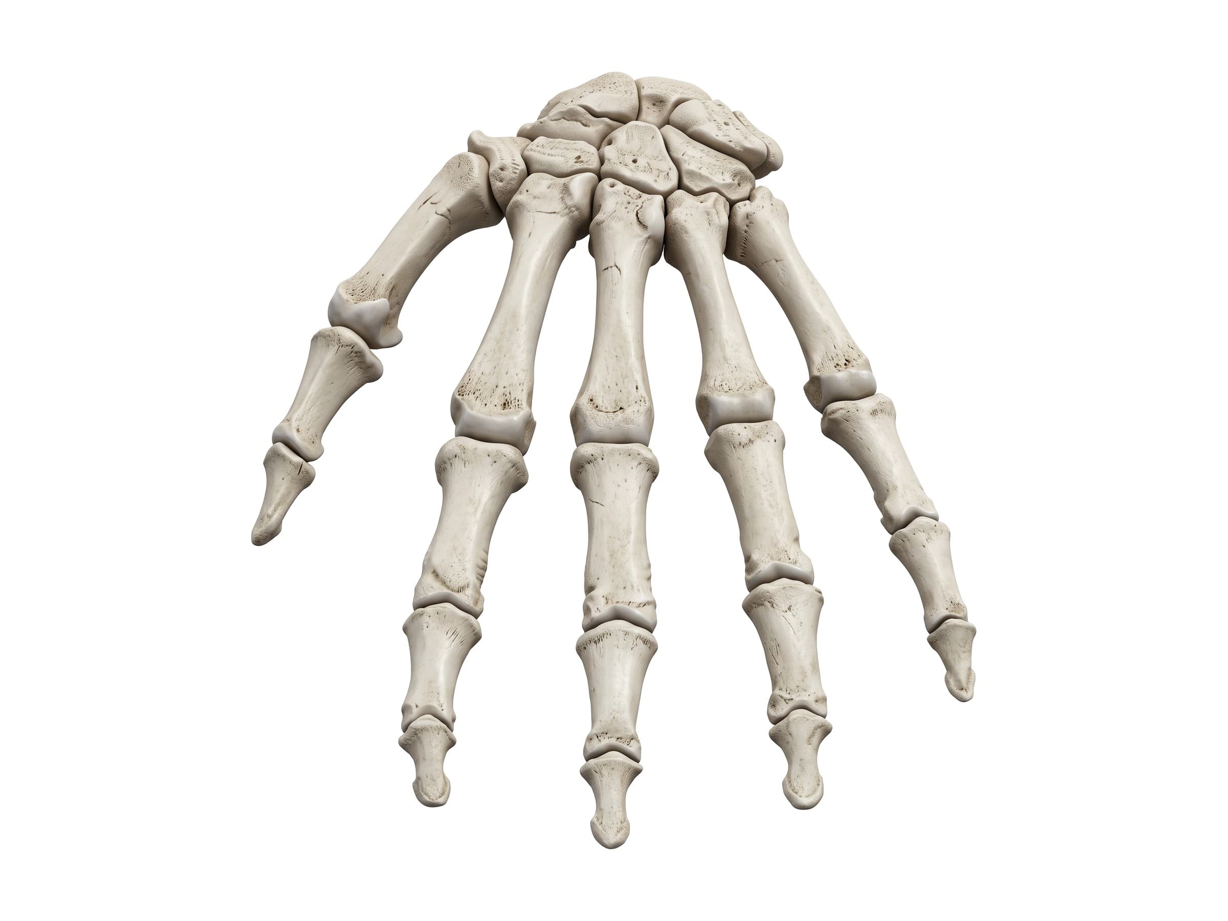
The hand and wrist are made of 27 bones, more than 30 muscles and three major nerves packed into a space small enough to be injured by a single fall. Hand and wrist anatomy comes down to the bones (8 carpals, 5 metacarpals and 14 phalanges, plus the ends of the radius and ulna), the joints and ligaments that hold them together, the long tendons from forearm muscles and the small muscles inside the hand, and the median, ulnar and radial nerves. Knowing how they fit together is the first step to reading a hand or wrist X-ray report, explaining a diagnosis to a patient, or illustrating a personal injury claim.
Bones of the hand and wrist
The hand has three groups of bones. In the view of the back of the hand above, the carpal bones form the base of the hand at the wrist, the five metacarpals run through the palm, and the phalanges make up the fingers and thumb.
- Carpal bones (8): two rows of four small bones that form the wrist and give it its flexibility.
- Metacarpals (5): the long bones of the palm, numbered 1 to 5 from the thumb. Each has a base at the wrist, a shaft, a neck and a head; the heads form the knuckles.
- Phalanges (14): three in each finger (proximal, middle and distal) and two in the thumb (proximal and distal).
Carpal bones (wrist bones)
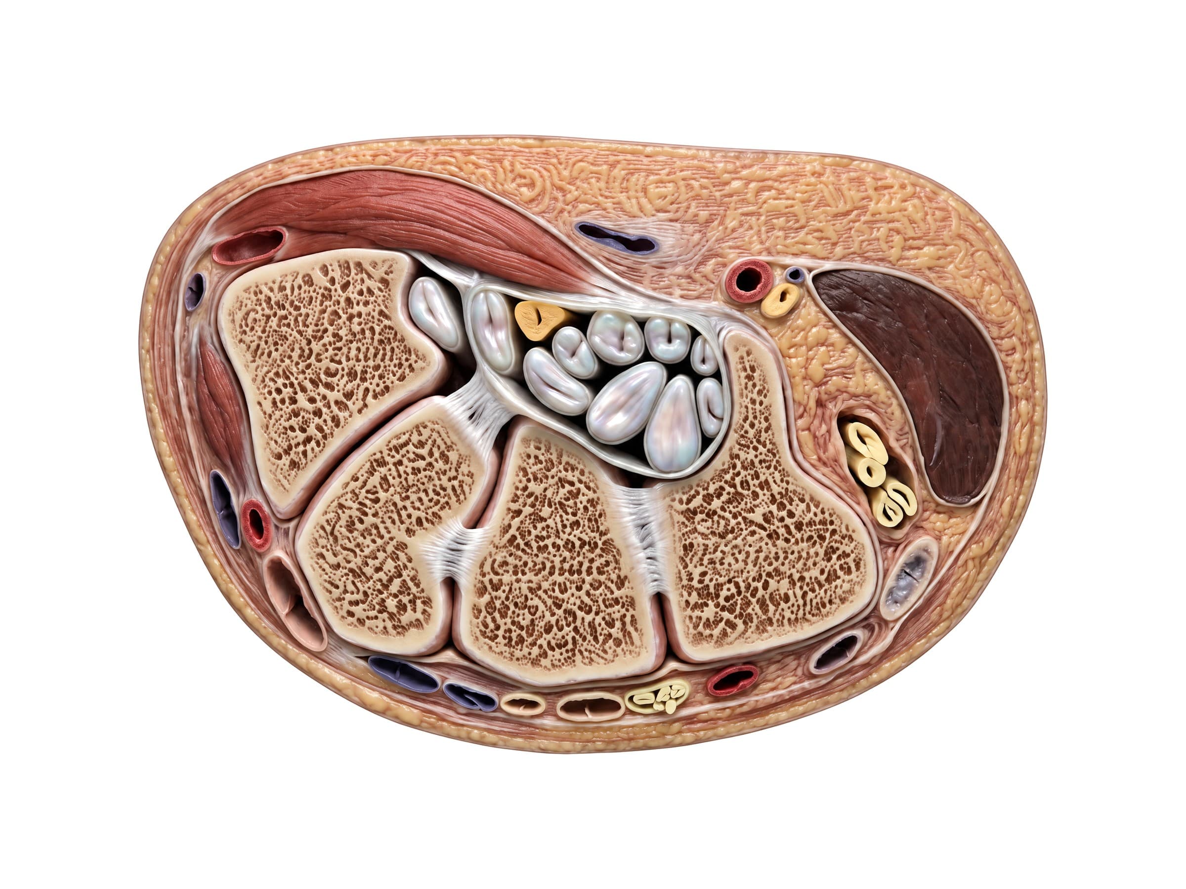
The eight carpal bones sit in two rows. The proximal row, from the thumb side, is the scaphoid, lunate, triquetrum and pisiform; the distal row is the trapezium, trapezoid, capitate and hamate. The proximal row meets the radius at the wrist joint, and the distal row meets the bases of the metacarpals. The section below, cut through the wrist from front to back, shows every carpal bone and the joints between them.
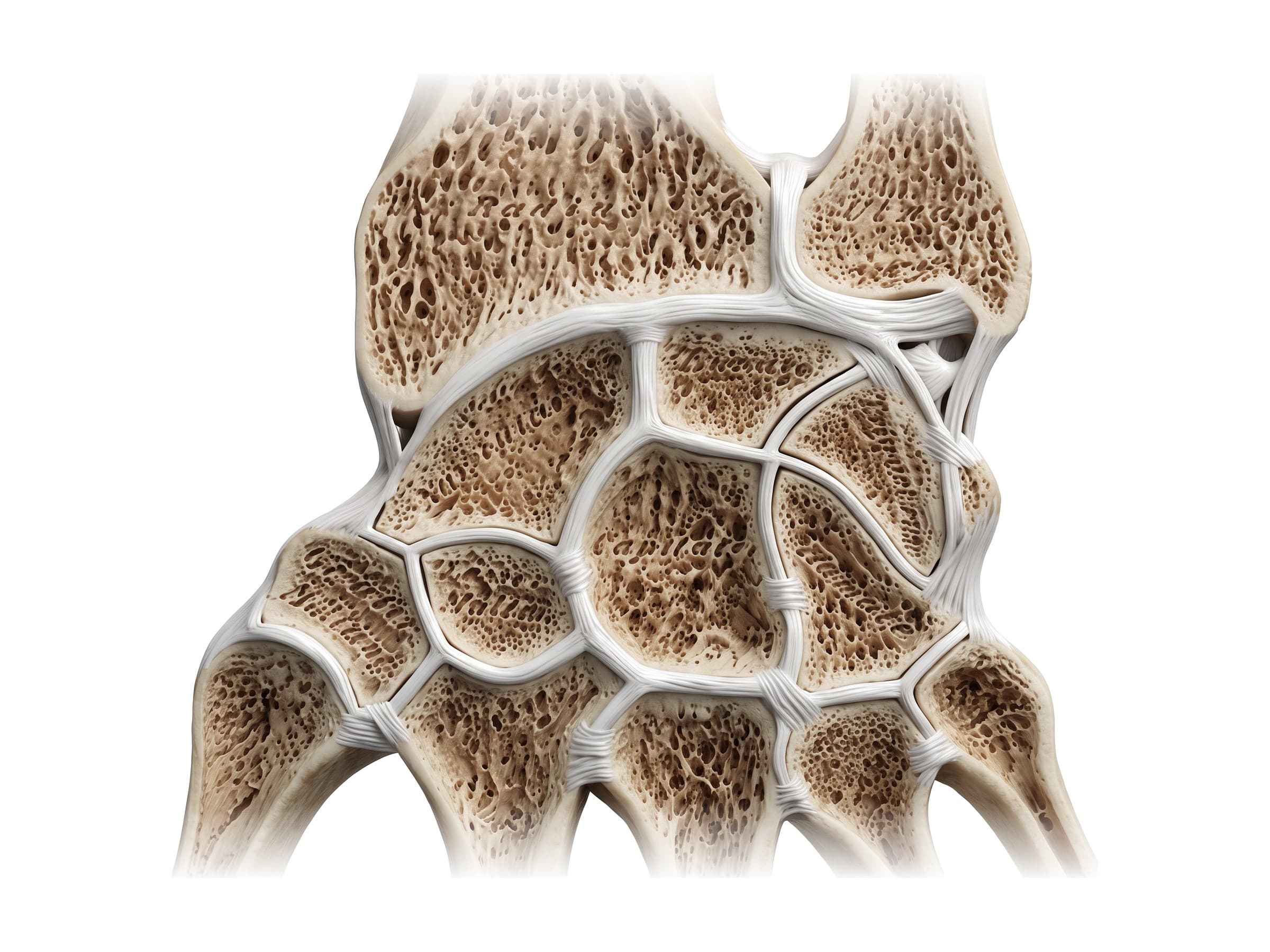
- Scaphoid: the boat-shaped bone on the thumb side that bridges both rows. It is the most commonly fractured carpal bone, and its blood supply enters from the far end, so a fracture through its waist can cut off blood to the near end.
- Lunate: the half-moon-shaped bone in the center of the proximal row. It is the carpal bone most often dislocated.
- Pisiform: a small pea-shaped bone sitting on the front of the triquetrum inside the tendon of flexor carpi ulnaris.
- Capitate: the largest carpal bone, in the center of the wrist.
- Hamate: the wedge-shaped bone on the little finger side, with a hook (the hook of hamate) that projects toward the palm. The hook forms a wall of both the carpal tunnel and Guyon's canal, and it can fracture in golf, baseball and racquet sports.
Radius and ulna at the wrist
The wrist joint is formed mainly by the radius, the forearm bone on the thumb side. Its broad lower end articulates directly with the scaphoid and lunate. The ulna, on the little finger side, does not touch the carpal bones: it is separated from them by the triangular fibrocartilage complex (TFCC), a cushion of cartilage and ligaments that also stabilizes the distal radioulnar joint (DRUJ), where the radius rotates around the ulna when you turn your palm up and down. All three are visible at the top of the wrist section above.
- Radial styloid: the bony point on the thumb side of the wrist.
- Ulnar styloid: the bony point on the little finger side, often broken off together with a distal radius fracture.
- Articular disc (TFCC): the triangular disc between the ulna and the carpus. Tears cause pain on the little finger side of the wrist.
Joints of the hand and thumb
- Radiocarpal joint: the wrist joint proper, between the radius and the proximal row of carpal bones. It bends the wrist forward and back and side to side.
- Midcarpal joint: between the two rows of carpal bones, adding to wrist motion.
- Carpometacarpal (CMC) joints: between the distal carpal row and the bases of the metacarpals. The thumb CMC joint is a saddle joint that allows opposition and is the most common site of hand arthritis; the CMC joints of the index and middle fingers barely move.
- Metacarpophalangeal (MCP) joints: the knuckles, where the metacarpal heads meet the proximal phalanges. They bend and straighten and also spread the fingers apart.
- Interphalangeal joints: hinge joints within the fingers, the proximal (PIP) and distal (DIP) interphalangeal joints. The thumb has a single interphalangeal joint.
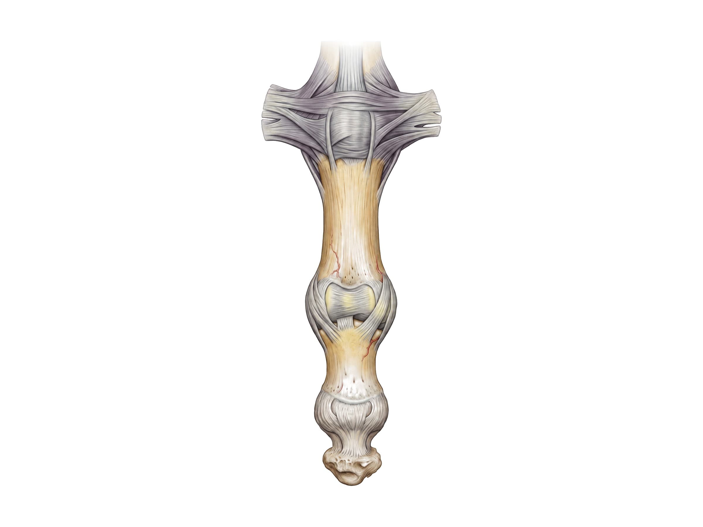
Each finger joint is stabilized by a collateral ligament on either side and a thick volar plate across its palm side, which stops the joint from bending backward.
Ligaments of the wrist
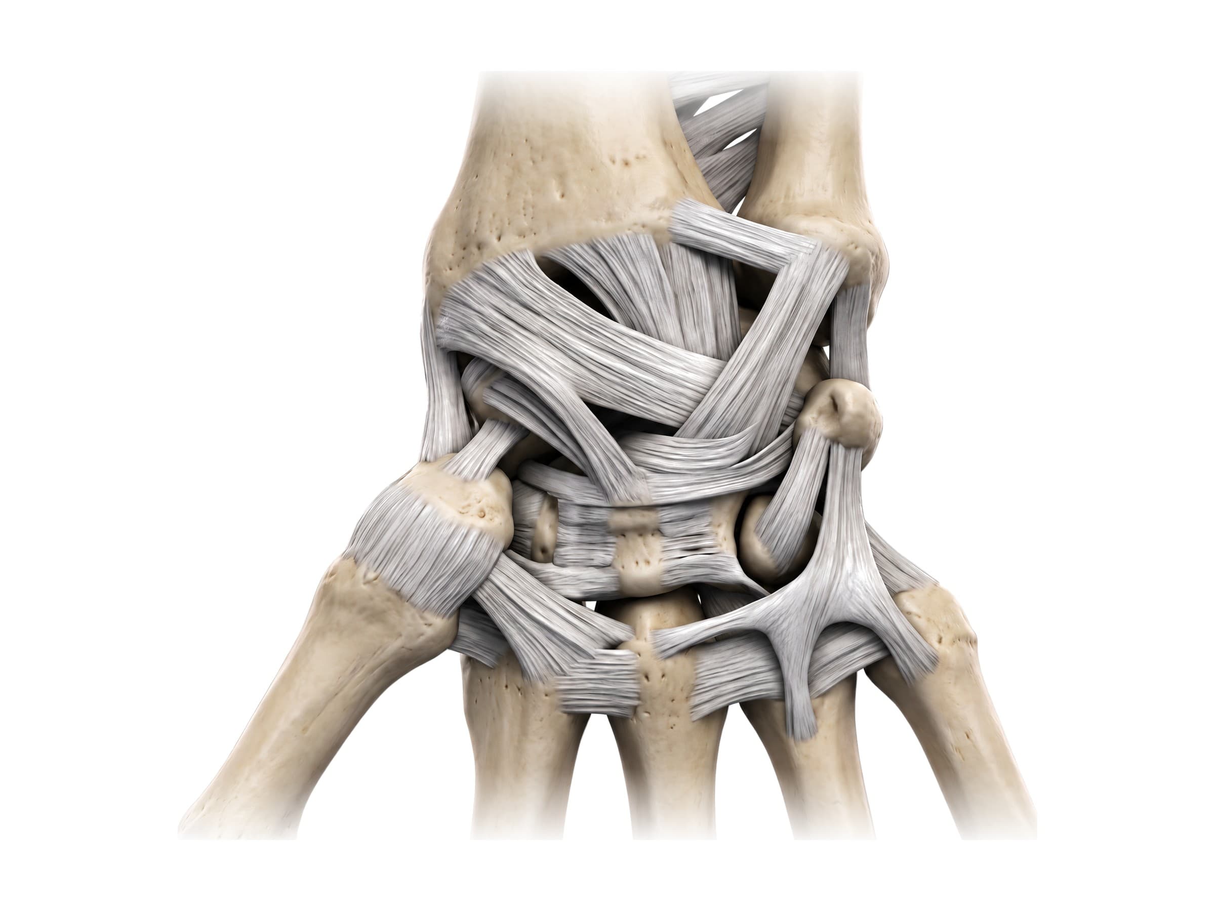
The carpal bones are held together by two sets of ligaments. Extrinsic ligaments connect the radius and ulna to the carpal bones; the strongest run across the palm side of the wrist, from the radius down to the scaphoid, lunate and capitate. Intrinsic ligaments connect the carpal bones to each other. The most important is the scapholunate ligament between the scaphoid and lunate, the most commonly injured wrist ligament: when it tears, the two bones separate, leaving a visible gap on X-ray.
Muscles of the hand
The muscles that work the hand fall into two groups. Extrinsic muscles start in the forearm and reach the hand through long tendons that cross the wrist; they provide most of the grip strength. Intrinsic muscles start and end inside the hand and handle fine movement.
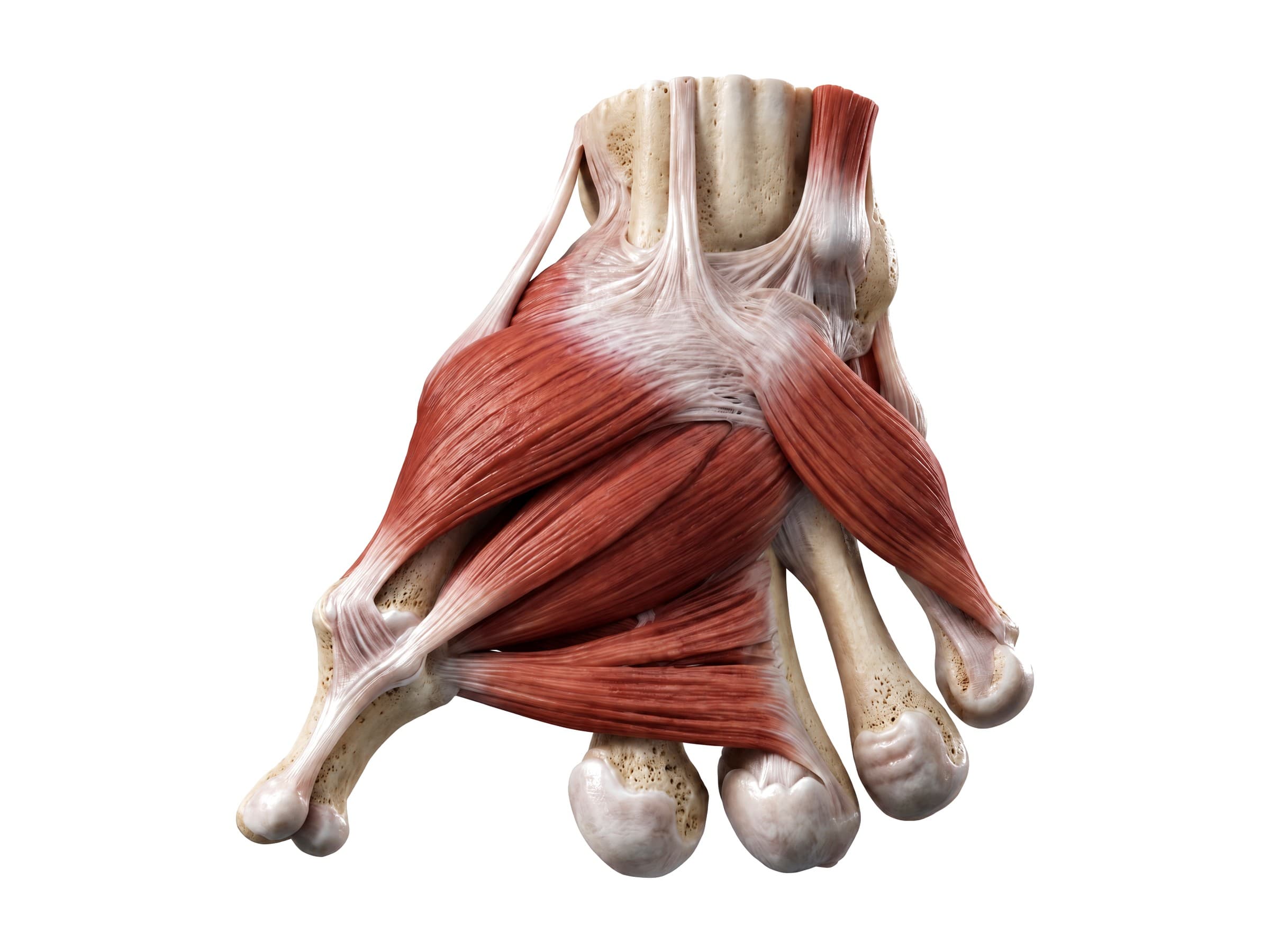
- Thenar muscles: the fleshy mound at the base of the thumb, made of abductor pollicis brevis, flexor pollicis brevis and opponens pollicis. They move the thumb away from the palm and across it to meet the fingers. Mostly supplied by the median nerve.
- Adductor pollicis: the deep muscle that pulls the thumb back against the palm, used in pinch grip. Supplied by the ulnar nerve.
- Hypothenar muscles: the mound at the base of the little finger, made of abductor digiti minimi, flexor digiti minimi brevis and opponens digiti minimi. Supplied by the ulnar nerve.
- Lumbricals: four small muscles attached to the flexor digitorum profundus tendons that bend the knuckles while straightening the finger joints.
- Interossei: muscles between the metacarpals. The palmar interossei bring the fingers together and the dorsal interossei spread them apart.
- Palmaris longus: a forearm muscle whose thin tendon runs over the front of the wrist into the palm. It is absent in about one in seven people and is a common donor tendon for reconstructive surgery.
Flexor and extensor tendons
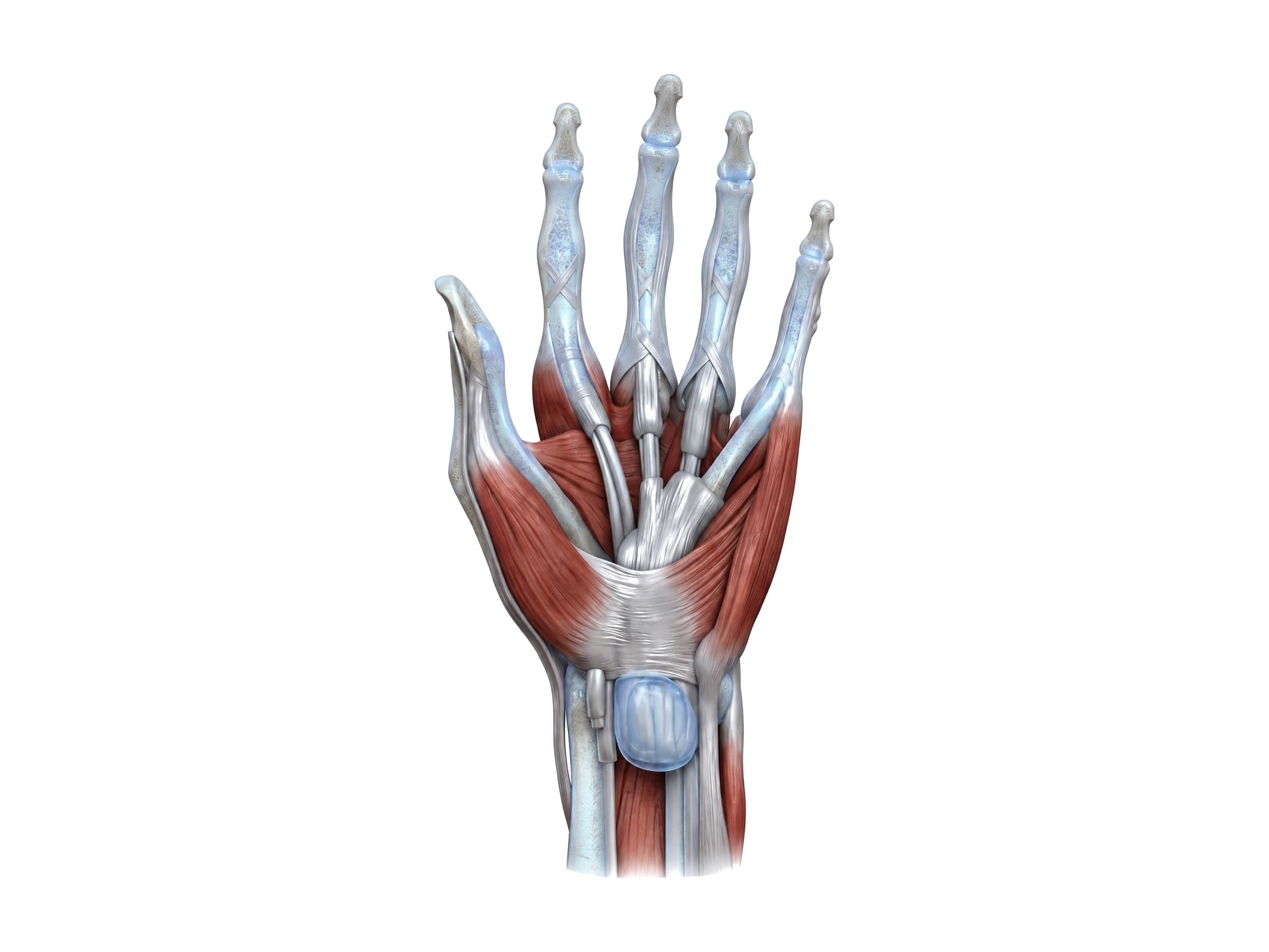
On the palm side, the tendons of flexor digitorum superficialis (which bends the middle finger joint) and flexor digitorum profundus (which bends the fingertip) run through the carpal tunnel and into the fingers inside lubricated synovial sheaths, held against the bone by a series of fibrous pulleys. The wrist itself is flexed by flexor carpi radialis and flexor carpi ulnaris.
On the back of the wrist, the extensor tendons pass under the extensor retinaculum in six separate tunnels called the extensor compartments, numbered from the thumb side:
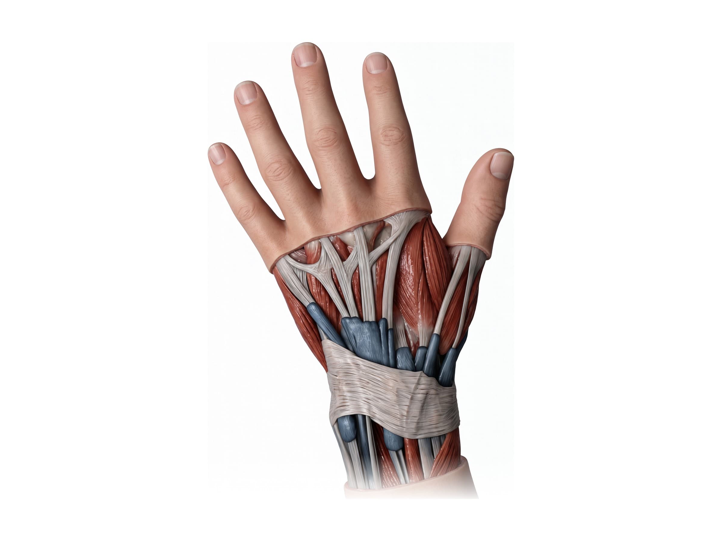
- Abductor pollicis longus and extensor pollicis brevis (inflamed in De Quervain's tenosynovitis)
- Extensor carpi radialis longus and brevis
- Extensor pollicis longus, which turns around a bony bump on the radius called Lister's tubercle
- Extensor digitorum and extensor indicis
- Extensor digiti minimi
- Extensor carpi ulnaris
The tendons of compartments 1 and 3 border the anatomical snuffbox, the hollow at the base of the thumb with the scaphoid in its floor.
Nerves and blood supply of the hand
Three nerves supply the hand, all branches of the brachial plexus:
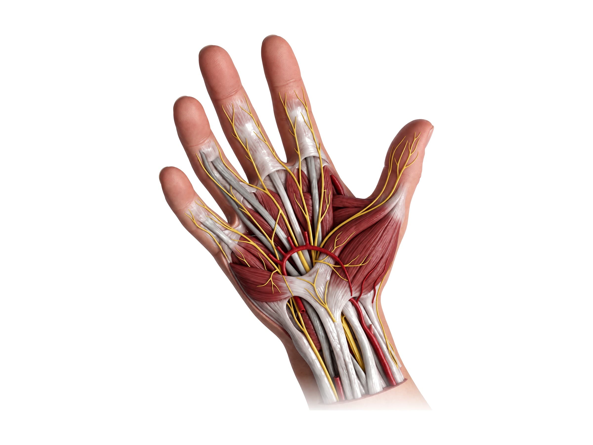
- Median nerve: passes through the carpal tunnel. It supplies most of the thenar muscles and the two lumbricals on the thumb side, and sensation to the palm side of the thumb, index and middle fingers and half of the ring finger. Compression in the carpal tunnel causes carpal tunnel syndrome.
- Ulnar nerve: passes through Guyon's canal, between the pisiform and the hook of hamate, outside the carpal tunnel. It supplies most of the intrinsic hand muscles and sensation to the little finger and half of the ring finger.
- Radial nerve: supplies no muscles in the hand. Its superficial branch carries sensation from the back of the hand on the thumb side; higher up, it supplies the forearm muscles that extend the wrist and fingers, which is why a radial nerve injury causes wrist drop.
Blood reaches the hand through the radial artery and ulnar artery, which join in the palm to form two arches: the superficial palmar arch, fed mainly by the ulnar artery, and the deeper deep palmar arch, fed mainly by the radial artery. Because the arches connect, either artery can usually supply the whole hand on its own; the Allen test checks this before the radial artery is used for a blood draw, a catheter, or a bypass graft.
The carpal tunnel
Seen in cross-section at the wrist, the carpal tunnel is a channel with the carpal bones as its floor and walls and the transverse carpal ligament as its roof. Through it run the median nerve and nine tendons: four of flexor digitorum superficialis, four of flexor digitorum profundus, and flexor pollicis longus. The ulnar nerve and artery pass outside the tunnel, on the little finger side.
Common hand and wrist injuries and how they appear on imaging
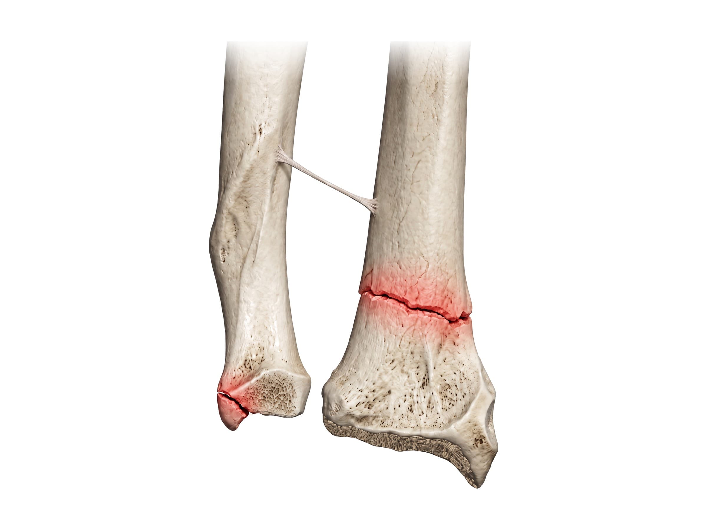
- Distal radius fracture: one of the most common fractures, usually from a fall on an outstretched hand (a classic mechanism of injury). A Colles fracture tilts the broken end toward the back of the hand; a Smith fracture tilts it toward the palm. Seen on standard wrist X-rays, with CT for fractures that enter the joint.
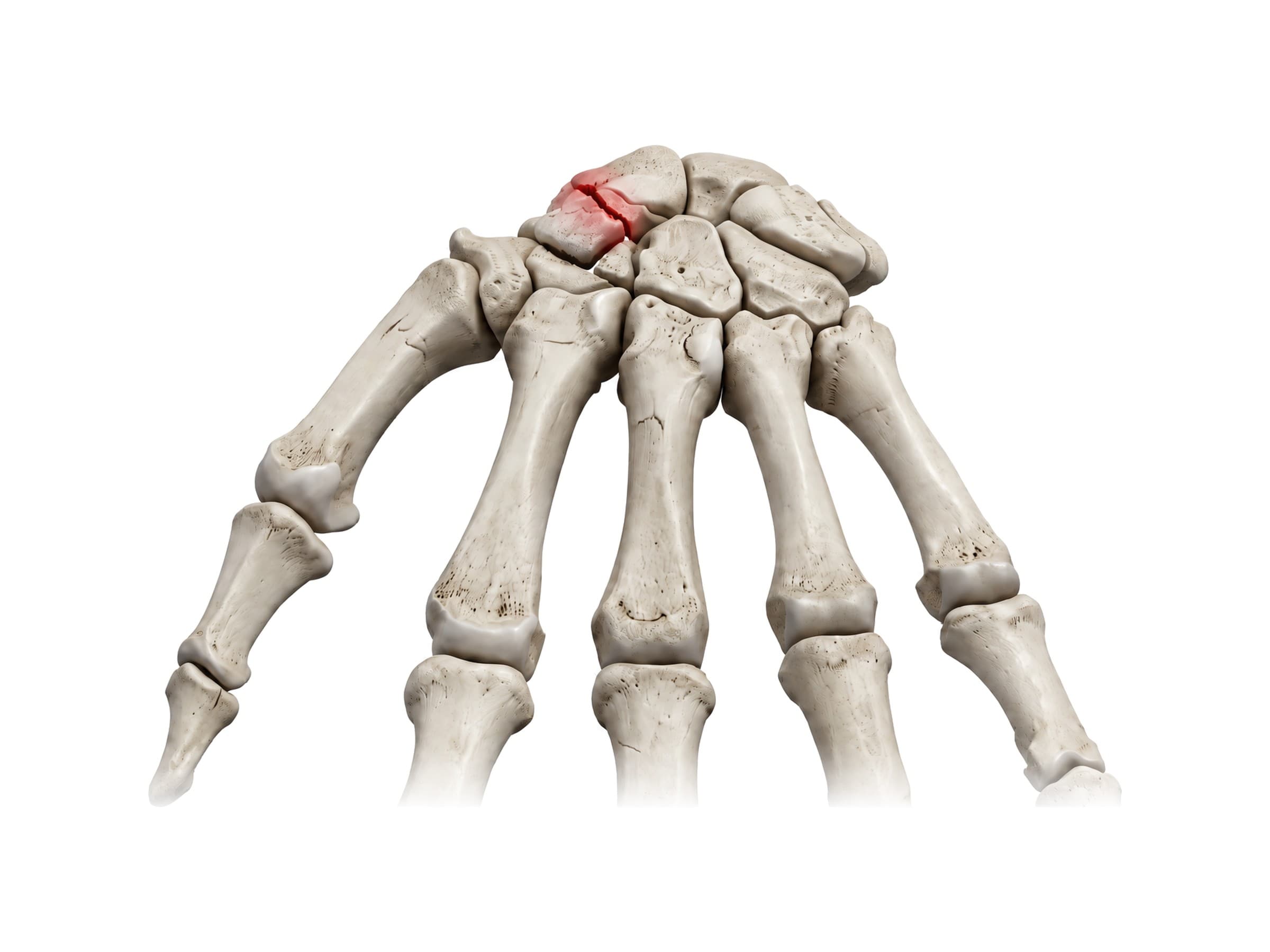
- Scaphoid fracture: tenderness in the anatomical snuffbox after a fall. It is often invisible on the first X-ray, so a suspected fracture is splinted and re-imaged, or evaluated with MRI. A missed scaphoid fracture can fail to heal or lose its blood supply (avascular necrosis).
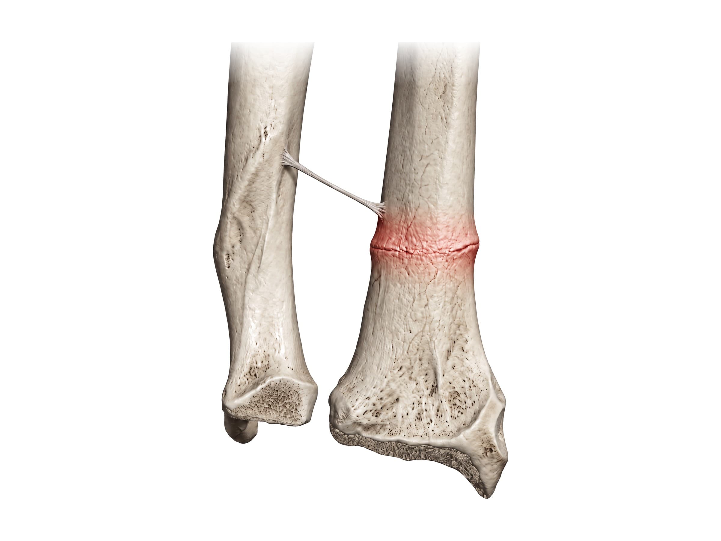
- Buckle (torus) fracture: a common childhood wrist fracture in which the soft bone of the distal radius bulges on one side without breaking through. Seen as a small bump in the bone's outline on X-ray.
- Boxer's fracture: a break of the neck of the fifth metacarpal from punching a hard object.
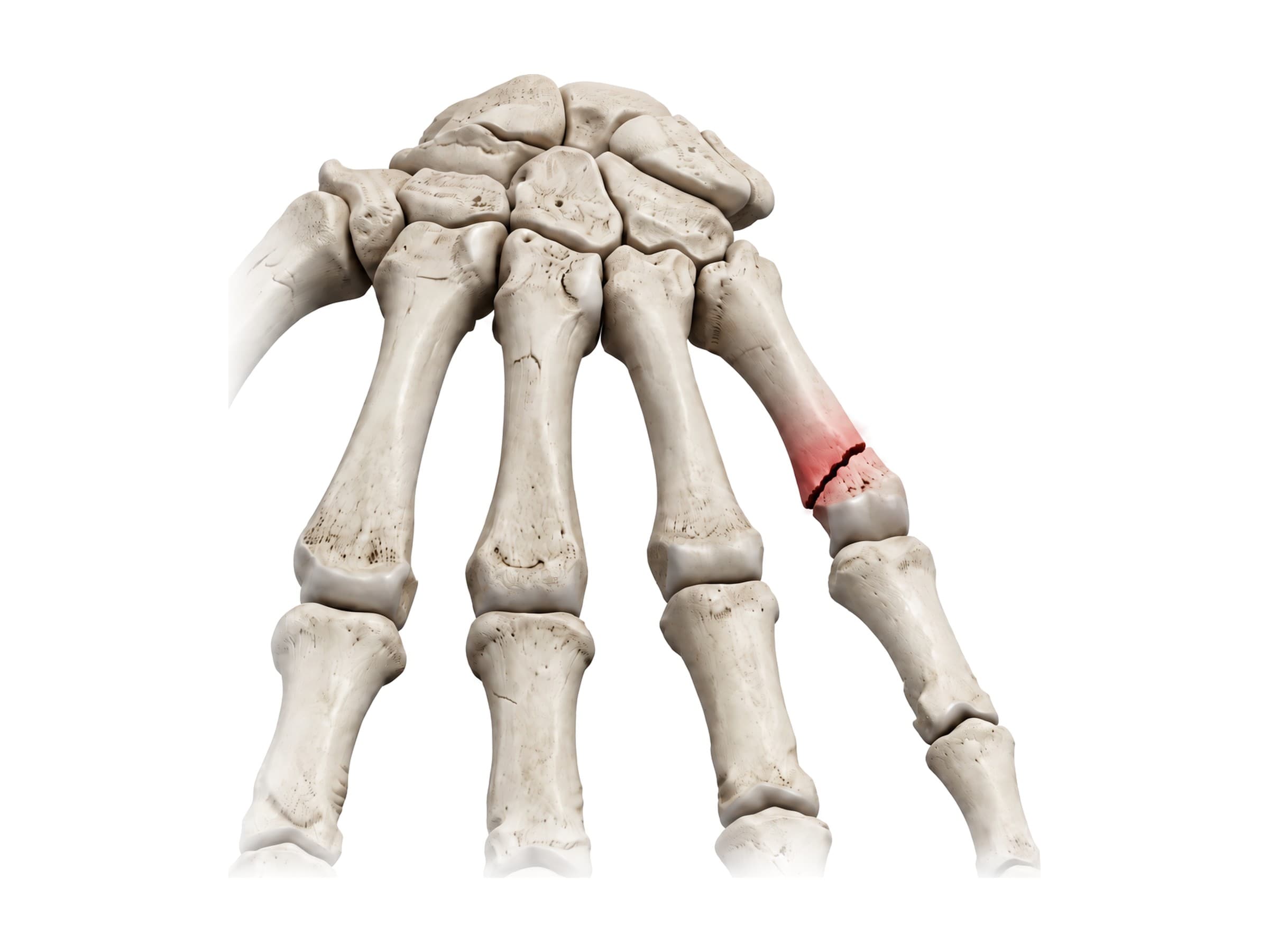
- Mallet finger: the extensor tendon is torn from the base of the fingertip, so the last joint droops and cannot be straightened.
- Scapholunate ligament tear: a widened gap between the scaphoid and lunate on X-ray, confirmed with MRI or arthroscopy.
- TFCC tear: pain on the little finger side of the wrist, best evaluated with MRI or MR arthrography.
- Carpal tunnel syndrome: compression of the median nerve, diagnosed by symptoms and nerve conduction studies; ultrasound can show a swollen nerve.
- Flexor tendon laceration: a cut on the palm side of the finger or palm that leaves the finger unable to bend. Injuries inside the fibrous pulleys of the finger are the hardest to repair.
- Degloving injury: skin and soft tissue torn away from the underlying tendons and bone, typically in machinery and crush accidents.
How the hand and wrist are shown in a personal injury case
A labeled illustration of normal hand and wrist anatomy is the baseline exhibit in most hand-injury claims: falls on an outstretched hand, crush and degloving injuries in the workplace, and lacerations. It shows a jury where the scaphoid, the distal radius or the injured tendon sits in an uninjured hand before the injury-specific exhibit (the fracture, the plate and screws, the tendon repair) is introduced. In wrist fracture cases, a side-by-side exhibit that pairs the normal anatomy with the patient's own X-rays helps separate an acute fracture from pre-existing arthritis, and in medical malpractice claims involving a missed scaphoid fracture, an illustration of the bone's blood supply explains why the delay in diagnosis mattered.
Natomy creates custom medical legal illustrations of hand and wrist injuries for demand packages and trial exhibits. For the joints above the hand, see elbow anatomy and shoulder anatomy; for the lower limb, hip anatomy, knee anatomy, ankle anatomy and foot bones; and for the trunk and head, back muscles anatomy and skull anatomy.
FAQ
How many bones are in the hand and wrist?+
Each hand and wrist has 27 bones: 8 carpal bones in the wrist, 5 metacarpal bones in the palm, and 14 phalanges in the fingers and thumb (three in each finger and two in the thumb). The lower ends of the two forearm bones, the radius and ulna, also form part of the wrist joint but are not counted among the hand bones. Small sesamoid bones near the thumb are usually not counted either.
What are the 8 carpal bones?+
The carpal bones sit in two rows. The proximal row, from the thumb side to the little finger side, is the scaphoid, lunate, triquetrum and pisiform. The distal row, in the same order, is the trapezium, trapezoid, capitate and hamate. A common mnemonic is "Some Lovers Try Positions That They Can't Handle", which runs through the proximal row and then the distal row from the thumb side.
Which bone is on the thumb side, the radius or the ulna?+
The radius is on the thumb side of the forearm and the ulna is on the little finger side. At the wrist, the radius is the larger of the two and carries most of the load: it articulates directly with the scaphoid and lunate. The ulna is separated from the carpal bones by the triangular fibrocartilage complex (TFCC).
What is the anatomical snuffbox?+
The anatomical snuffbox is the small hollow on the thumb side of the back of the wrist that appears when you extend your thumb. It is bordered by the tendons of extensor pollicis longus on one side and abductor pollicis longus and extensor pollicis brevis on the other, and the scaphoid bone lies in its floor. Tenderness in the snuffbox after a fall is a classic sign of a scaphoid fracture, even when the first X-ray looks normal.
What are the joints of the thumb?+
The thumb has three joints. The carpometacarpal (CMC) joint at its base, between the trapezium and the first metacarpal, is a saddle joint that lets the thumb swing across the palm to touch the other fingers (opposition), and it is the most common site of arthritis in the hand. Above it are the metacarpophalangeal (MCP) joint and a single interphalangeal (IP) joint. The other fingers each have an MCP joint and two interphalangeal joints, the PIP and DIP.
What nerve causes carpal tunnel syndrome?+
Carpal tunnel syndrome is caused by compression of the median nerve as it passes through the carpal tunnel, a narrow passage at the front of the wrist formed by the carpal bones and roofed by the transverse carpal ligament. The tunnel also carries the nine flexor tendons to the fingers and thumb, so swelling of the tendon sheaths or the wrist can squeeze the nerve, causing numbness and tingling in the thumb, index, middle and part of the ring finger.
Which muscles move the fingers?+
Most of the strength in the fingers comes from extrinsic muscles in the forearm, whose long tendons cross the wrist: flexor digitorum superficialis and profundus bend the fingers, and extensor digitorum straightens them. The small intrinsic muscles inside the hand (the thenar and hypothenar muscles, the lumbricals and the interossei) handle fine control: spreading and closing the fingers, opposing the thumb, and bending the knuckles while keeping the fingers straight.
What is a Colles fracture?+
A Colles fracture is a break of the distal radius, just above the wrist, in which the broken end of the radius tilts and shifts toward the back of the hand. It usually happens in a fall on an outstretched hand and gives the wrist a characteristic "dinner fork" shape from the side. A Smith fracture is the reverse pattern, with the fragment displaced toward the palm, typically from a fall onto a flexed wrist.
Need a custom illustration of this joint?
Generate a labeled or exhibit-ready figure for a specific patient, injury, or case in minutes.
Try Natomy →