Hip Anatomy: Bones, Muscles & Ligaments
Labeled illustrations of the hip bones, acetabulum, labrum, ligaments, hip flexors, gluteal muscles and common hip fractures and labral tears.
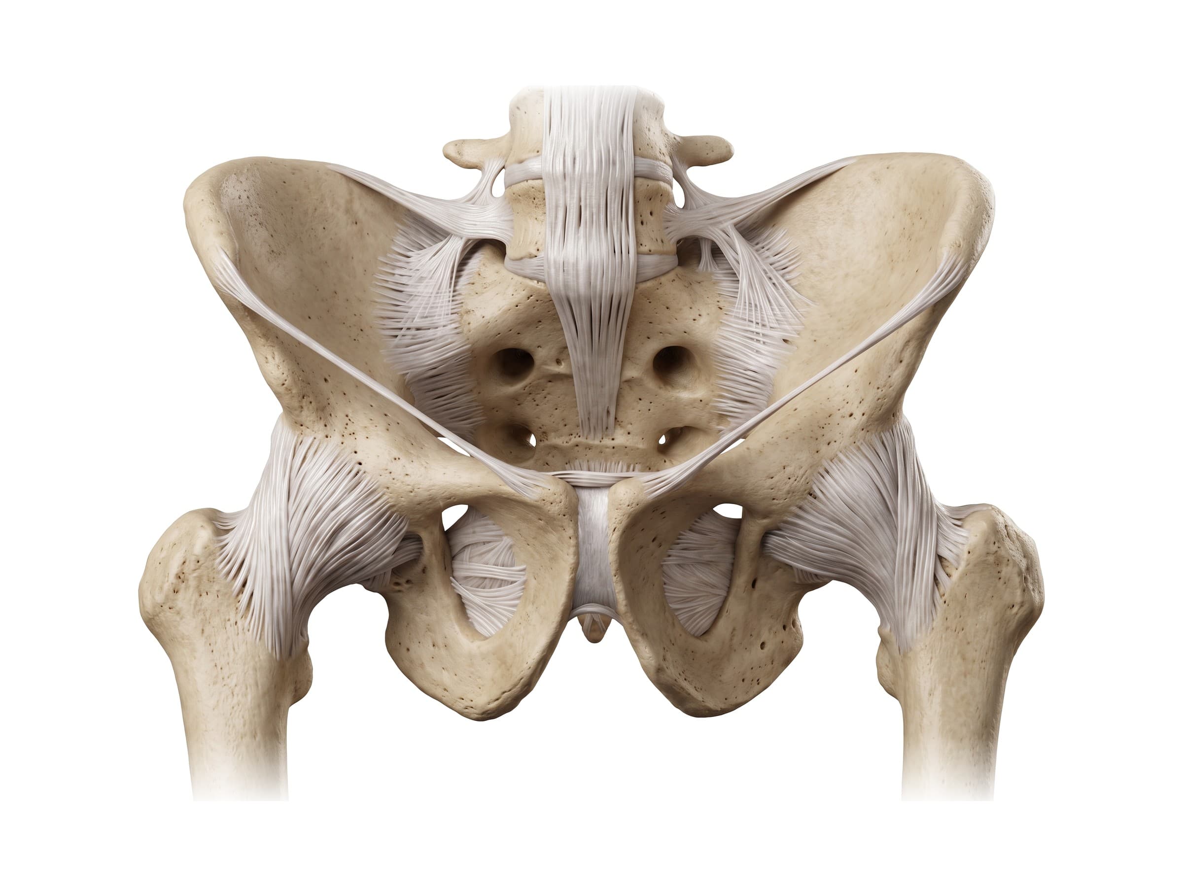
The hip is the ball-and-socket joint where the thighbone meets the pelvis. Hip anatomy comes down to the femoral head (the ball) sitting deep in the acetabulum of the hip bone (the socket), a rim of cartilage called the labrum that deepens the socket, a thick capsule reinforced by three strong ligaments, and the large muscle groups around it: the hip flexors in front, the gluteal muscles on the side and back, and the adductors on the inside. It carries the weight of the upper body and is one of the most stable joints in the body, which is why hip fractures and dislocations usually take either a fall in an older person or a high-energy injury such as a car crash, and why the anatomy matters for reading a hip X-ray report, explaining a diagnosis to a patient, or illustrating a personal injury claim.
Bones of the hip and pelvis
The pelvis is a ring made of the two hip bones and the sacrum at the back, joined in front at the pubic symphysis and behind at the two sacroiliac joints. In the front view above, each hip joint sits on the outer side of the pelvis, with the head of the femur fitted into the socket of the hip bone.
- Hip bone (innominate bone): the large flat bone on each side of the pelvis, made of the ilium, ischium and pubis fused together.
- Sacrum: the triangular bone at the base of the spine, wedged between the two hip bones.
- Femur: the thighbone, the longest and strongest bone in the body. Its upper end forms the ball of the hip joint.
Bony landmarks you can feel through the skin include the iliac crest along the top of the pelvis, the anterior superior iliac spine (ASIS) at its front end, the greater trochanter on the outer side of the upper thigh, and the ischial tuberosity, the sitting bone.
Ilium, ischium and pubis
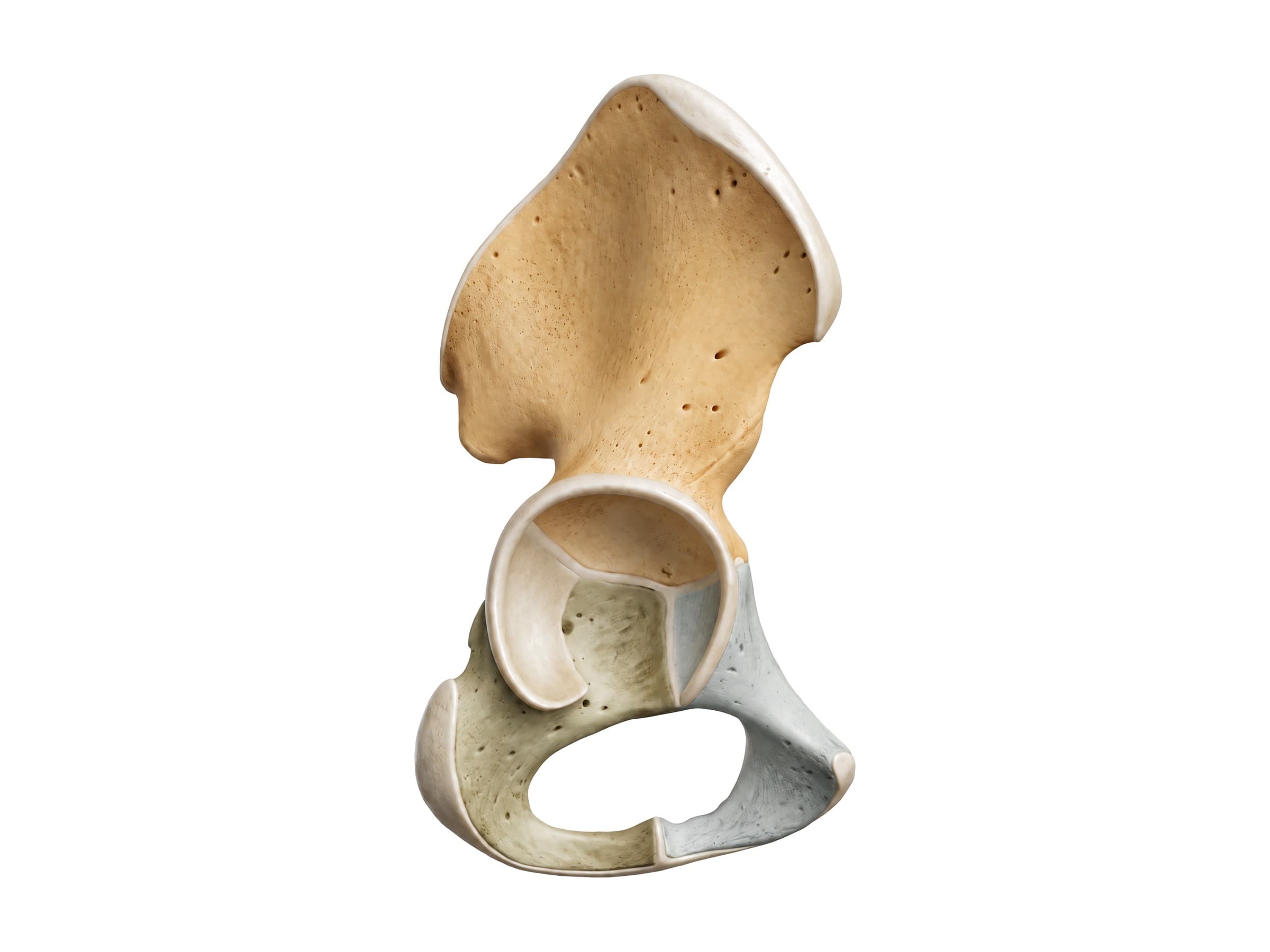
Each hip bone starts as three separate bones that meet in a Y-shaped growth plate (the triradiate cartilage) in the floor of the socket and fuse in the mid-teens.
- Ilium: the broad upper wing that forms the flare of the hip. Its upper edge is the iliac crest, which ends in front at the anterior superior iliac spine. Behind, it forms the greater sciatic notch, through which the sciatic nerve and piriformis leave the pelvis.
- Ischium: the lower back part of the hip bone. Its thick lower end, the ischial tuberosity, carries the body's weight when sitting and is where the hamstring muscles attach.
- Pubis: the lower front part. The two pubic bones meet at the pubic symphysis, and the pubis and ischium together surround the obturator foramen, a large opening closed by a membrane.
The acetabulum and labrum
The acetabulum is the hip socket, a deep cup on the outer side of the hip bone formed by all three of its parts. Only its horseshoe-shaped outer rim, the lunate surface, is covered with cartilage and bears weight; the center (the acetabular fossa) is filled with fat. The acetabular labrum, a ring of fibrocartilage attached around the rim, deepens the socket and creates a suction seal around the femoral head that helps keep it in place.
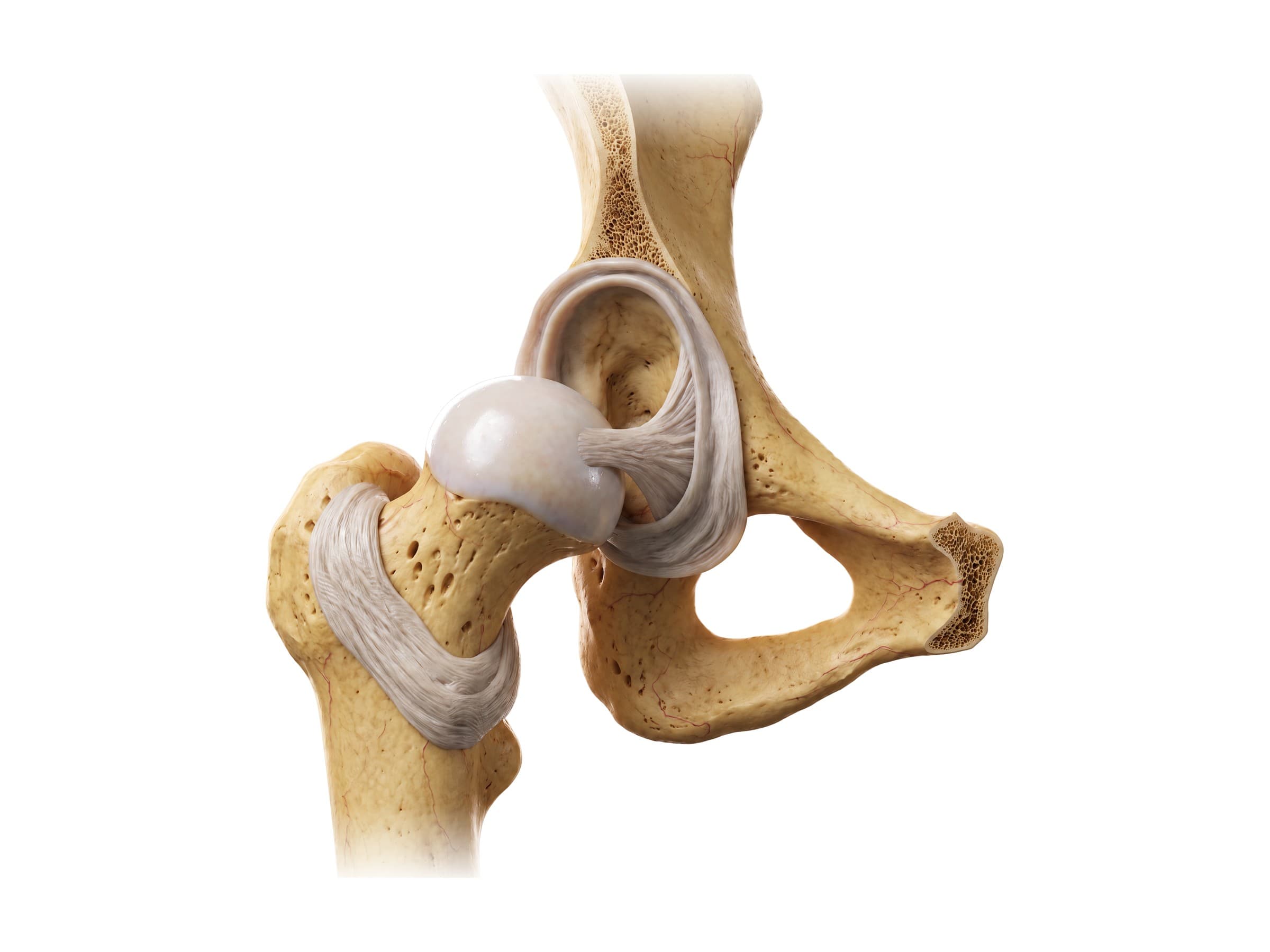
The ligamentum teres runs from the floor of the socket to a small pit on the femoral head, the fovea. It carries a small artery that matters in children but supplies little blood to the head in adults.
The femoral head, neck and trochanters
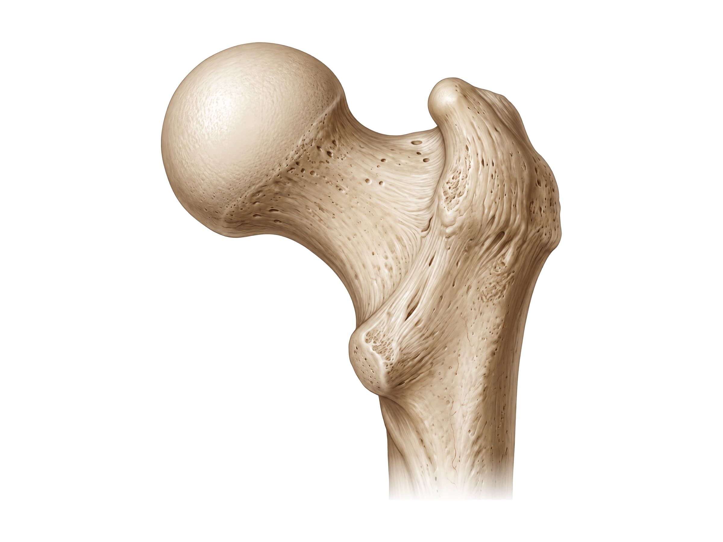
- Femoral head: the ball of the joint, about two-thirds of a sphere and covered in cartilage everywhere except the fovea.
- Femoral neck: the narrow bridge between the head and the shaft, set at an angle of about 125 to 130 degrees to the shaft. Most of the blood supply to the head runs up along the neck, mainly from the medial femoral circumflex artery, which is why a displaced femoral neck fracture can leave the head without blood (avascular necrosis).
- Greater trochanter: the large bony prominence on the outer side, where the gluteus medius, gluteus minimus and the deep hip rotators attach. It is the bony point you feel on the side of the hip and the site of trochanteric pain.
- Lesser trochanter: the smaller knob on the inner back side, where the iliopsoas attaches.
- Intertrochanteric line and crest: the ridges joining the two trochanters in front and behind. The joint capsule attaches along the front line, which separates fractures inside the capsule (femoral neck) from those outside it (intertrochanteric).
Hip ligaments
The joint capsule of the hip is thick and strong, running from the rim of the acetabulum to the femoral neck. It is reinforced by three ligaments that spiral around the joint and tighten when the hip straightens, screwing the femoral head into the socket.
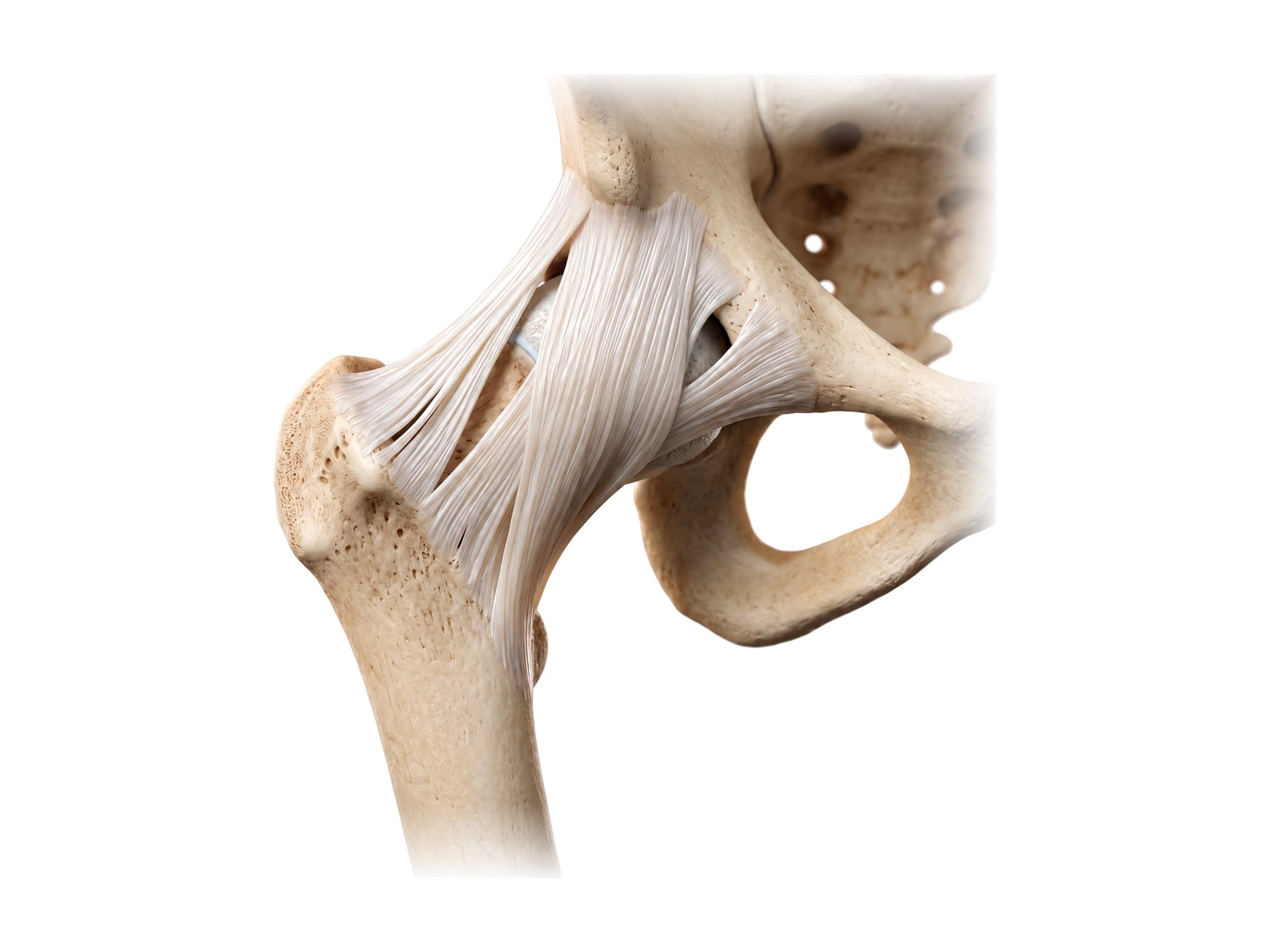
- Iliofemoral ligament (Y-ligament of Bigelow): on the front, from the anterior inferior iliac spine to the intertrochanteric line, shaped like an upside-down Y. It is the strongest ligament in the body and stops the hip from extending too far, so you can stand upright with little muscle effort.
- Pubofemoral ligament: on the front and underside, from the pubis to the femoral neck. It limits spreading the legs apart (abduction) and outward rotation.
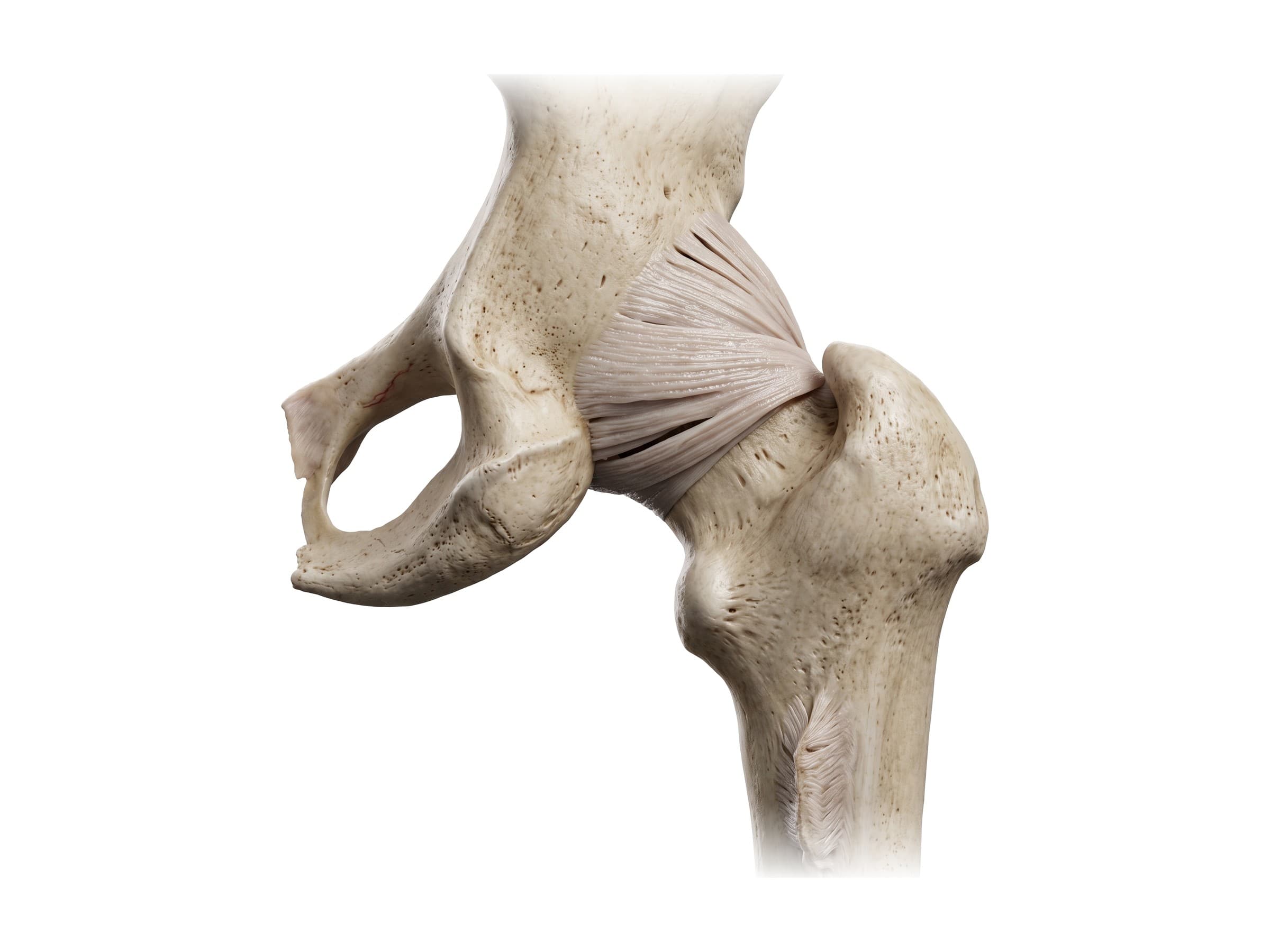
- Ischiofemoral ligament: on the back, from the ischium spiraling up and outward to the femoral neck. It limits inward rotation. It is the thinnest of the three, which is one reason most hip dislocations happen toward the back.
- Zona orbicularis: a ring of circular capsule fibers around the femoral neck that holds the head in the socket.
Hip flexors and the iliopsoas
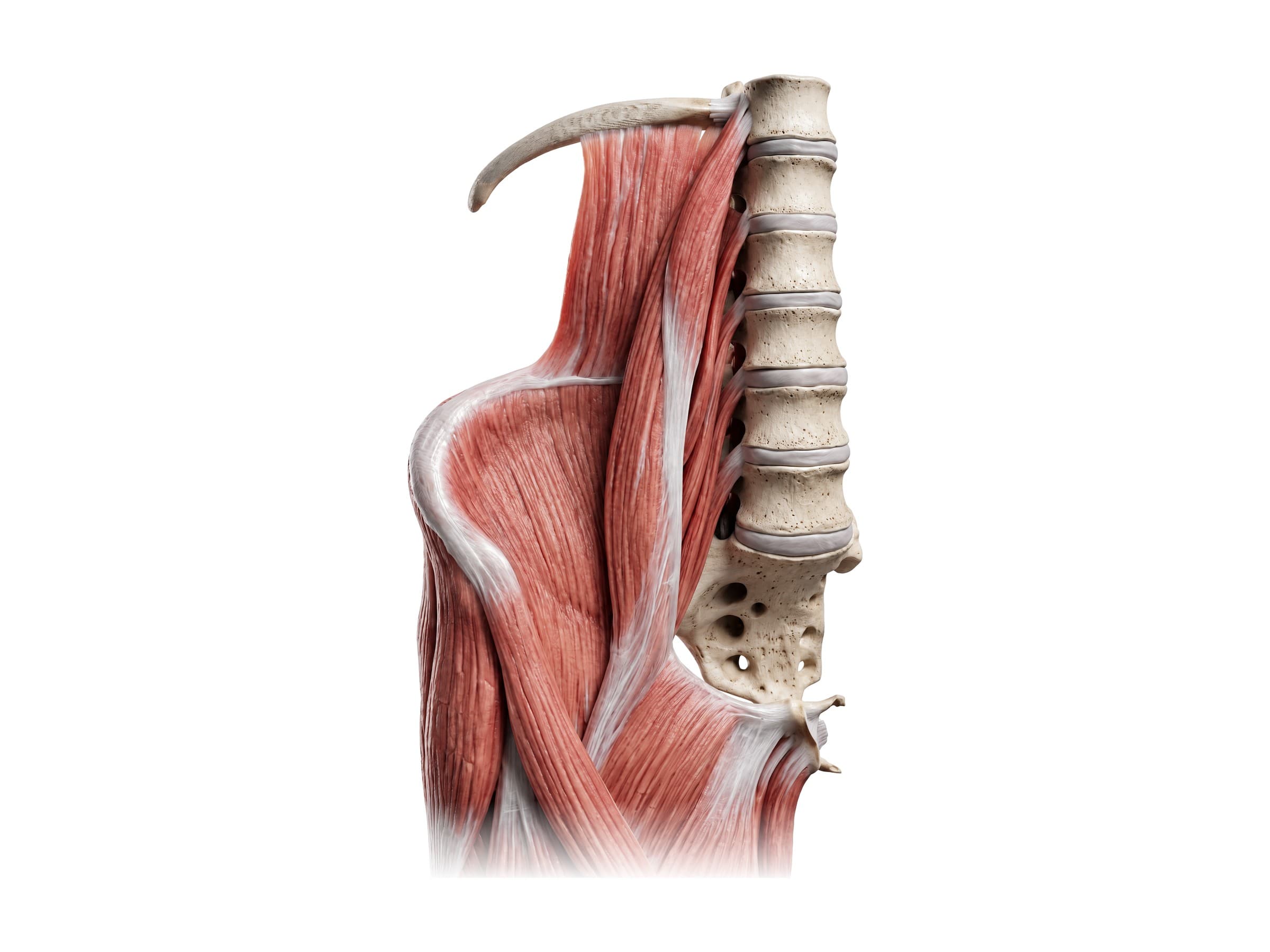
The hip flexors lift the thigh toward the trunk, for walking, climbing stairs and sitting up.
- Iliopsoas: the strongest hip flexor, made of two muscles. The psoas major starts on the sides of the lumbar vertebrae (T12 to L5) and runs down along the brim of the pelvis; the iliacus fills the inside of the iliac wing. They join into a single tendon that passes in front of the hip joint to the lesser trochanter. The psoas also connects the spine directly to the leg, which is why a tight or inflamed psoas can cause both hip and lower back pain.
- Rectus femoris: the only quadriceps muscle that crosses the hip, from the anterior inferior iliac spine to the kneecap. It flexes the hip and straightens the knee and is a common site of strains in kicking and sprinting.
- Sartorius: the longest muscle in the body, running diagonally across the front of the thigh from the anterior superior iliac spine to the inside of the knee.
- Tensor fasciae latae: a small muscle on the front and side of the hip that tightens the iliotibial band.
- Pectineus: a flat muscle at the top of the inner thigh that flexes and adducts the hip.
Gluteal muscles and deep hip rotators
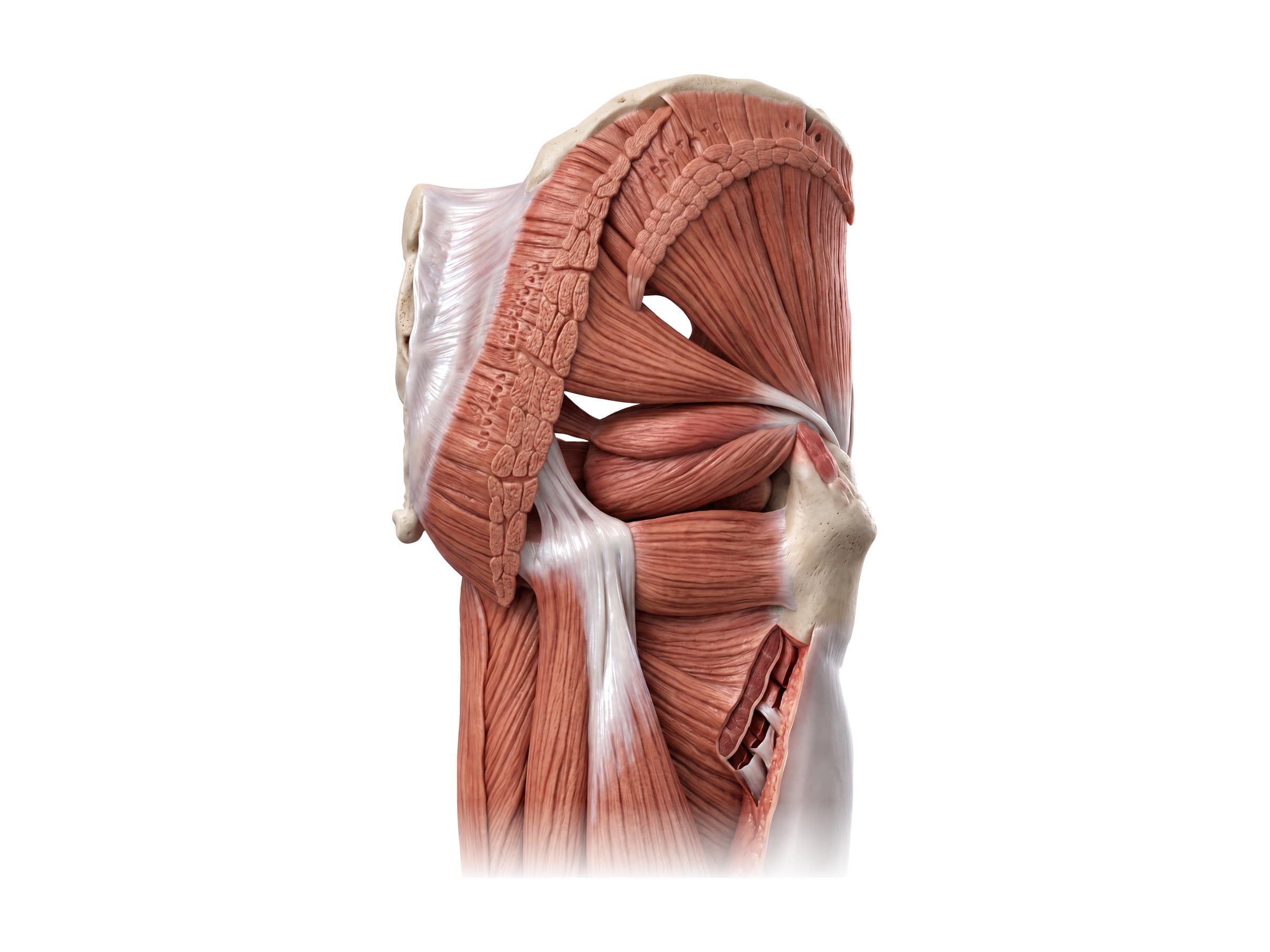
- Gluteus maximus: the largest muscle of the buttock and the main hip extender, used for climbing, running and rising from a chair. Supplied by the inferior gluteal nerve.
- Gluteus medius: a fan-shaped muscle under the gluteus maximus on the side of the pelvis, attaching to the greater trochanter. It is the main hip abductor and holds the pelvis level each time you stand on one leg. When it is weak or its nerve (the superior gluteal nerve) is injured, the opposite side of the pelvis drops with each step, the Trendelenburg sign.
- Gluteus minimus: the smallest and deepest gluteal muscle, under the gluteus medius, with the same job.
- Deep hip rotators: six small muscles behind the joint that turn the thigh outward and hold the femoral head in the socket: the piriformis, gemellus superior, obturator internus, gemellus inferior, quadratus femoris and obturator externus. The sciatic nerve usually leaves the pelvis just below the piriformis, and compression there can cause buttock pain and sciatica-like symptoms (piriformis syndrome).
Below the gluteal muscles, the hamstrings start on the ischial tuberosity and extend the hip, and the adductors (adductor longus, brevis and magnus, gracilis and pectineus) on the inner thigh pull the leg inward. A strain of the adductors is a groin pull.
Hip bursae
A bursa is a small fluid-filled sac that reduces friction where tendons pass over bone. The hip has three that matter clinically:
- Trochanteric bursa: over the greater trochanter, under the iliotibial band. Pain on the outer side of the hip, worse lying on that side, is called greater trochanteric pain syndrome; it was long blamed on trochanteric bursitis but most often comes from tendinopathy of the gluteus medius and minimus.
- Iliopsoas bursa: between the iliopsoas tendon and the front of the hip joint. Inflammation causes groin pain, and the tendon can snap over the bone (internal snapping hip).
- Ischial bursa: over the ischial tuberosity, irritated by prolonged sitting on hard surfaces.
Nerves and blood supply of the hip
- Femoral nerve: passes in front of the hip beside the iliopsoas, supplying the hip flexors and quadriceps and sensation to the front of the thigh.
- Obturator nerve: leaves the pelvis through the obturator foramen to supply the adductors. Because it also supplies the hip joint and the skin of the inner knee, hip problems are sometimes felt as knee pain, especially in children.
- Sciatic nerve: the largest nerve in the body, leaving the pelvis through the greater sciatic notch behind the hip. It is injured in roughly 10 to 20 percent of posterior hip dislocations.
- Superior and inferior gluteal nerves: supply the gluteus medius and minimus and the gluteus maximus.
- Lateral femoral cutaneous nerve: passes near the anterior superior iliac spine; compression causes numbness and burning on the outer thigh (meralgia paresthetica).
The femoral head is supplied mainly by the medial and lateral femoral circumflex arteries, branches of the deep femoral artery that wrap around the femoral neck and send small vessels up along it to the head, with a minor contribution from the artery of the ligamentum teres.
Movements of the hip
- Flexion: bringing the thigh forward and up, about 120 degrees with the knee bent (iliopsoas, rectus femoris).
- Extension: moving the thigh backward, about 10 to 20 degrees (gluteus maximus, hamstrings).
- Abduction: moving the leg out to the side, about 45 degrees (gluteus medius and minimus).
- Adduction: bringing the leg back across the midline, about 30 degrees (adductor group).
- Internal and external rotation: turning the thigh inward and outward (gluteus minimus and tensor fasciae latae inward; the deep rotators and gluteus maximus outward).
Common hip injuries and how they appear on imaging
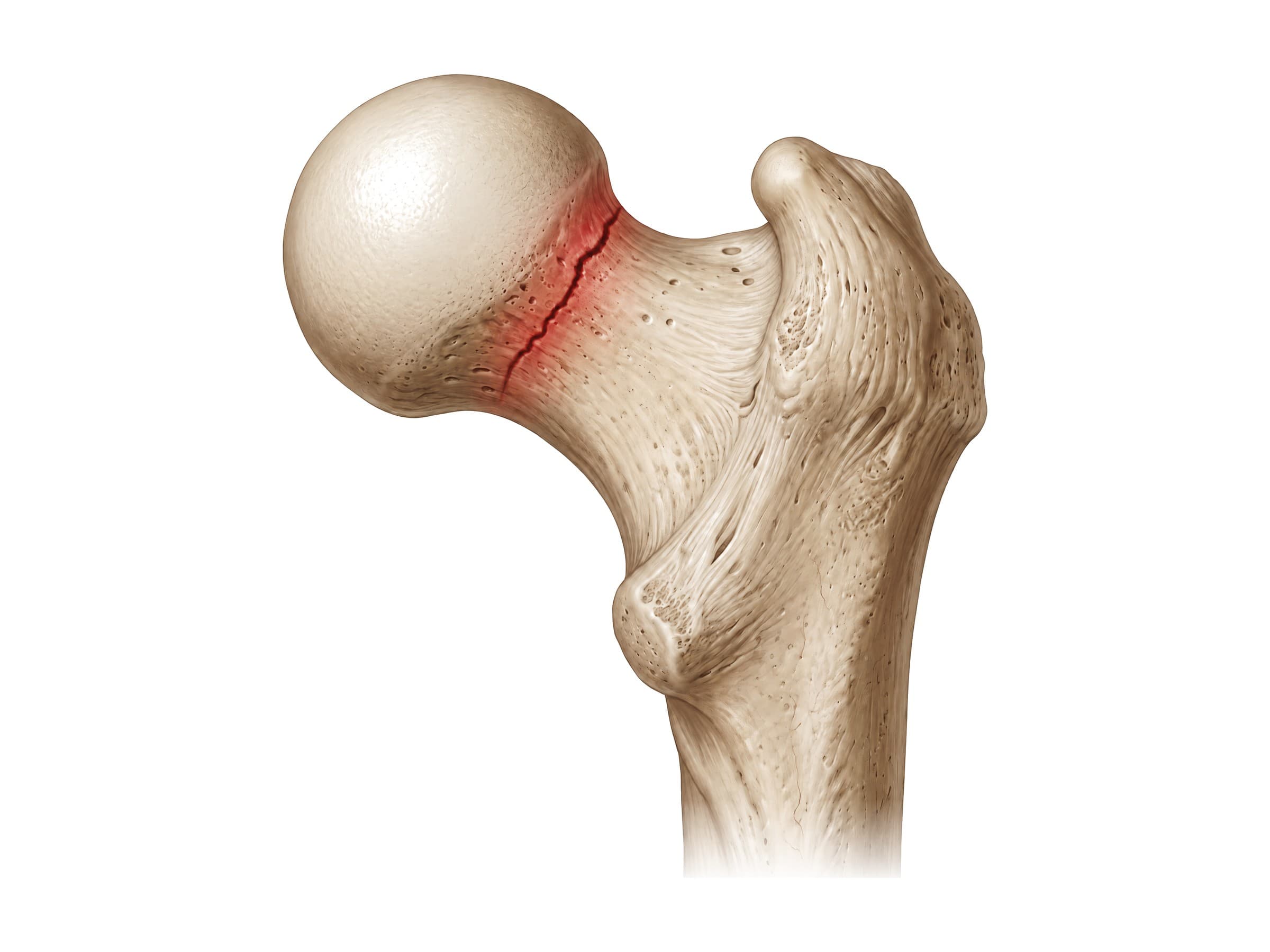
- Femoral neck fracture: a break through the femoral neck, inside the joint capsule, usually from a fall in an older adult with osteoporosis. Because the fracture can tear the vessels running up the neck, displaced fractures risk avascular necrosis and nonunion and are usually treated with a hemiarthroplasty or total hip replacement; undisplaced fractures can be fixed with screws. Seen on an AP pelvis and lateral hip X-ray; MRI finds fractures that the X-ray misses.
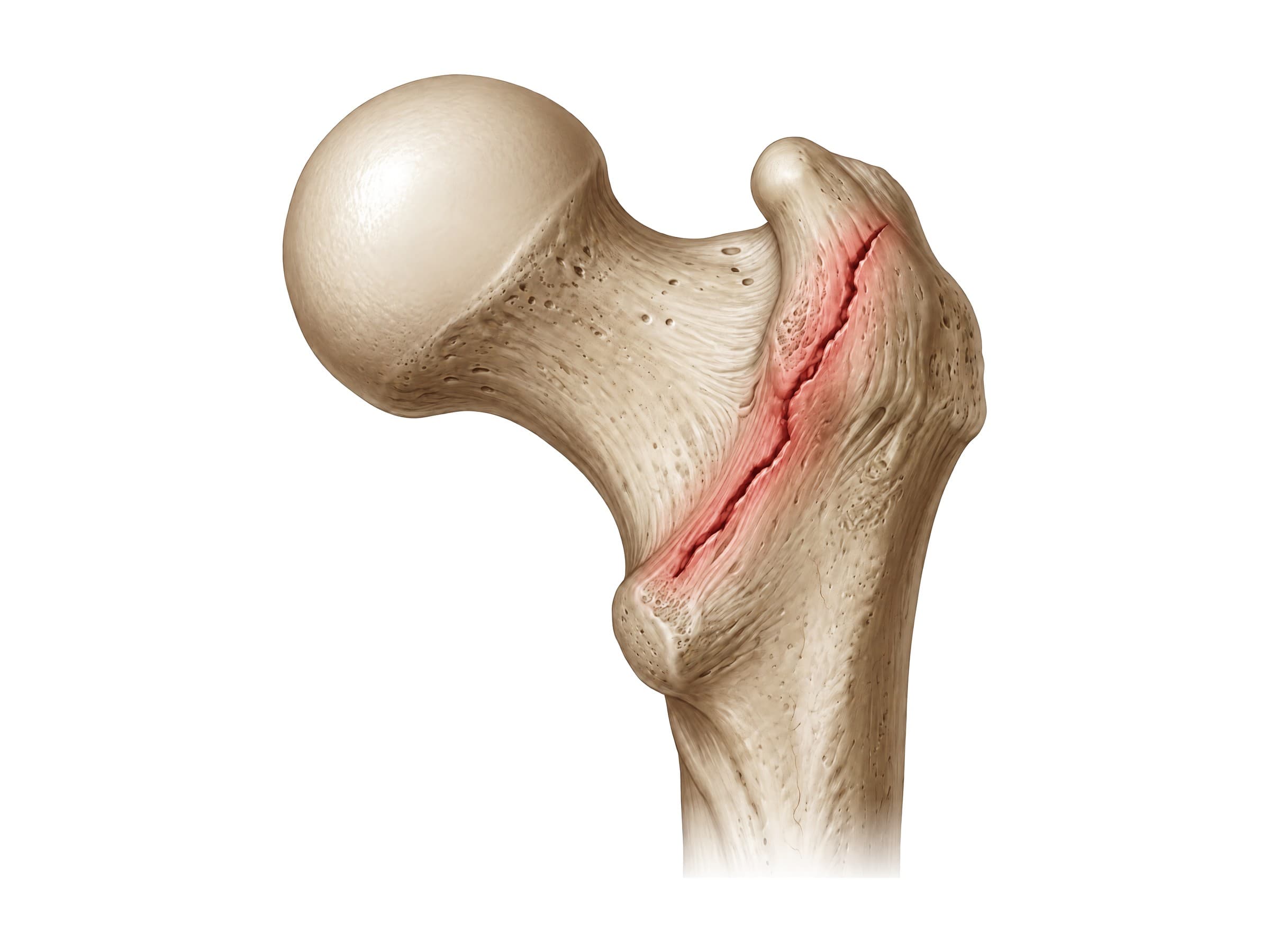
- Intertrochanteric fracture: a break between the greater and lesser trochanters, outside the capsule. The blood supply is good, so the bone usually heals once it is fixed with a sliding hip screw and plate or an intramedullary (cephalomedullary) nail. Hip fractures of either type are serious in older adults: roughly 20 to 30 percent of older patients die within a year of the injury.
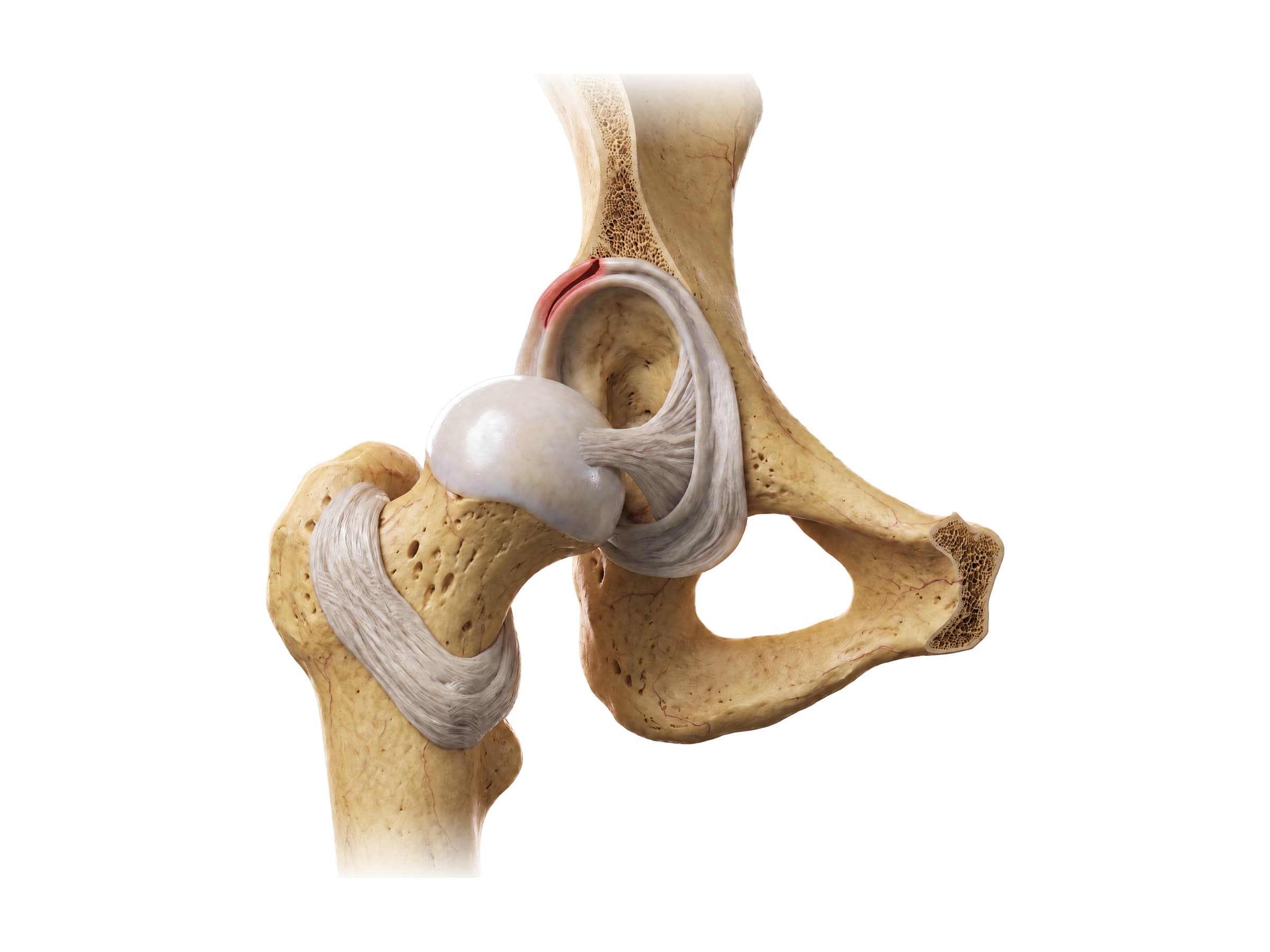
- Hip labral tear: a tear of the labrum, causing deep groin pain, clicking or catching. Tears follow falls, car crashes, dislocations and twisting sports injuries, or develop from femoroacetabular impingement (FAI). Best seen on an MR arthrogram.
- Posterior hip dislocation: the femoral head is forced out of the back of the socket, classically when the knee strikes the dashboard in a car crash and drives the femur backward (a mechanism of injury known as a dashboard injury). About 90 percent of hip dislocations are posterior. The leg looks shortened and turned inward. It is an emergency: the hip should be put back in place quickly, ideally within about 6 hours, to reduce the risk of avascular necrosis, and the sciatic nerve may be injured. Often combined with a fracture of the back wall of the acetabulum.
- Acetabular fracture: a break of the socket, usually from a high-energy impact such as a car crash or a fall from height. Evaluated with CT, often with 3D reconstructions.
- Pelvic fracture: a break of the pelvic ring, ranging from a stable pubic ramus fracture after a fall to unstable fractures with heavy bleeding in high-energy trauma.
- Hip flexor and adductor strains: tears of the iliopsoas, rectus femoris or adductor muscles in sprinting, kicking and sudden changes of direction. Seen on MRI or ultrasound.
- Greater trochanteric pain syndrome: pain over the outer hip from gluteal tendinopathy or trochanteric bursitis. Ultrasound or MRI shows thickened or torn tendons and fluid in the bursa.
- Hip osteoarthritis: wear of the joint cartilage causing groin pain and stiffness. X-rays show a narrowed joint space, bone spurs and cysts. Post-traumatic arthritis can develop years after an acetabular fracture, dislocation or labral injury.
How the hip is shown in a personal injury case
Hip injuries appear in slip-and-fall claims, nursing home and premises liability cases involving older adults, and motor vehicle collisions, where a dashboard injury can dislocate the hip or fracture the acetabulum. A labeled illustration of the normal hip, showing the femoral head in the acetabulum, the labrum and the femoral neck with its blood supply, establishes the anatomy before the injury exhibit is introduced: the fracture line, the dislocated femoral head, or the screws, nail or prosthesis placed at surgery. Illustrations are especially useful for explaining why a femoral neck fracture led to a hip replacement (the blood supply was cut off), why a dislocated hip developed avascular necrosis months later, and why a labral tear after a crash, rather than pre-existing arthritis, explains a plaintiff's pain. A side-by-side exhibit pairing the normal hip with the patient's own X-rays or MRI makes these points concrete for a jury.
Natomy creates custom medical legal illustrations of hip injuries for demand packages and trial exhibits. For more on how normal anatomy is used in court, see demonstrative evidence. Related atlas pages: knee anatomy, ankle anatomy, foot bones, back muscles anatomy, shoulder anatomy, elbow anatomy, hand and wrist anatomy and skull anatomy.
FAQ
What bones make up the hip joint?+
The hip joint is formed by two bones: the femur (thighbone), whose rounded head is the ball, and the hip bone, whose cup-shaped acetabulum is the socket. Each hip bone (also called the innominate or pelvic bone) is itself made of three bones, the ilium, ischium and pubis, which fuse together at the acetabulum during the teenage years. The two hip bones and the sacrum together form the pelvis.
What is the acetabulum?+
The acetabulum is the deep, cup-shaped socket on the outer side of the hip bone that holds the head of the femur. All three bones of the hip, the ilium, ischium and pubis, contribute to it. Only its horseshoe-shaped outer part (the lunate surface) is covered with cartilage and bears weight; the center is a fat-filled hollow where the ligamentum teres attaches. A ring of fibrocartilage, the acetabular labrum, runs around its rim and deepens the socket.
What part of my hip is hurting?+
The location of hip pain is a rough guide to its source. Pain in the groin or the front of the hip usually comes from the hip joint itself, such as arthritis, a labral tear or a hip fracture, or from the hip flexors. Pain on the outer side of the hip, over the bony point of the greater trochanter, usually comes from the gluteal tendons or the trochanteric bursa. Pain in the buttock is often referred from the lower back or the sacroiliac joint, or comes from the piriformis and the sciatic nerve. Persistent hip pain should be examined by a doctor.
What are the hip flexor muscles?+
The hip flexors are the muscles that lift the thigh toward the trunk. The strongest is the iliopsoas, formed by the psoas major, which starts on the lumbar spine, and the iliacus, which lines the inside of the pelvis; they join and attach to the lesser trochanter of the femur. The rectus femoris (one of the quadriceps), sartorius, tensor fasciae latae and pectineus also flex the hip.
What is the hip bursa?+
A bursa is a small fluid-filled sac that lets tendons glide over bone. The hip has several, but the one people usually mean is the trochanteric bursa, which sits over the greater trochanter on the outer side of the hip, under the iliotibial band. Pain there, often called trochanteric bursitis, is now usually described as greater trochanteric pain syndrome because it most often comes from the gluteus medius and minimus tendons rather than the bursa itself. The iliopsoas bursa at the front of the hip and the ischial bursa under the sitting bone can also become inflamed.
What is the strongest ligament in the hip?+
The iliofemoral ligament, also called the Y-ligament of Bigelow, is the strongest ligament in the hip and one of the strongest in the body. It runs from the anterior inferior iliac spine to the intertrochanteric line on the front of the femur and stops the hip from bending too far backward, which lets a person stand upright with little muscle effort. The pubofemoral ligament below it and the ischiofemoral ligament behind complete the capsule.
What is the difference between a femoral neck fracture and an intertrochanteric fracture?+
Both are hip fractures of the upper femur. A femoral neck fracture breaks the narrow neck just below the femoral head, inside the joint capsule, where it can tear the vessels that supply the head; displaced neck fractures are usually treated with a partial or total hip replacement. An intertrochanteric fracture runs between the greater and lesser trochanters, outside the capsule, where the blood supply is good, so it is usually fixed with a sliding hip screw and plate or an intramedullary nail.
What is a hip labral tear?+
A hip labral tear is a tear of the acetabular labrum, the ring of fibrocartilage around the rim of the hip socket. It causes groin pain, clicking, catching or locking, especially when bending or twisting the hip. Tears can follow a single injury, such as a fall, a car crash or a dislocation, or develop over time from femoroacetabular impingement, where an irregular femoral head or socket rim pinches the labrum. They are best seen on an MR arthrogram and are treated with physical therapy or hip arthroscopy.
Need a custom illustration of this joint?
Generate a labeled or exhibit-ready figure for a specific patient, injury, or case in minutes.
Try Natomy →