Foraminal Stenosis: Causes, Symptoms & Treatment
Foraminal stenosis is narrowing of the opening where a nerve root leaves the spine, often at L4-L5 or L5-S1. See it illustrated beside normal anatomy.
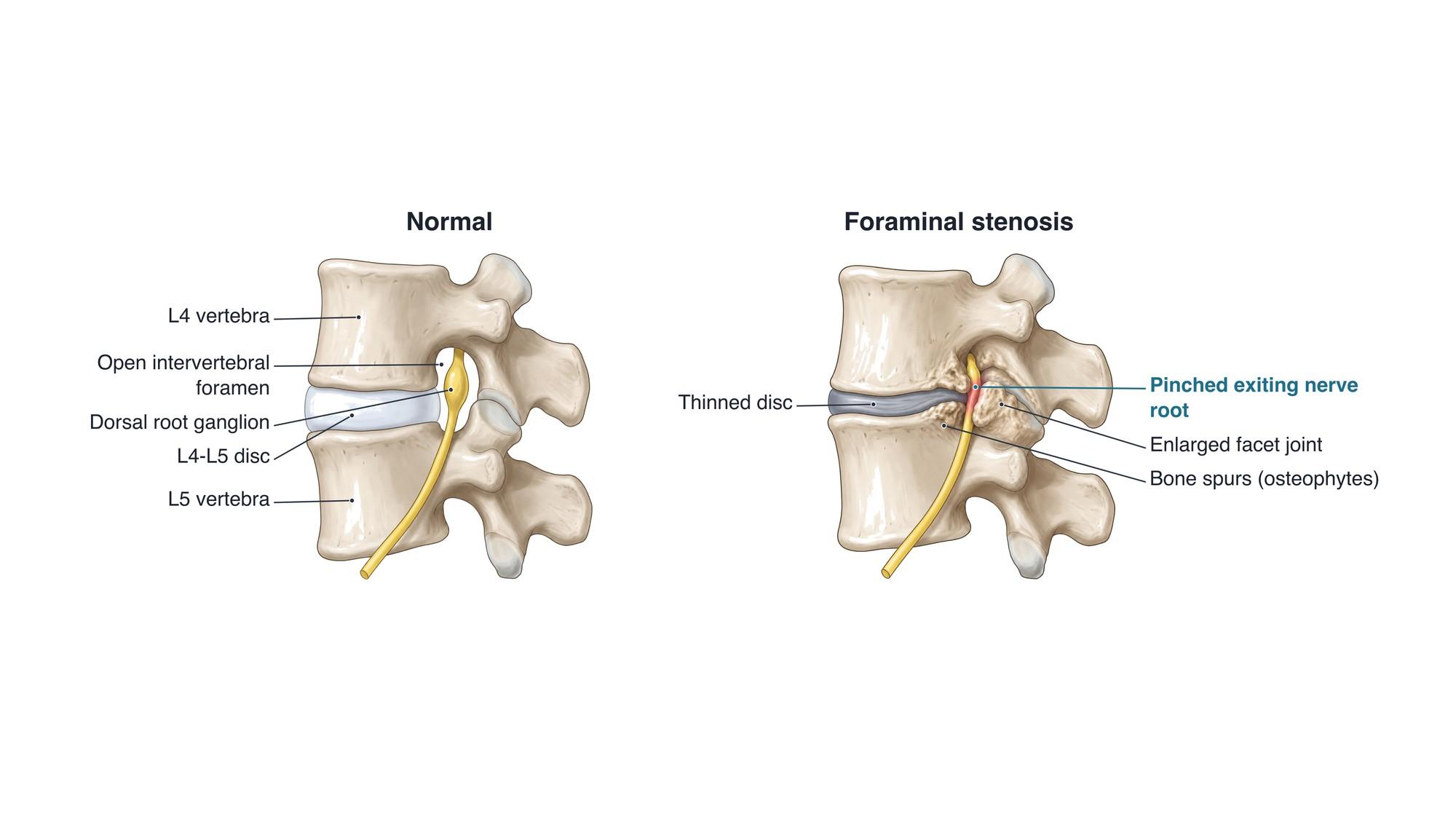
Foraminal stenosis is narrowing of the intervertebral foramen, the bony opening on each side of the spine where a nerve root leaves the spinal canal. When the foramen narrows, it can pinch the exiting nerve root. This causes pain, numbness, tingling or weakness along that nerve (radiculopathy). It is most common in the lower back at L4-L5 and L5-S1, and in the neck at C5-C6 and C6-C7. The illustration above shows a normal L4-L5 foramen beside one narrowed by a thinned disc, bone spurs and an enlarged facet joint.
The anatomy
Each pair of vertebrae forms two foramina, one on the left and one on the right. Each foramen is bounded by the pedicle of the vertebra above and the pedicle below. In front lie the back of the vertebral body and the intervertebral disc. Behind lies the facet joint. In the lower back, the nerve root that exits at a level is named for the vertebra above it. So the L4 root leaves through the L4-L5 foramen. Its dorsal root ganglion, a swelling that holds the sensory nerve cells, sits inside the foramen and is very sensitive to pressure. For the full anatomy, see lumbar spine anatomy.
Grades
Radiologists often grade foraminal narrowing on MRI:
| Grade | What the MRI shows |
|---|---|
| Normal | Fat surrounds the nerve root on all sides |
| Mild | Fat is reduced on one side (top and bottom, or front and back) |
| Moderate | Fat is reduced on all sides, but the root keeps its shape |
| Severe | No fat around the root, and the root is flattened or deformed |
The grade on the report does not always match the symptoms. Some people with severe narrowing have no pain, and some with moderate narrowing have a lot.
Causes
- Disc degeneration: the disc loses height, so the vertebra above sinks and the foramen gets shorter. The back of the disc can also bulge into the foramen.
- Bone spurs (osteophytes): extra bone that grows at the edges of the vertebral bodies and the facet joints as the spine wears.
- Facet joint arthritis (facet arthropathy): the facet joint enlarges and pushes forward into the back of the foramen.
- Herniated disc: a foraminal or far lateral herniation sits right in the opening. See lumbar disc herniation.
- Spondylolisthesis: a vertebra slipping forward narrows the foramen, especially with a pars defect. See spondylolisthesis.
- Thickened ligaments, cysts and scoliosis: less common contributors.
Symptoms
- Pain that travels down one leg (sciatica) or one arm, following a single nerve
- Numbness, tingling or a burning feeling in the same area
- Weakness of the muscles that nerve controls, such as foot drop at L5 or a weak grip at C7
- Back or neck pain, often milder than the limb pain
- Symptoms worse with standing, walking, arching back or tilting toward the painful side
- Relief when sitting, bending forward or lying with the knees bent
Diagnosis
The doctor tests strength, reflexes and sensation to find which nerve root is involved. Tests that stretch or close the foramen, such as the straight leg raise in the low back or the Spurling test in the neck, may reproduce the pain. X-rays show disc height, spurs and any slip. MRI shows the foramen best. It shows whether fat still surrounds the nerve root, or whether the root is flattened. CT shows bone spurs in more detail. Nerve conduction studies and EMG can confirm which nerve is affected. A selective nerve root block can confirm the pain source before surgery.
Treatment
- Activity changes: avoiding long periods of back arching and heavy lifting during flare-ups
- Physical therapy: flexion exercises, core strength, nerve gliding and posture work
- Medicines: anti-inflammatory drugs and, for nerve pain, medicines such as gabapentin
- Epidural or transforaminal steroid injection: medicine placed next to the inflamed nerve root
- Surgery: a foraminotomy widens the foramen by removing bone spurs, part of the facet and disc material. It is often done through a small incision. When the level is unstable or there is a slip, the surgeon may add a fusion. For a disc fragment in the foramen, a microdiscectomy removes it.
Recovery
Many flare-ups ease within 6 to 12 weeks of therapy and medicine. After a foraminotomy, most people walk the same day and go home within a day. Desk work often resumes in about 2 to 4 weeks. Leg pain often improves quickly, while numbness and weakness can take months to recover.
Complications
- Chronic nerve root pain and numbness
- Lasting muscle weakness when compression goes on too long
- Narrowing at more than one level, or narrowing of the central canal as well
- After surgery: recurrent narrowing, nerve irritation, dural tear, and instability if too much facet is removed
How foraminal stenosis is shown in a personal injury case
Foraminal stenosis is common in MRI reports after car crashes, falls and work injuries. The legal question is often whether the narrowing came from the injury or was already present from age-related wear. Bone spurs and facet enlargement take years to form, while a new disc herniation or a new slip can appear suddenly. Earlier imaging, the timing of symptoms, and a nerve exam that matches the narrowed level all matter. A side view illustration like the one above shows a jury what the opening normally looks like and how the nerve is pinched. For how these images are used at trial, see demonstrative evidence and medical legal illustration.
Related pages
- Lumbar spine anatomyThe normal anatomy of the lumbar spine.
- AnterolisthesisAnterolisthesis is a vertebra slipped forward over the one below, most often L4 on L5. See it illustrated beside normal anatomy.
- Facet arthropathyFacet arthropathy is arthritis of the small joints at the back of the spine, most often at L4-L5 and L5-S1. See it illustrated beside normal facet joints.
- Lumbar disc herniationA herniated disc is disc gel pushed through a tear in its outer ring, often pressing on a nerve root. See protrusion, extrusion and sequestration illustrated.
- SpondylolisthesisSpondylolisthesis is a vertebra that has slipped forward over the one below, often L5 on S1 after a pars fracture. See it illustrated beside normal anatomy.
FAQ
Is foraminal stenosis serious?+
Mild or moderate foraminal stenosis is common with age and often causes no symptoms. It matters when it pinches the nerve root enough to cause leg or arm pain, numbness or weakness. Severe narrowing with muscle weakness that keeps getting worse needs prompt evaluation.
What are the symptoms of foraminal stenosis?+
Pain that runs along one nerve, from the back into the buttock and leg, or from the neck into the arm. It often comes with numbness, tingling or weakness in the same area. Symptoms are usually on one side and are often worse when standing, walking or bending backward.
What are the symptoms of C5-C6 foraminal stenosis?+
Narrowing at C5-C6 pinches the C6 nerve root. It can cause pain in the neck, shoulder and outer forearm, numbness of the thumb and index finger, and weakness of the biceps and wrist extensors. Turning or tilting the head toward the painful side often makes it worse.
What exercises help foraminal stenosis?+
Flexion-based exercises that open the foramen, such as knee-to-chest stretches, often help. Core and hip strengthening, walking or cycling, and nerve gliding exercises are common in physical therapy. A therapist will tailor the plan and may limit exercises that arch the back.
What should you not do with foraminal stenosis?+
Movements that close down the foramen often aggravate it: arching the back for long periods, heavy overhead lifting, and repeated twisting toward the painful side. In the neck, looking up for long periods or tilting the head toward the painful side can do the same.
What is the treatment for severe foraminal stenosis at L5-S1?+
Treatment starts with physical therapy, medicine and often an epidural steroid injection. If pain or weakness persists, a surgeon can widen the foramen (foraminotomy) to free the L5 nerve root. Fusion is added when the level is unstable or the slip needs to be held.
What is the best sleeping position for spinal stenosis?+
Many people are most comfortable on their side with the knees slightly bent and a pillow between them, or on their back with a pillow under the knees. Both positions flex the spine a little, which opens the foramen.
Can foraminal stenosis be caused by an injury?+
Most foraminal stenosis develops slowly from wear. An injury can add a herniated disc or a vertebral slip that narrows the foramen further, or make a narrowed foramen start to cause symptoms. Imaging over time helps separate new changes from old ones.
Need this illustration for a patient or a case?
Describe the injury or upload a scan and get a clear, labeled medical illustration for a consult, a chart or a case exhibit.
Try Natomy →