Ankle Anatomy: Bones, Ligaments & Tendons
Labeled illustrations of the ankle bones, ankle mortise, lateral and deltoid ligaments, tendons and common ankle sprains and fractures.
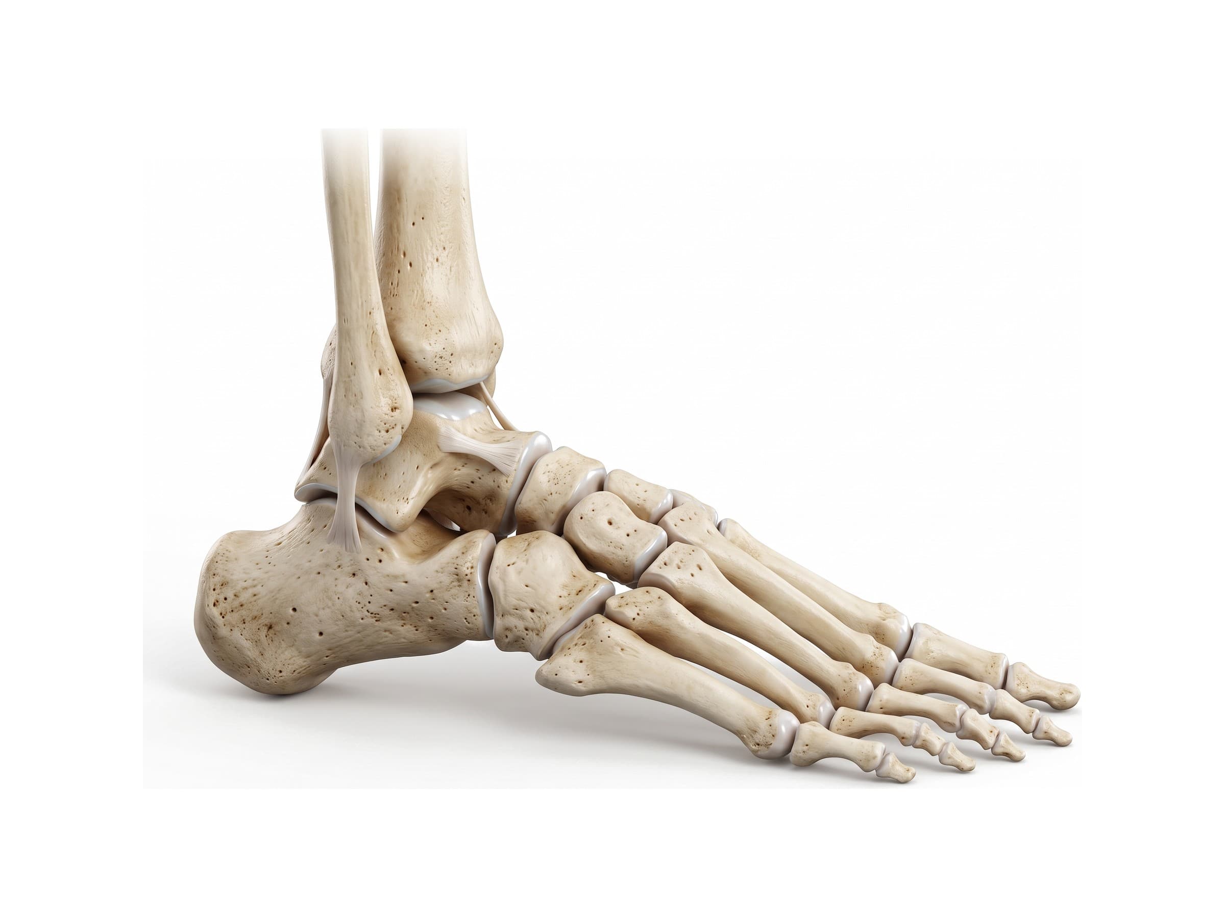
The ankle is the joint where the leg meets the foot. Ankle anatomy comes down to three bones (the tibia, fibula and talus) that lock together in a hinge called the ankle mortise, the calcaneus below them, three groups of ligaments that hold the joint together, and the tendons that wrap around the ankle bones on their way to the foot. It carries the full weight of the body with every step, which is why ankle sprains and fractures are among the most common injuries treated in emergency departments, and why the anatomy matters for reading an ankle X-ray report, explaining a diagnosis to a patient, or illustrating a personal injury claim.
Bones of the ankle
The ankle joint, also called the talocrural joint, is made of three bones. In the view of the outer side of the ankle above:
- Tibia: the shinbone, the main weight-bearing bone of the leg. Its flat lower surface (the plafond) rests on top of the talus, and its inner edge extends downward as the medial malleolus.
- Fibula: the thin bone on the outer side of the leg. It carries little weight but forms the outer wall of the ankle joint, ending in the lateral malleolus.
- Talus: the top bone of the foot, sitting between the malleoli. Its rounded upper surface, the trochlea or talar dome, is covered with cartilage. The talus has no muscles attached to it, and its blood supply enters through a few small vessels, so a talar neck fracture can cut off blood to the bone (avascular necrosis).
Below the talus is the calcaneus, the heel bone and the largest bone of the foot. In front of the talus are the navicular and, on the outer side, the cuboid, which lead to the five metatarsals and the phalanges of the toes. The talus, calcaneus, navicular, cuboid and three cuneiform bones together make up the seven tarsal bones, the bones most people mean when they search for "ankle bones". All 26 bones of the foot are labeled on the foot bones page.
Medial and lateral malleolus
The two bumps you can feel on either side of the ankle are the malleoli.
- Medial malleolus: the inner ankle bone, the lower end of the tibia. The deltoid ligament attaches to it.
- Lateral malleolus: the outer ankle bone, the lower end of the fibula. It sits lower and further back than the medial malleolus, which is one reason the ankle turns inward (inversion) more easily than outward: the outer wall extends further down and blocks outward movement.
- Posterior malleolus: not a separate bump but the back edge of the lower tibia. It matters mainly in fractures, where it can break off together with the other two.
The ankle joint and the ankle mortise
Seen in a coronal section, cut from side to side through the ankle, the tibia and the two malleoli form an upside-down U that wraps around the talar dome. This socket is the ankle mortise. It works as a hinge, allowing the foot to move up (dorsiflexion) and down (plantarflexion). Because the front of the talar dome is wider than the back, the mortise grips the talus most tightly when the foot is pulled up and is loosest when the foot is pointed, which is the position in which most ankle sprains happen.
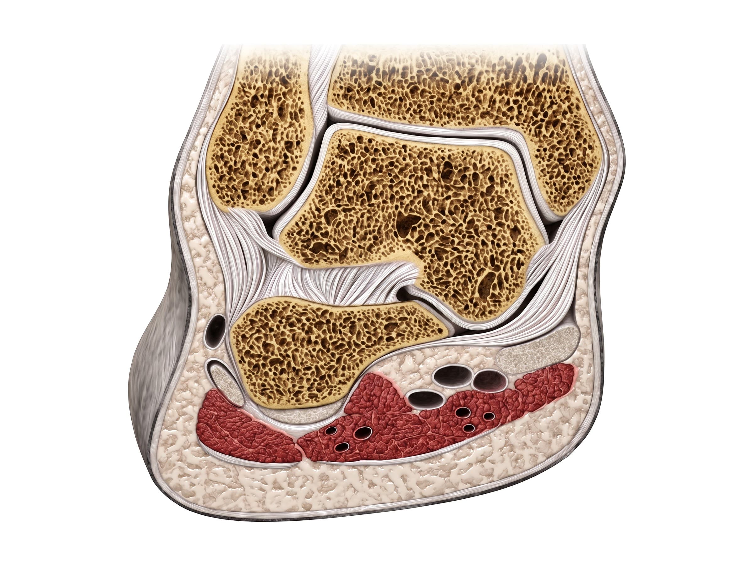
The ankle region contains three joints:
- Talocrural joint: the true ankle joint, between the tibia, fibula and talus. It handles up-and-down movement.
- Subtalar joint: between the talus and the calcaneus, below the ankle joint. It tilts the heel inward and outward (inversion and eversion), which lets the foot adapt to uneven ground. The interosseous talocalcaneal ligament sits in the middle of this joint, in a channel called the sinus tarsi.
- Distal tibiofibular joint (syndesmosis): where the lower ends of the tibia and fibula are bound together by strong ligaments just above the ankle joint. It barely moves, but it keeps the mortise the right width.
Ankle ligaments
The ankle is held together by three groups of ligaments: the lateral ligaments on the outer side, the deltoid ligament on the inner side, and the syndesmotic ligaments between the tibia and fibula.
Lateral ankle ligaments
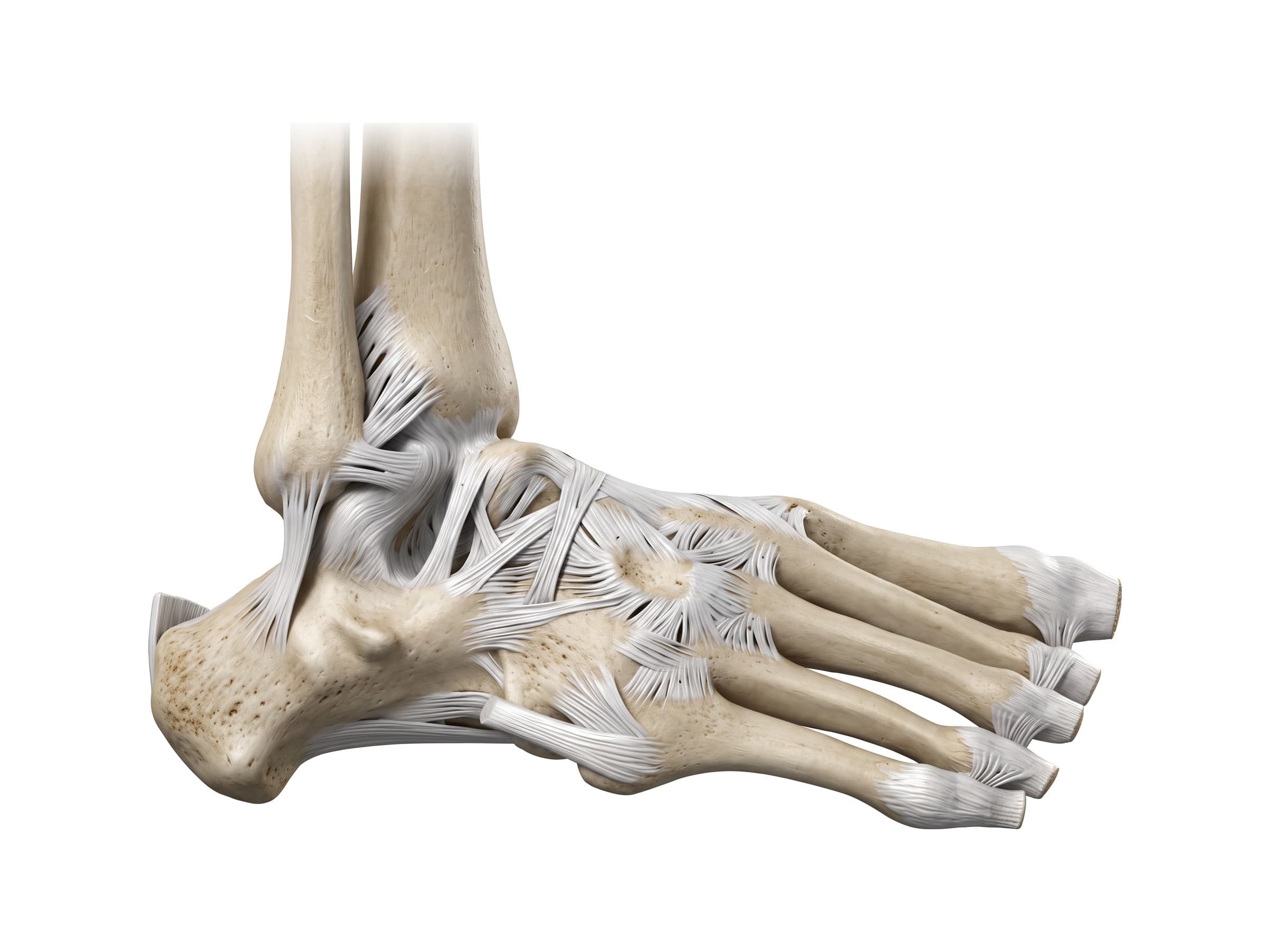
Three ligaments run from the lateral malleolus to the bones of the foot:
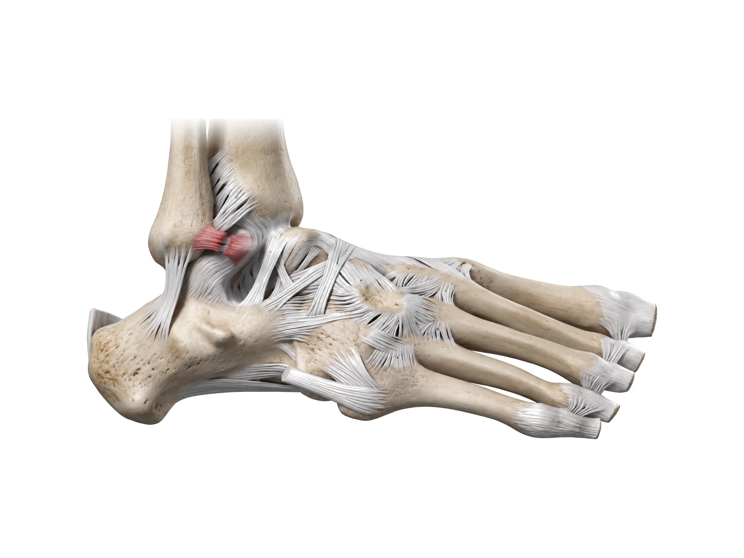
- Anterior talofibular ligament (ATFL): a short, flat band from the front of the lateral malleolus forward to the neck of the talus. It is the weakest of the three and is tight when the foot is pointed, so it is the ligament torn in most ankle sprains.
- Calcaneofibular ligament (CFL): a cord-like ligament from the tip of the lateral malleolus down and back to the outer side of the calcaneus. It crosses both the ankle and subtalar joints and is the second ligament to tear in a severe sprain.
- Posterior talofibular ligament (PTFL): the strongest of the three, running from the inner back surface of the lateral malleolus straight back to the talus. It is torn only in severe injuries such as dislocations.
Deltoid ligament
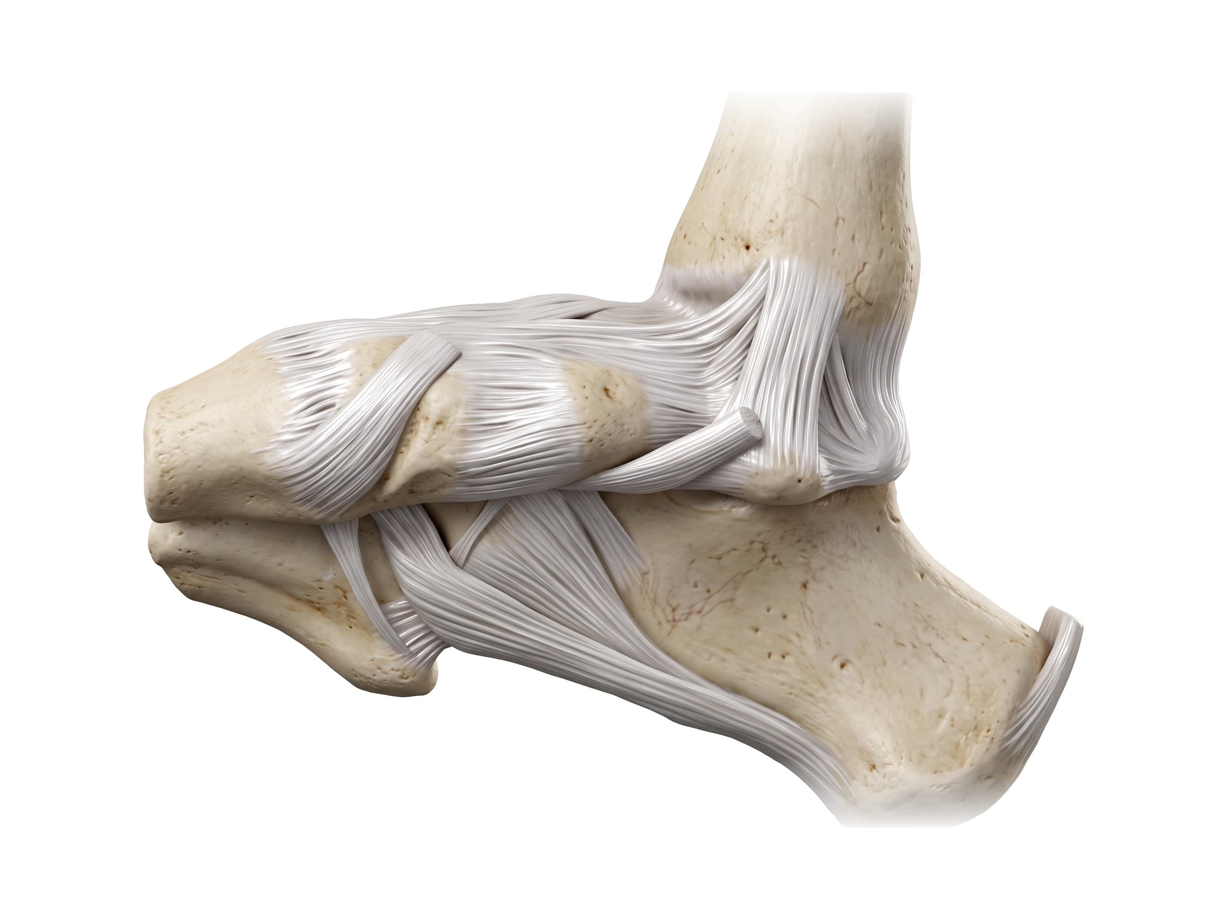
On the inner side, the deltoid ligament (also called the medial collateral ligament of the ankle) fans out from the medial malleolus in a triangle, which is where it gets its name. It has a superficial layer, whose fibers run to the navicular, the spring ligament and the sustentaculum tali of the calcaneus, and a deep layer that runs short and thick straight to the talus. The deltoid ligament is much stronger than the lateral ligaments and is rarely torn on its own; when it is injured, it is usually in combination with a fracture of the fibula.
Below it, the spring ligament (plantar calcaneonavicular ligament) runs from the calcaneus to the navicular and supports the head of the talus, holding up the inner arch of the foot. The long plantar ligament runs along the sole from the calcaneus to the cuboid and the bases of the metatarsals.
Syndesmosis and posterior ligaments
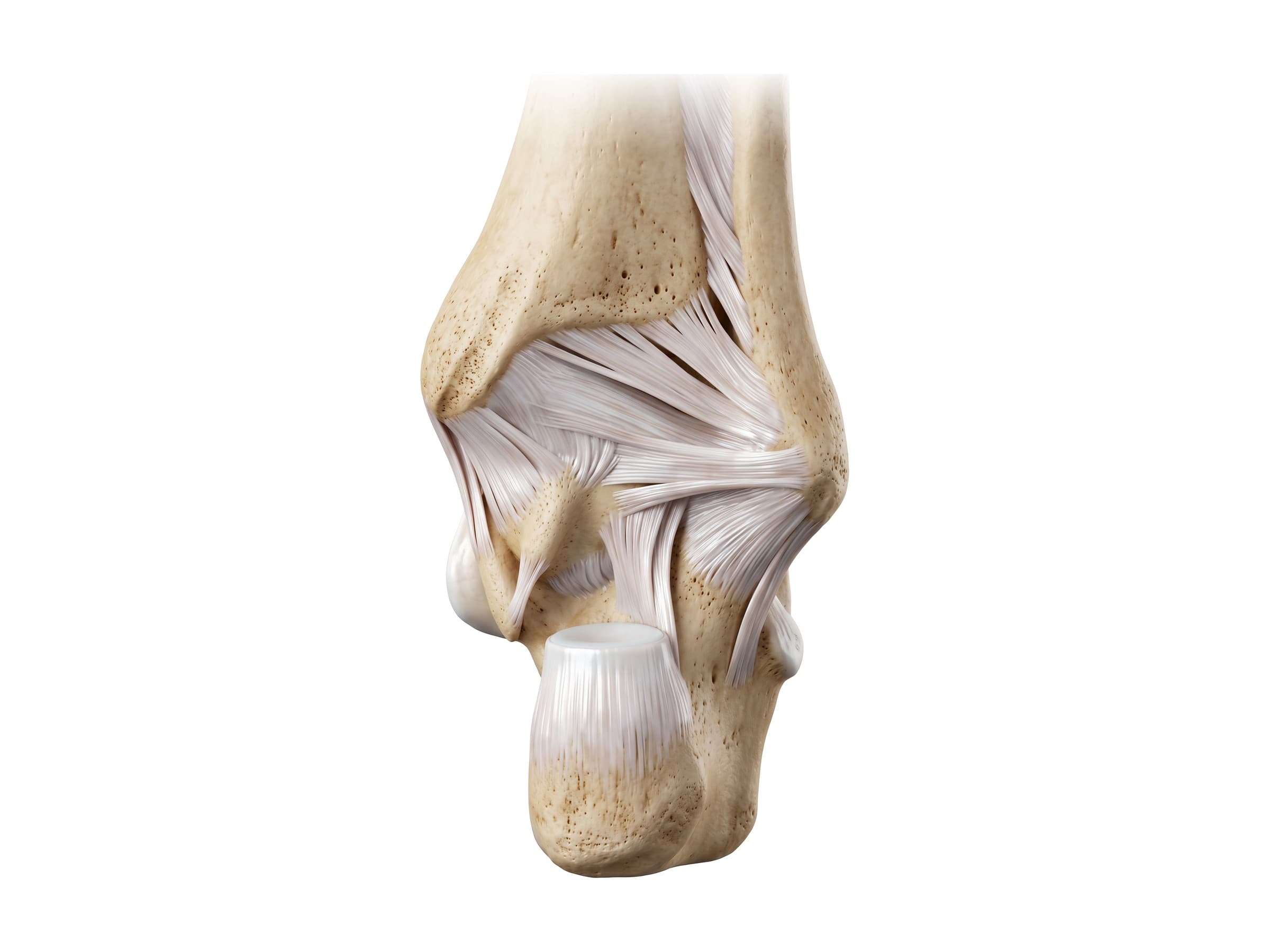
The lower tibia and fibula are held together by the anterior inferior tibiofibular ligament (AITFL) in front, the posterior inferior tibiofibular ligament (PITFL) behind, the interosseous ligament between them, and the deep transverse tibiofibular ligament. Together these are the syndesmosis ligaments. They are torn in a high ankle sprain, usually when the foot is forced to twist outward, and they are often injured together with fractures of the fibula above the ankle joint.
Tendons and muscles of the ankle
No muscles start or end at the ankle itself. The muscles that move the ankle are in the lower leg, and their tendons cross the joint in four groups, held in place by bands of thickened tissue called retinacula.
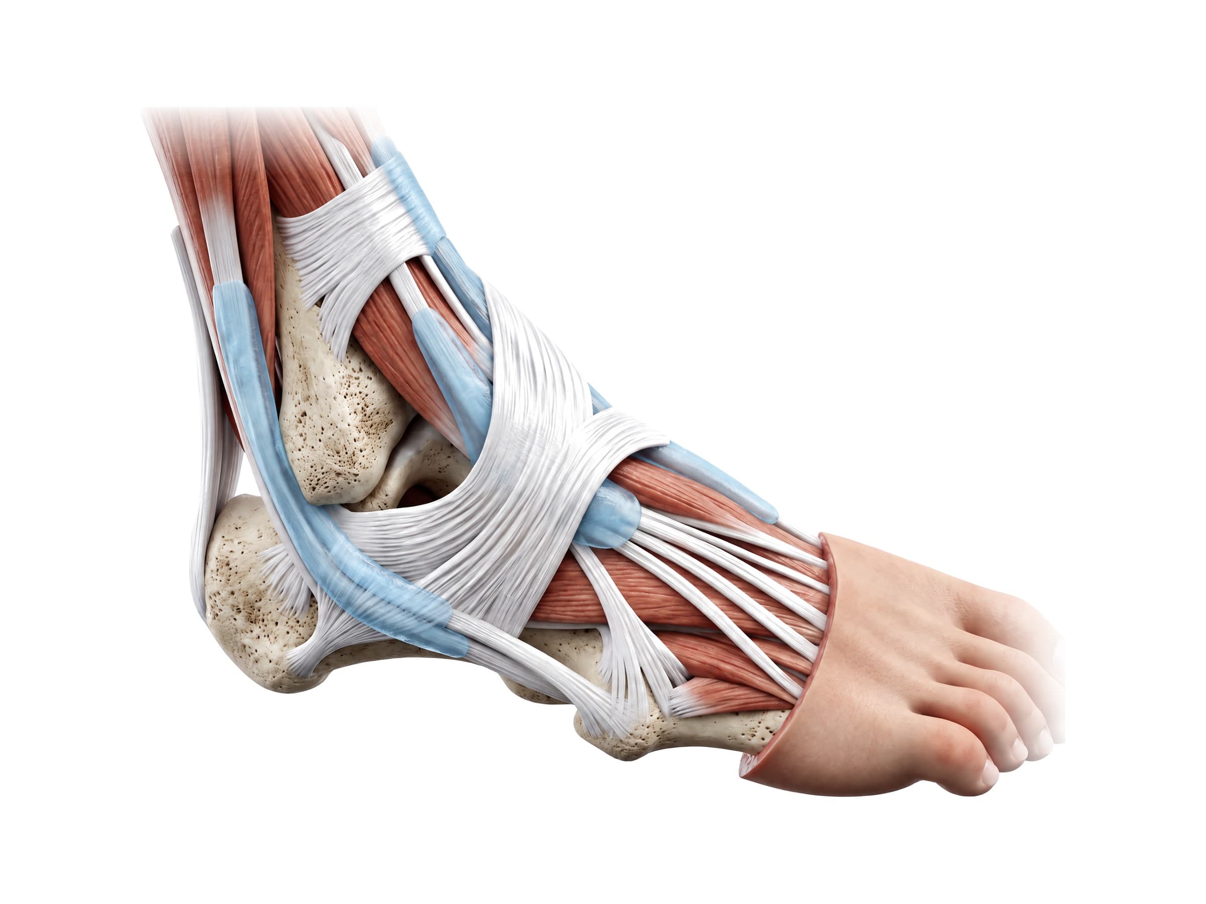
- Front (anterior) group: the tibialis anterior, extensor hallucis longus and extensor digitorum longus run under the extensor retinaculum across the front of the ankle. They lift the foot (dorsiflexion) and the toes. Weakness in this group causes foot drop.
- Outer (lateral) group: the peroneus longus and peroneus brevis (also called the fibularis longus and brevis) run in a groove behind the lateral malleolus, held down by the superior peroneal retinaculum. They turn the foot outward (eversion) and protect against sprains. The brevis attaches to the base of the fifth metatarsal; the longus crosses under the sole to the base of the first metatarsal. Peroneal tendon tears and dislocations often follow ankle sprains.
- Back (posterior) group: the Achilles tendon (calcaneal tendon), formed by the gastrocnemius and soleus muscles of the calf, attaches to the back of the calcaneus. It is the thickest and strongest tendon in the body and points the foot (plantarflexion) for walking, running and jumping.
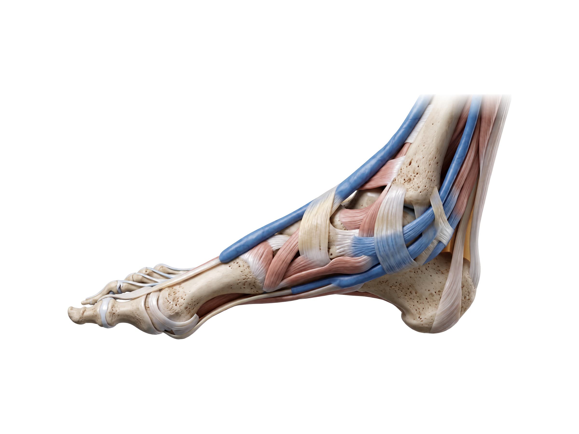
- Inner (medial) group: the tibialis posterior, flexor digitorum longus and flexor hallucis longus pass behind the medial malleolus. The tibialis posterior is the main support of the inner arch of the foot; when it fails, the arch collapses (adult-acquired flatfoot).
The tarsal tunnel
Behind the medial malleolus, the three medial tendons travel with the posterior tibial artery, its veins and the tibial nerve through the tarsal tunnel, a channel roofed by the flexor retinaculum. From front to back, the contents are the tibialis posterior, flexor digitorum longus, artery and veins, nerve, and flexor hallucis longus, remembered as "Tom, Dick And Very Nervous Harry". Compression of the tibial nerve in this tunnel causes tarsal tunnel syndrome, with burning and numbness in the sole of the foot.
Nerves and blood supply of the ankle
Five nerves cross the ankle, all branches of the sciatic and femoral nerves:
- Tibial nerve: through the tarsal tunnel to the sole of the foot, where it divides into the medial and lateral plantar nerves.
- Deep peroneal (fibular) nerve: across the front of the ankle with the anterior tibial artery, supplying the extensor muscles and a small patch of skin between the first and second toes.
- Superficial peroneal nerve: over the front and outer side of the ankle, supplying skin on the top of the foot. It is at risk during ankle surgery and arthroscopy.
- Sural nerve: behind the lateral malleolus, supplying the outer edge of the foot.
- Saphenous nerve: in front of the medial malleolus, supplying skin on the inner side of the ankle.
Blood reaches the ankle and foot through three arteries: the anterior tibial artery, which becomes the dorsalis pedis artery on top of the foot (the pulse felt there), the posterior tibial artery behind the medial malleolus (the pulse felt there), and the peroneal (fibular) artery on the outer side. Checking these two pulses is part of every ankle trauma exam.
Movements of the ankle
- Dorsiflexion: pulling the foot up toward the shin, about 20 degrees, by the tibialis anterior and the toe extensors.
- Plantarflexion: pointing the foot down, about 50 degrees, by the calf muscles through the Achilles tendon, helped by the tibialis posterior, the long toe flexors and the peroneal muscles.
- Inversion: tilting the sole inward, mostly at the subtalar joint, by the tibialis posterior and tibialis anterior. Forced inversion is the mechanism of the common ankle sprain.
- Eversion: tilting the sole outward, by the peroneus longus and brevis.
Common ankle injuries and how they appear on imaging
- Lateral ankle sprain: the foot rolls inward (inversion), tearing the ATFL and, in more severe cases, the CFL. It is the single most common sports injury. Sprains are graded from I (stretched) to III (completely torn). X-rays are normal or show soft-tissue swelling; MRI or ultrasound shows the torn ligament. Repeated sprains can leave the ankle chronically unstable.
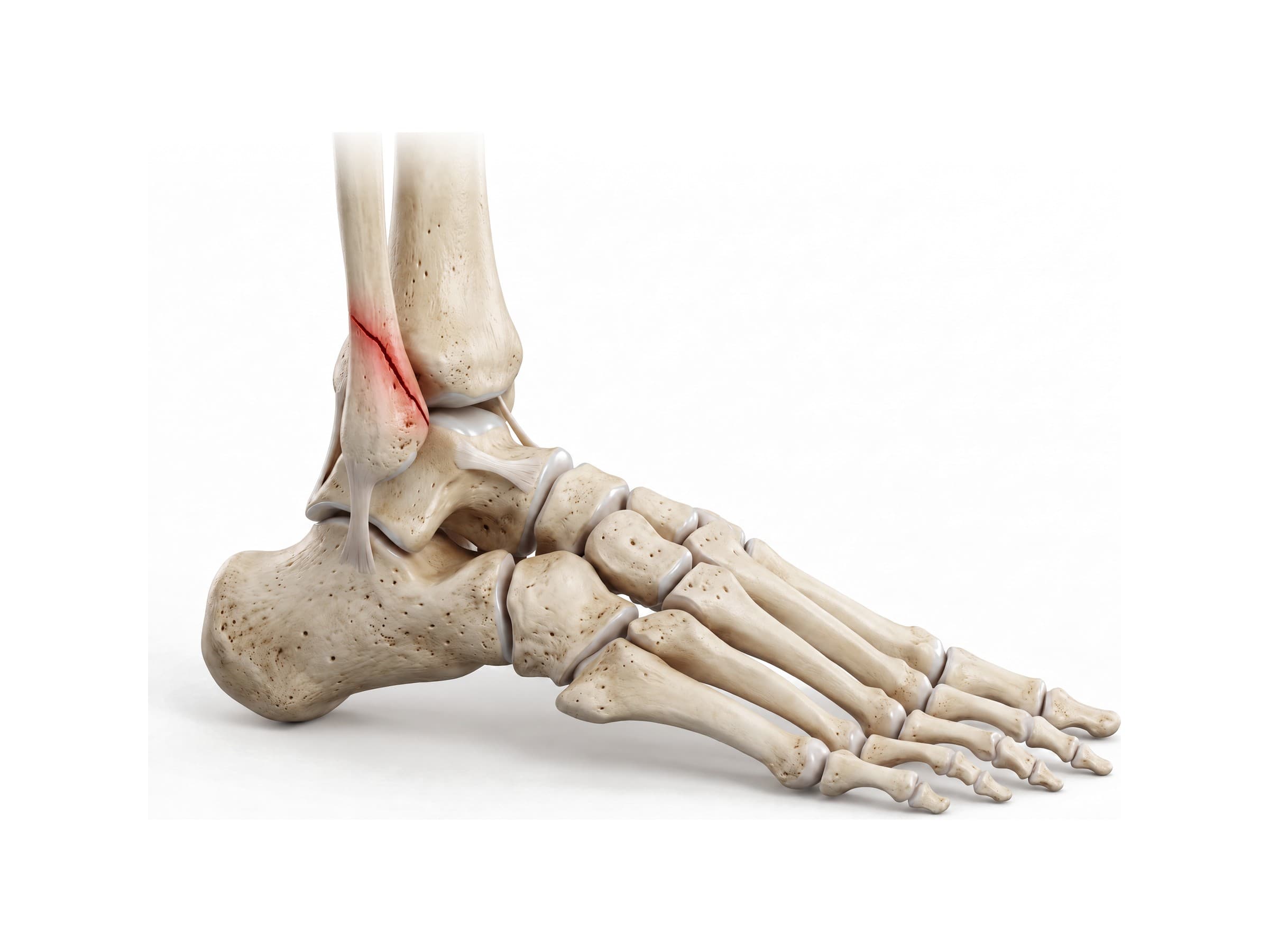
- Lateral malleolus fracture: the most common ankle fracture, a break of the lower fibula. Fibula fractures are described with the Weber classification by their level relative to the syndesmosis: Weber A below the joint line, Weber B at the level of the joint (the most common), and Weber C above it, where the syndesmosis is usually torn too. Seen on the standard three ankle X-ray views.
- Bimalleolar and trimalleolar fractures: both malleoli are broken (bimalleolar), or both malleoli plus the posterior malleolus (trimalleolar). These fractures make the mortise unstable and usually need open reduction and internal fixation (ORIF), with a plate on the fibula and screws across the medial and posterior malleoli. CT is often added to plan fixation of a posterior malleolus fragment.
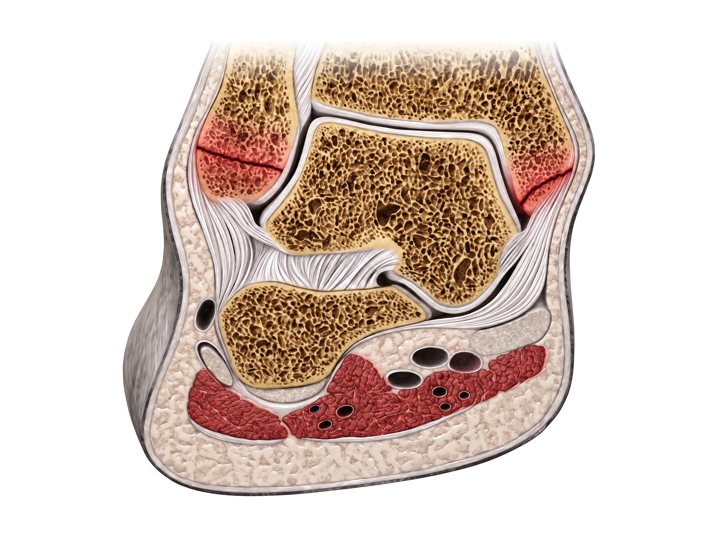
- High ankle sprain (syndesmosis injury): a tear of the tibiofibular ligaments from an outward twist. On X-ray or CT, the gap between the tibia and fibula widens; stress views or MRI confirm it.
- Deltoid ligament injury: usually combined with a fibula fracture. On the mortise view, the space between the medial malleolus and the talus (the medial clear space) widens beyond about 4 mm.
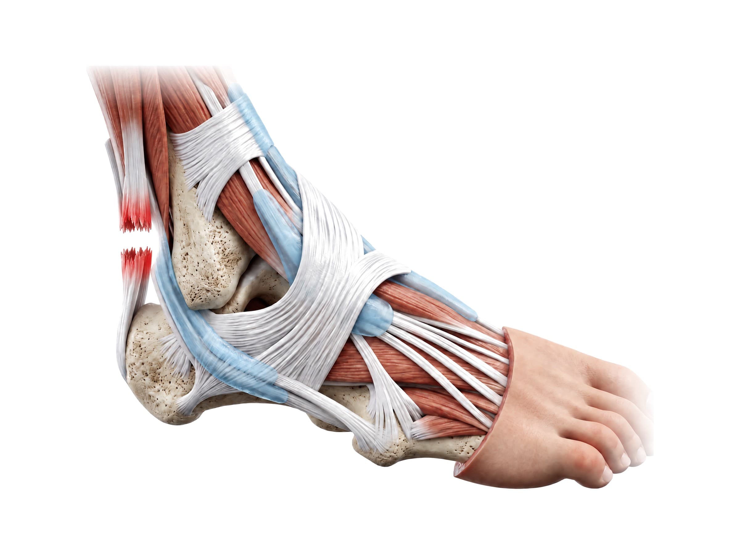
- Achilles tendon rupture: a sudden pop at the back of the ankle during a push-off, often described as feeling kicked in the heel. Most ruptures happen 2 to 6 cm above the heel, where the tendon's blood supply is poorest. Diagnosed with the Thompson test (squeezing the calf does not point the foot) and confirmed with ultrasound or MRI.
- Osteochondral lesion of the talus: damage to the cartilage and underlying bone of the talar dome, often after a sprain, causing deep ankle pain that persists for months. Seen best on MRI or CT.
- Talus fracture: usually from high-energy trauma such as a car crash or a fall from height. Fractures through the talar neck carry a high risk of avascular necrosis.
- Ankle arthritis: most ankle arthritis is post-traumatic, developing years after a fracture or repeated sprains, rather than from age alone. X-rays show narrowing of the joint space and bone spurs.
The way an injury happened, its mechanism of injury, largely predicts which of these structures fails: an inward roll tears the lateral ligaments, an outward twist tears the syndesmosis and breaks the fibula higher up, and a fall from height drives the talus into the tibial plafond (a pilon fracture) or breaks the calcaneus.
How the ankle is shown in a personal injury case
Ankle fractures and ligament tears are among the most common injuries in slip-and-fall, trip-and-fall and motor vehicle claims, and the anatomy is simple enough for a jury to follow if it is shown clearly. A labeled illustration of the normal ankle, showing the mortise, the malleoli and the lateral ligaments, establishes what an uninjured ankle looks like before the injury exhibit is introduced: the fractured malleoli, the torn ATFL, or the plate and screws placed during ORIF. Hardware matters in these cases because it is permanent and may need a second surgery to remove, and because post-traumatic ankle arthritis can develop years later, which an illustration of the damaged joint surface helps a jury understand. In a medical malpractice claim over a missed syndesmosis injury or a missed talus fracture, an illustration of how the mortise widens, or how the talus loses its blood supply, explains why the delay mattered.
Natomy creates custom medical legal illustrations of ankle injuries for demand packages and trial exhibits. For more on how normal anatomy is used in court, see demonstrative evidence. Related atlas pages: foot bones, knee anatomy, hip anatomy, elbow anatomy, hand and wrist anatomy, shoulder anatomy, back muscles anatomy and skull anatomy.
FAQ
How many bones are in the ankle?+
The ankle joint itself is formed by three bones: the tibia (shinbone), the fibula (the thin outer bone of the lower leg) and the talus, the bone of the foot that sits between them. The calcaneus (heel bone) sits directly below the talus and forms the subtalar joint, which is often counted as part of the ankle. The foot and ankle together contain 26 bones: 7 tarsal bones (including the talus and calcaneus), 5 metatarsals and 14 phalanges, plus small sesamoid bones under the big toe.
What is the bone that sticks out on the side of my ankle?+
The bumps on either side of the ankle are the malleoli. The bump on the inside is the medial malleolus, the lower end of the tibia. The bump on the outside is the lateral malleolus, the lower end of the fibula. The lateral malleolus sits lower and slightly further back than the medial malleolus, which is why the outer ankle bone looks lower when you look at your own ankle from the front.
What are the three most common ligaments damaged in the ankle?+
The three ligaments on the outer side of the ankle are the most commonly injured: the anterior talofibular ligament (ATFL), the calcaneofibular ligament (CFL) and the posterior talofibular ligament (PTFL). The ATFL is the weakest and tears first when the foot rolls inward; most ankle sprains are an ATFL tear alone. More severe sprains also tear the CFL, and the PTFL is torn only in severe injuries such as ankle dislocations.
What is the ankle mortise?+
The ankle mortise is the bony socket formed by the lower end of the tibia and the two malleoli, which wraps around the top of the talus like the mortise of a woodworking joint holds a tenon. Its tight fit is what makes the ankle stable. On an X-ray, the mortise view is taken with the leg turned about 15 to 20 degrees inward so the joint space can be seen evenly all the way around the talus; a widened gap suggests a fracture or a torn ligament.
What is the difference between a high ankle sprain and a regular ankle sprain?+
A regular (lateral) ankle sprain tears the ligaments on the outer side of the ankle, usually the ATFL, when the foot rolls inward. A high ankle sprain tears the syndesmosis, the group of ligaments that binds the lower tibia and fibula together just above the ankle joint, usually when the foot is forced to twist outward. High ankle sprains are less common but take longer to heal, and an unstable syndesmosis may need surgical fixation with screws or a suture button.
What tendons are in the ankle?+
Four groups of tendons cross the ankle. In front, the tibialis anterior, extensor hallucis longus and extensor digitorum longus lift the foot and toes. Behind the outer ankle bone, the peroneus longus and brevis turn the foot outward. Behind the inner ankle bone, the tibialis posterior, flexor digitorum longus and flexor hallucis longus support the arch and point the foot. At the back, the Achilles tendon, the strongest tendon in the body, attaches the calf muscles to the heel bone.
What is a trimalleolar fracture?+
A trimalleolar fracture is a break of all three malleoli: the lateral malleolus of the fibula, the medial malleolus of the tibia, and the posterior malleolus, the back edge of the lower tibia. It is the most unstable of the common ankle fractures and is usually treated with open reduction and internal fixation (ORIF), using plates and screws to rebuild the ankle mortise.
Which X-rays are taken for an ankle injury?+
A standard ankle series has three views: an AP (front-to-back) view, a lateral (side) view, and a mortise view taken with the leg rotated about 15 to 20 degrees inward. The Ottawa ankle rules help decide whether X-rays are needed at all: imaging is recommended if there is bone tenderness along the back edge or tip of either malleolus, or if the person cannot take four steps. MRI is used for ligament, tendon and cartilage injuries that do not show on X-ray.
Need a custom illustration of this joint?
Generate a labeled or exhibit-ready figure for a specific patient, injury, or case in minutes.
Try Natomy →