Knee Anatomy: Bones, Ligaments, Muscles & Nerves (Illustrated)
A labeled illustration of the knee joint's bones, ligaments, muscles, and nerves, plus common injuries and how it's shown in a demonstrative exhibit.
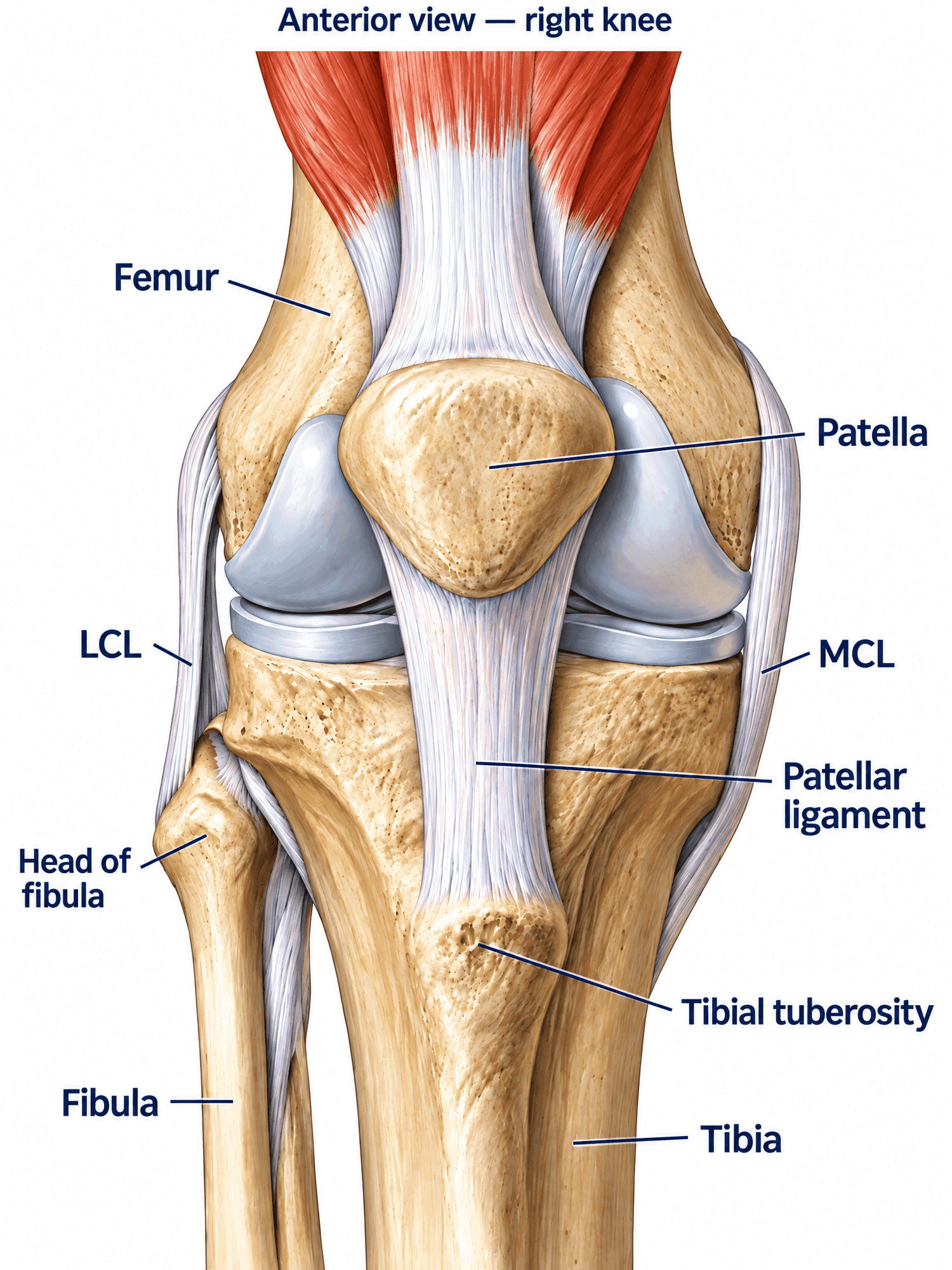
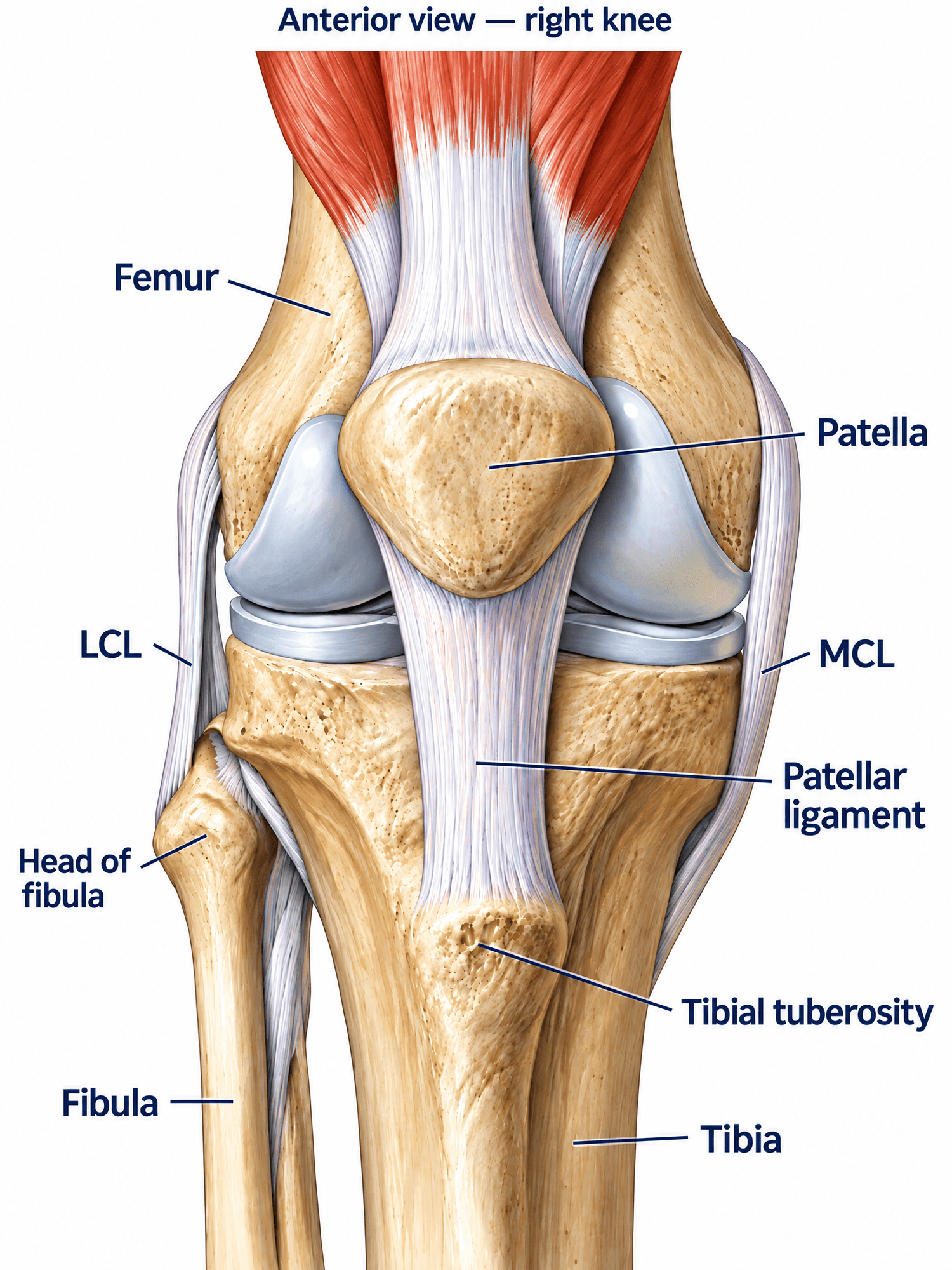
Image is free to use. Please cite natomy.com.
The knee is the largest joint in the body and the one most frequently imaged after a fall, sports injury, or motor vehicle collision. Understanding its bones, ligaments, menisci, muscles, and nerves is the first step to reading an MRI report, explaining a diagnosis to a patient, or building a demonstrative exhibit for a personal injury claim.
Bones of the knee
- Femur — the distal (lower) end forms the femoral condyles, the rounded surfaces that articulate with the tibia.
- Tibia — the proximal (upper) end forms the tibial plateau, which bears the femoral condyles.
- Patella — the kneecap, a sesamoid bone embedded in the quadriceps tendon that glides in the trochlear groove of the femur.
- Fibula — runs alongside the tibia and does not bear significant weight, but its head is a key landmark for the LCL and the common peroneal nerve.
Ligaments and menisci
- Anterior cruciate ligament (ACL) — prevents the tibia from sliding forward relative to the femur; the most commonly torn knee ligament in sports.
- Posterior cruciate ligament (PCL) — prevents the tibia from sliding backward; often injured in dashboard-impact motor vehicle collisions.
- Medial collateral ligament (MCL) — resists valgus (inward) stress on the knee.
- Lateral collateral ligament (LCL) — resists varus (outward) stress on the knee.
- Medial and lateral menisci — C-shaped cartilage pads that cushion and stabilize the joint.
 Image is free to use. Please cite natomy.com.
Muscles of the knee joint
- Quadriceps (rectus femoris, vastus lateralis, vastus medialis, vastus intermedius) — extend the knee via the patellar tendon.
- Hamstrings (biceps femoris, semitendinosus, semimembranosus) — flex the knee and stabilize it during rotation.
- Popliteus — a small muscle behind the knee that unlocks the joint from full extension.
- Gastrocnemius — crosses the back of the knee and contributes to flexion.
Nerves of the knee
- Femoral nerve — supplies the quadriceps and sensation to the front of the thigh and knee.
- Tibial nerve — passes behind the knee and supplies the hamstrings and lower leg.
- Common peroneal nerve — wraps around the fibular head just below the knee and is especially vulnerable to lateral trauma or dislocation.
Common knee injuries and how they appear on imaging
- ACL tear — on MRI, a discontinuous or wavy ligament signal on sagittal views; often paired with bone bruising on the lateral femoral condyle and posterior tibia.
- Meniscus tear — linear or complex increased signal extending to the articular surface on MRI; may be accompanied by a joint effusion.
- Patellar fracture — visible as a lucent fracture line on plain radiographs, often from a direct blow to a flexed knee.
- Tibial plateau fracture — best seen on CT, frequently from axial loading in falls or MVA; associated with joint depression and effusion.
How the knee is shown in a demonstrative exhibit
A labeled anatomy illustration of the knee is the baseline exhibit in most knee-injury claims — it establishes for a jury what a normal, uninjured joint looks like before the injury-specific exhibit (the torn ligament, the fracture line, the surgical hardware) is introduced. Pairing a normal-anatomy figure with the patient's own MRI or CT findings, side by side, is typically clearer to a lay jury than either image alone.
Need a custom illustration of this joint?
Generate a labeled or exhibit-ready figure for a specific patient, injury, or case in minutes.
Try Natomy →