Subarachnoid Hemorrhage: Causes, Symptoms & Treatment
A subarachnoid hemorrhage is bleeding into the fluid-filled space around the brain, from a head injury or a burst aneurysm. See it beside normal anatomy.
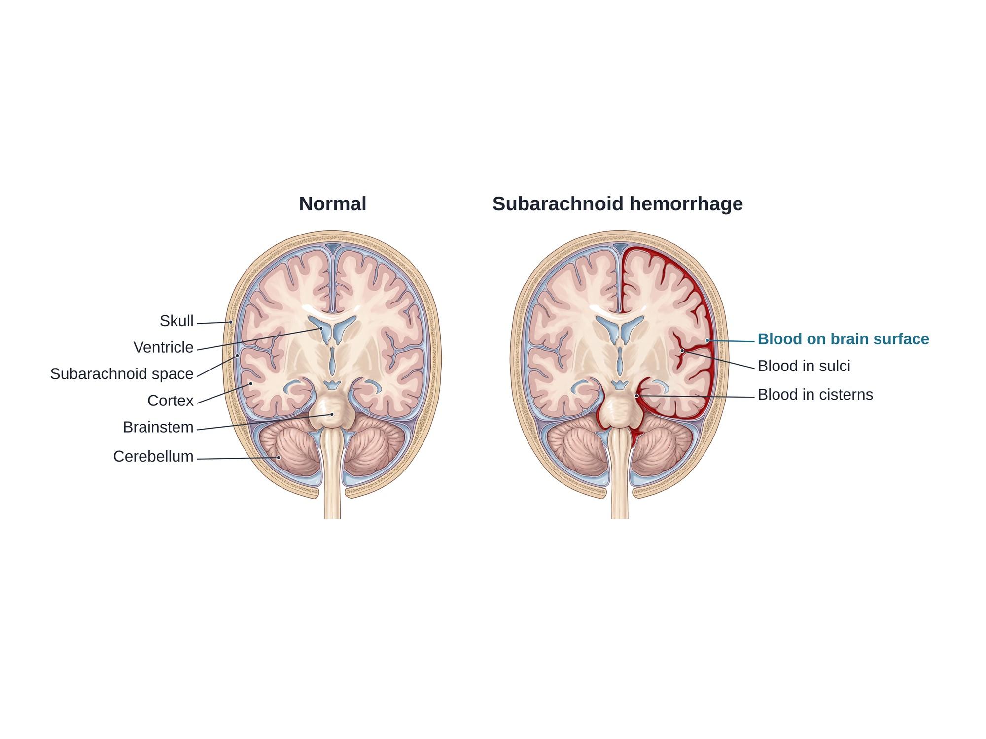
A subarachnoid hemorrhage (SAH) is bleeding into the subarachnoid space, the thin, fluid-filled layer that surrounds the brain. Blood mixes with the clear cerebrospinal fluid and spreads over the brain surface and into its folds. The most common cause is head trauma. The most common cause without injury is a burst brain aneurysm, which is a medical emergency. A subarachnoid hemorrhage can raise pressure in the skull, narrow the brain's arteries and block fluid flow. The illustration above shows the space filled with clear fluid in a normal brain and with blood after a hemorrhage.
The anatomy
The brain is covered by three thin layers called the meninges. See brain anatomy for more.
- The dura mater is the tough outer layer, just inside the skull.
- The arachnoid mater is a thin, web-like layer under the dura.
- The pia mater is a delicate layer that hugs the brain surface and dips into every groove.
- The subarachnoid space lies between the arachnoid and the pia. It is filled with cerebrospinal fluid (CSF) and holds the large arteries and veins on the brain surface.
- The space widens into pools called cisterns, mainly around the base of the brain and the brainstem (the basal cisterns).
In a subarachnoid hemorrhage, blood enters this space. It follows the fluid into the sulci (the grooves between folds) and the cisterns. This is different from an epidural or subdural hematoma, where blood collects outside the arachnoid.
Types
| Type | Cause | Where blood usually collects |
|---|---|---|
| Traumatic SAH | Head injury from a crash, fall or assault | Sulci over the brain surface, often near a bruise or skull fracture |
| Aneurysmal SAH | A burst aneurysm (a weak, bulging spot on an artery) | Basal cisterns and the Sylvian fissures, spreading widely |
| Perimesencephalic SAH | Often a small vein bleed; cause not always found | Cisterns in front of the brainstem only; usually a milder course |
| Other | Arteriovenous malformation, vessel tear (dissection), blood thinners, drug use | Varies |
Doctors grade aneurysmal SAH by the patient's condition, such as the Hunt and Hess or World Federation of Neurosurgical Societies (WFNS) scales. They grade the amount of blood on CT with the modified Fisher scale. Higher grades carry more risk.
Causes
- Head trauma, the most common cause overall. The blow tears small surface vessels.
- Ruptured brain aneurysm, the cause of most non-traumatic cases.
- Arteriovenous malformation, a tangle of abnormal vessels.
- Blood thinners and bleeding disorders, which make any bleed larger.
Risk factors for aneurysm rupture include high blood pressure, smoking, heavy alcohol use, cocaine or amphetamine use, a family history of aneurysms, and some inherited conditions such as polycystic kidney disease.
Symptoms
- Sudden, severe headache, often called a thunderclap headache or "the worst headache of my life," reaching full strength within a minute
- Stiff neck, because blood irritates the meninges
- Nausea and vomiting
- Sensitivity to light
- Confusion, drowsiness or loss of consciousness
- Seizures
- Weakness, numbness or trouble speaking, in some cases
After a head injury, symptoms often blend with those of a concussion: headache, confusion, dizziness and memory loss. Some people have a smaller "warning" headache days or weeks before a large aneurysm bleed. A sudden headache with weakness can look like other conditions, such as a hemiplegic migraine, so it always needs urgent imaging.
Diagnosis
- CT scan of the head without contrast is the first test. Fresh blood looks bright white in the sulci and cisterns. CT finds most bleeds in the first hours.
- Lumbar puncture (spinal tap) is done if the CT is normal but the history suggests a bleed. It looks for blood or yellow-tinged fluid (xanthochromia).
- CT angiography, MR angiography or catheter angiography looks for an aneurysm or other vessel problem.
- MRI can show smaller or older bleeds.
Treatment
Emergency care comes first: protecting the airway, controlling blood pressure, and reversing blood thinners.
For aneurysmal SAH:
- Endovascular coiling: a catheter is passed through an artery to the aneurysm, and soft platinum coils fill it so blood no longer enters.
- Surgical clipping: a surgeon opens the skull and places a small metal clip across the neck of the aneurysm.
- Nimodipine, a medicine given for 21 days to reduce brain injury from vasospasm.
- Intensive care monitoring, including checks for vasospasm with ultrasound of the brain arteries.
- External ventricular drain, a tube that drains spinal fluid if hydrocephalus develops.
- Decompressive surgery for severe brain swelling, such as a decompressive craniectomy.
For traumatic SAH, small bleeds are often watched with repeat CT scans. Treatment focuses on the overall brain injury, such as pressure control, seizure prevention and surgery for any other bleed.
Recovery
- A hospital stay of about 2 weeks or longer is common after an aneurysm bleed, mostly in intensive care.
- The risk of vasospasm is highest about 3 to 14 days after the bleed.
- Many people need rehabilitation: physical, occupational, speech and cognitive therapy.
- Headaches, tiredness, memory and attention problems, low mood and anxiety are common for months.
- Recovery after traumatic SAH depends on the other brain injuries present.
Complications
- Rebleeding of an untreated aneurysm, which is often fatal
- Vasospasm and delayed cerebral ischemia, when narrowed arteries cut off blood flow and cause a stroke days later
- Hydrocephalus, a buildup of spinal fluid when blood blocks its drainage
- Seizures
- Low sodium (hyponatremia)
- Heart and lung strain from the stress of the bleed
- Lasting brain injury, including memory, thinking, mood and movement problems
How subarachnoid hemorrhage is shown in a personal injury case
Traumatic subarachnoid hemorrhage is one of the most common findings on a head CT after a car crash, a fall or an assault. It is evidence that the brain itself was injured, not just bruised skin. In medical malpractice cases, the question is often whether a thunderclap headache or a warning bleed was missed, or whether vasospasm was treated in time. A medical illustration of a normal brain beside one with blood in the subarachnoid space shows a jury where the blood went, why it spread into the brain's folds, and how it can cause problems days later. For how these images are used at trial, see demonstrative evidence and medical legal illustration.
Related pages
- Brain anatomyThe normal anatomy of the brain.
- Hemiplegic migraineHemiplegic migraine is a rare migraine with aura that causes weakness on one side of the body, often mistaken for a stroke. See its symptoms illustrated.
- Horner syndromeHorner syndrome is a droopy eyelid, small pupil and less sweating on one side of the face from a damaged nerve pathway. See the pathway illustrated.
FAQ
Can a fall cause a subarachnoid hemorrhage?+
Yes. Head trauma, including falls, car crashes and assaults, is the most common cause of subarachnoid hemorrhage overall. A blow to the head tears small vessels on the brain surface, and blood spreads into the fluid around the brain. Older adults and people on blood thinners are at higher risk after even a minor fall.
Is a subarachnoid hemorrhage a stroke?+
A subarachnoid hemorrhage from a burst aneurysm is a type of hemorrhagic stroke. It accounts for a small share of all strokes, often quoted as about 5 in 100. A traumatic subarachnoid hemorrhage from a head injury is usually called a brain injury rather than a stroke.
What is a brain bleed stroke?+
A brain bleed stroke, or hemorrhagic stroke, happens when a blood vessel breaks and bleeds in or around the brain. The two main types are intracerebral hemorrhage, bleeding into the brain tissue, and subarachnoid hemorrhage, bleeding into the space around the brain.
Can a brain bleed be seen on a CT scan?+
Usually, yes. A CT scan of the head without contrast shows fresh blood as bright white in the spaces around the brain. It is most accurate in the first 6 hours. If the CT is normal but the story strongly suggests a bleed, doctors may do a lumbar puncture to look for blood in the spinal fluid.
What are the treatment options for subarachnoid hemorrhage?+
Treatment depends on the cause. A burst aneurysm is sealed, either by coiling through a catheter in the blood vessels or by clipping in open surgery. Patients are watched in an intensive care unit for vasospasm, high brain pressure and hydrocephalus. Small traumatic bleeds are often watched with repeat scans.
What are the symptoms of a subarachnoid hemorrhage?+
The classic symptom of an aneurysm bleed is a sudden, severe 'thunderclap' headache, often called the worst headache of one's life. Neck stiffness, nausea, vomiting, light sensitivity, confusion, a seizure or passing out may follow. After a head injury, headache and confusion are common.
Can you fully recover from a subarachnoid hemorrhage?+
Many people do, especially after a small traumatic bleed. Recovery after an aneurysm bleed varies widely. Some people return to normal life, while others have lasting problems with memory, attention, mood, tiredness or movement. Recovery often continues for a year or more.
What is the survival rate of a subarachnoid hemorrhage?+
It depends on the cause and severity. Small traumatic bleeds rarely cause death by themselves. Aneurysmal subarachnoid hemorrhage is much more serious: a large share of people die before or soon after reaching the hospital, and many survivors have lasting problems. Fast treatment improves the outlook.
Need this illustration for a patient or a case?
Describe the injury or upload a scan and get a clear, labeled medical illustration for a consult, a chart or a case exhibit.
Try Natomy →