Bones of the Foot: 26 Foot Bones Labeled
Foot bones labeled: the 26 bones of the foot from above, below and the inner side, with the tarsals, metatarsals, phalanges, arches and foot fractures.
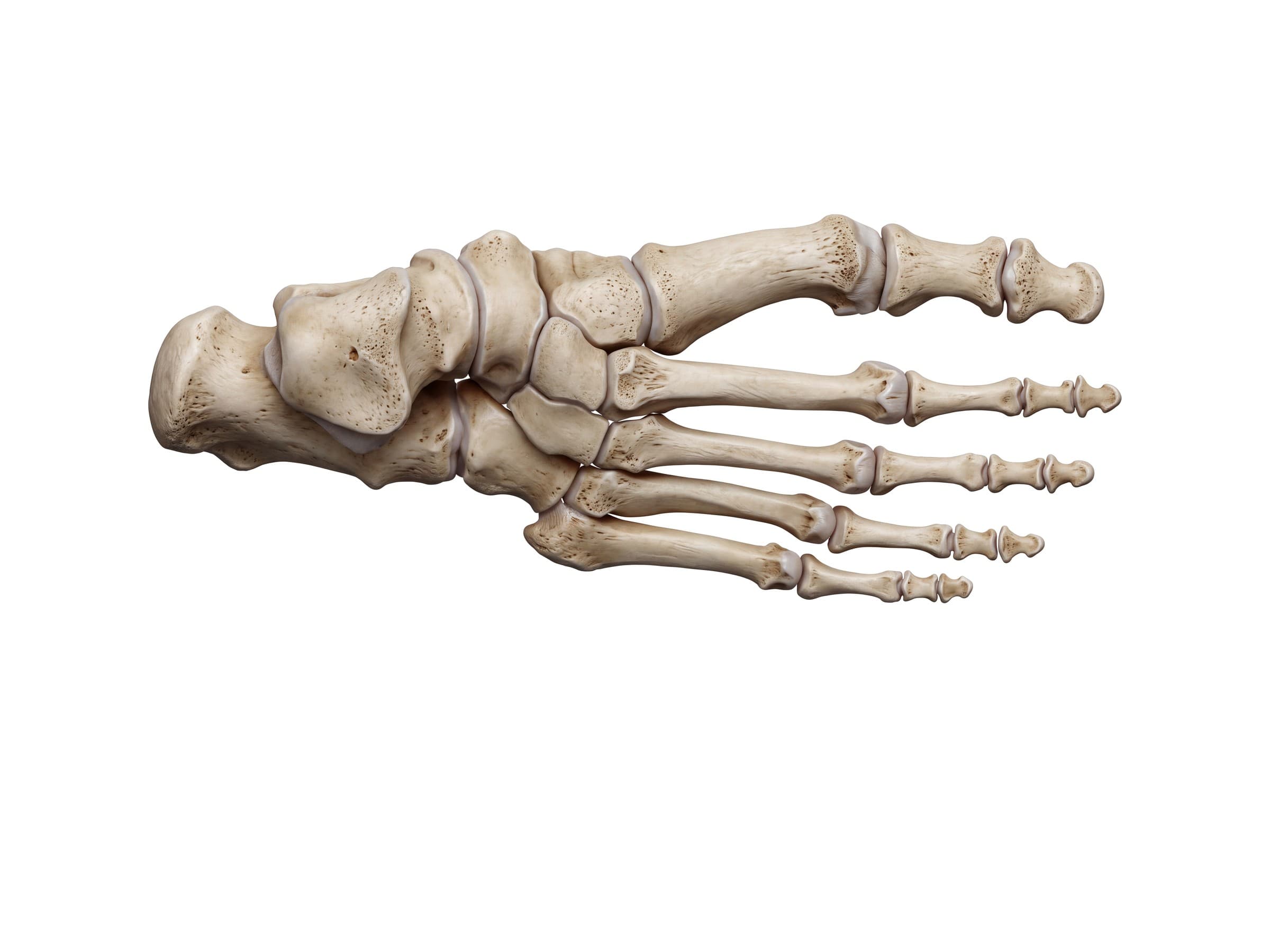
The foot has 26 bones that work together as a flexible arch, carrying the full weight of the body with every step. The bones of the foot fall into three groups: 7 tarsal bones in the heel and midfoot, 5 metatarsals in the middle of the foot, and 14 phalanges in the toes, plus two small sesamoid bones under the big toe. Knowing where each bone sits, and how the joints between them move, is the first step to reading a foot X-ray report, explaining a fracture to a patient, or illustrating a personal injury claim.
Bones of the foot
The labeled view from above shows a right foot with the heel on the left and the toes on the right. Counting from the heel:
- Tarsal bones (7): the talus, calcaneus, navicular, cuboid and three cuneiform bones. They form the heel, the ankle connection and the arched midfoot.
- Metatarsals (5): the long bones in the middle of the foot, numbered 1 to 5 from the big toe. Each has a base near the midfoot, a shaft, and a head that forms the ball of the foot.
- Phalanges (14): the toe bones. The big toe (hallux) has two, a proximal and a distal phalanx; each of the other four toes has three: proximal, middle and distal.
Together with the two sesamoids, that is 28 bones in most adults. The foot and ankle also contain 33 joints and more than 100 muscles, tendons and ligaments.
Hindfoot, midfoot and forefoot
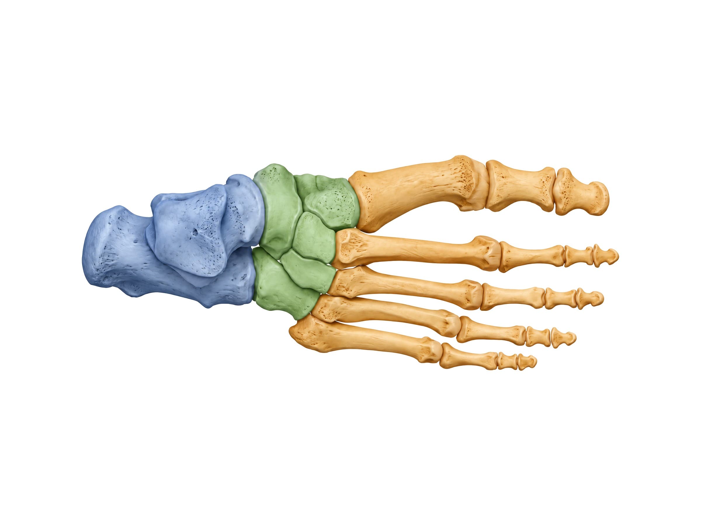
Doctors divide the foot into three regions, and most foot injuries are described by which region they involve.
- Hindfoot: the talus and calcaneus. The talus connects the foot to the leg at the ankle joint, and the calcaneus forms the heel.
- Midfoot: the navicular, cuboid and three cuneiforms. These five bones lock together to form the top of the arch.
- Forefoot: the five metatarsals and fourteen phalanges, which spread the load across the ball of the foot and push off when you walk.
The hindfoot meets the midfoot at the Chopart joint (the transverse tarsal joint), and the midfoot meets the forefoot at the Lisfranc joint (the tarsometatarsal joint). Both joint lines are common sites of injury.
Tarsal bones
The seven tarsal bones are short, blocky bones that fit together like a puzzle. A common way to remember them is "Tiger Cubs Need MILC": Talus, Calcaneus, Navicular, Medial, Intermediate and Lateral cuneiforms, and Cuboid.
Talus
The talus is the top bone of the foot and the link between the leg and the foot. Its rounded upper surface (the trochlea) fits into the socket formed by the tibia and fibula at the ankle, its underside sits on the calcaneus at the subtalar joint, and its head meets the navicular in front. More than half of the talus is covered with cartilage and no muscles attach to it, so its blood supply is limited. A fracture through the neck of the talus can cut off that supply and lead to avascular necrosis.
Calcaneus
The calcaneus, or heel bone, is the largest bone of the foot. Its back end forms the heel and anchors the Achilles tendon, and its underside takes the first impact of every step. A shelf on its inner side, the sustentaculum tali, props up the talus. The calcaneus is the most commonly fractured tarsal bone.
Navicular
The navicular is the boat-shaped bone on the inner side of the midfoot, between the head of the talus and the three cuneiforms. Its tuberosity, the bump you can feel on the inner side of the foot in front of the ankle, is the main attachment of the tibialis posterior tendon, which holds up the arch. Stress fractures of the navicular are a known problem in sprinters and jumpers and are easily missed on plain X-rays.
Cuboid
The cuboid is the cube-shaped bone on the outer side of the midfoot, in front of the calcaneus. It connects to the fourth and fifth metatarsals. A groove on its underside carries the peroneus longus tendon as it crosses the sole of the foot.
Cuneiform bones
The three cuneiforms (medial, intermediate and lateral) are wedge-shaped bones in a row between the navicular and the first three metatarsals. The medial cuneiform is the largest and connects to the first metatarsal. The intermediate cuneiform is the smallest and sits slightly further back than the other two, which creates a slot that locks the base of the second metatarsal in place. This keystone arrangement makes the midfoot stable, and it is why injuries at the base of the second metatarsal (Lisfranc injuries) are so important.
Metatarsal bones
The five metatarsals are long bones that connect the midfoot to the toes. They are numbered from the inner side of the foot:
- First metatarsal: the shortest and thickest, carrying about a third of the body's weight during push-off. Its head sits on the two sesamoid bones.
- Second metatarsal: the longest and the most rigidly held, locked in by the cuneiforms. It is the most common site of a metatarsal stress fracture.
- Third and fourth metatarsals: slender bones that move slightly more than the second.
- Fifth metatarsal: on the outer edge of the foot, with a large tuberosity at its base where the peroneus brevis tendon attaches. It is the most commonly fractured metatarsal.
The heads of the metatarsals form the ball of the foot and meet the toes at the metatarsophalangeal (MTP) joints.
Phalanges of the foot
The 14 phalanges are the bones of the toes. The big toe has two (proximal and distal), and the second through fifth toes each have three (proximal, middle and distal), although the middle and distal phalanges of the little toe are often fused. The joints between the phalanges are the interphalangeal joints: one in the big toe and two in each of the other toes.
Bones of the bottom of the foot
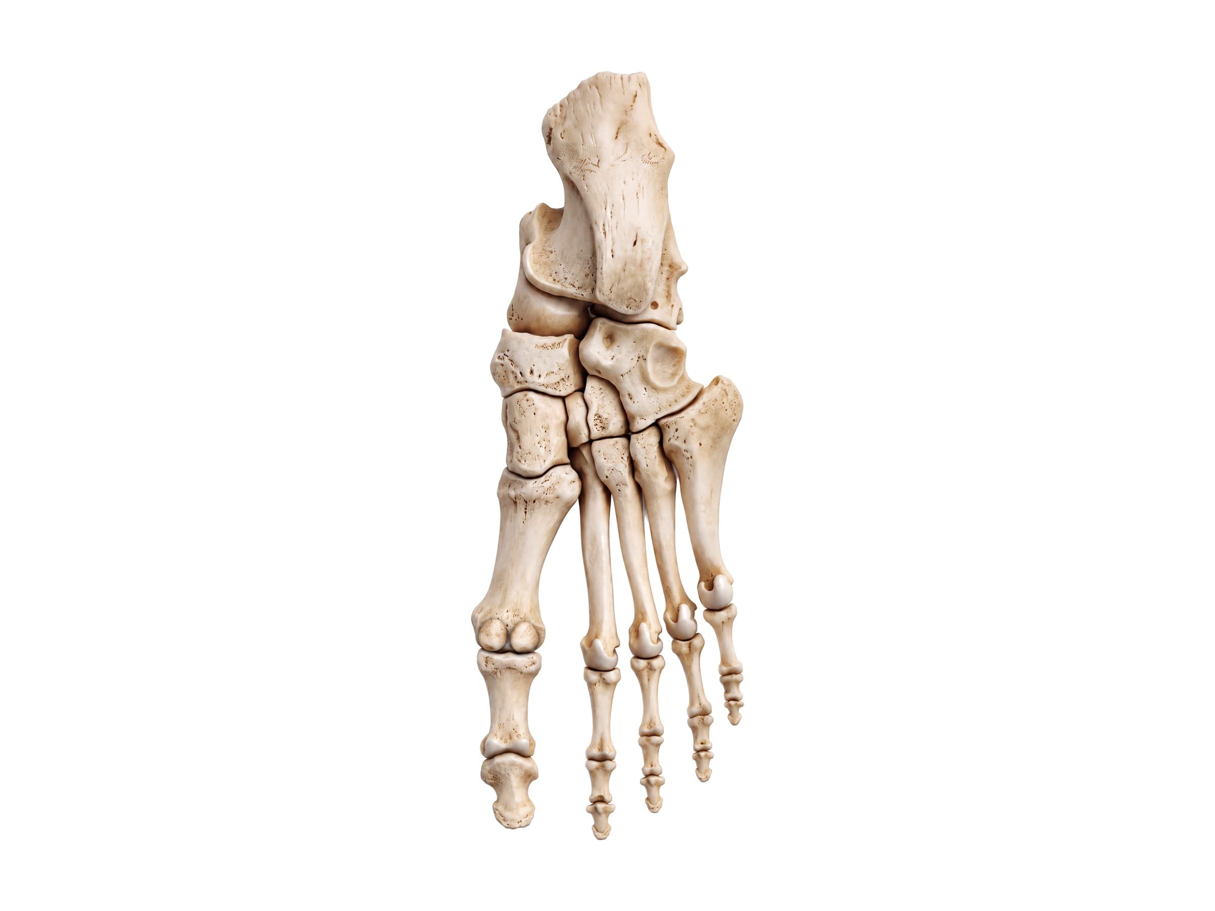
Seen from the sole, the calcaneus dominates the back of the foot, and the head of the talus is visible on its inner side. Further forward, the groove for the peroneus longus tendon runs across the underside of the cuboid, and the tuberosity of the fifth metatarsal stands out on the outer edge.
Sesamoid bones
Under the head of the first metatarsal sit two pea-sized sesamoid bones, embedded in the tendons of the flexor hallucis brevis muscle. They work like pulleys for those tendons and absorb force when you push off from the big toe. Sesamoiditis and sesamoid fractures cause pain under the ball of the foot. One of the sesamoids is sometimes in two pieces (a bipartite sesamoid), which can look like a fracture on X-ray.
Arches of the foot
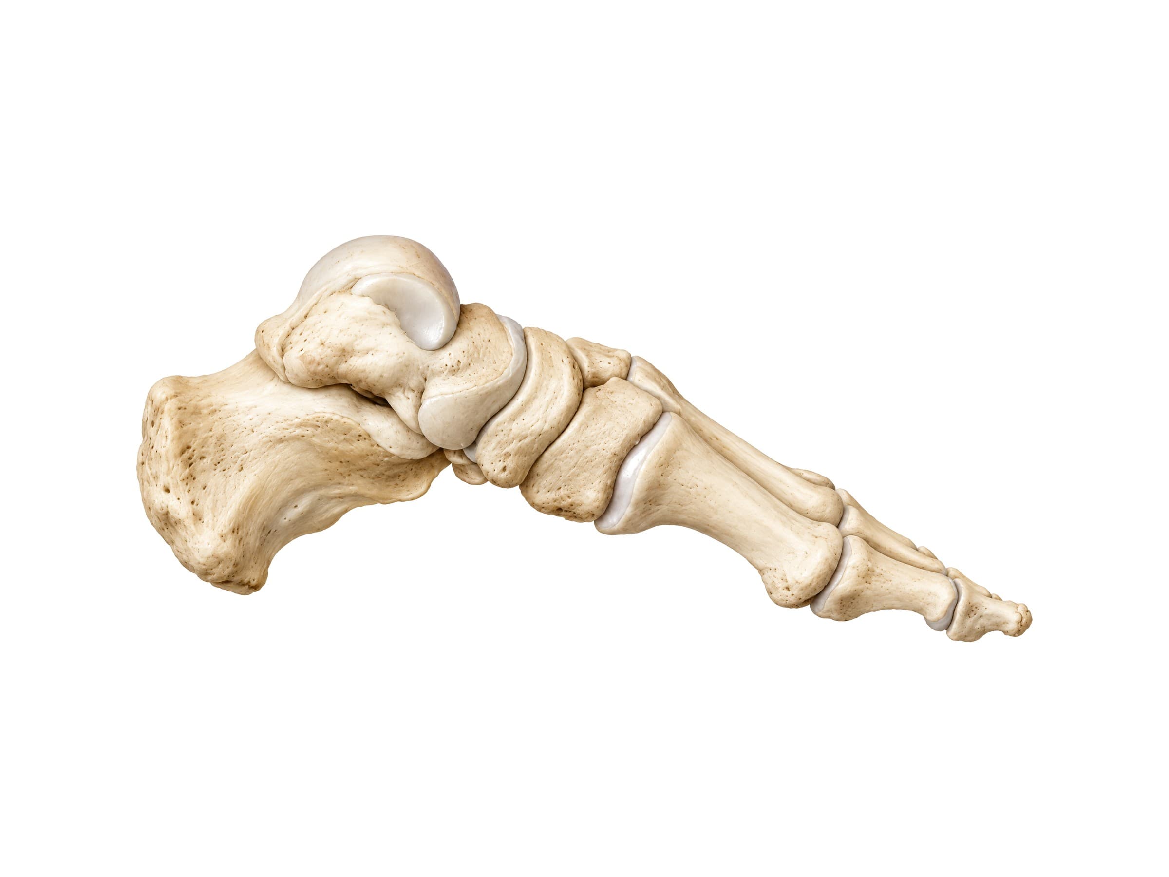
The bones of the foot are not flat. They form three arches, held up by the shape of the bones, the ligaments and the plantar fascia, and supported by muscles and tendons.
- Medial longitudinal arch: the highest arch, along the inner side of the foot. It runs from the calcaneus through the talus, navicular, the three cuneiforms and the first three metatarsals. When it collapses, the result is a flat foot.
- Lateral longitudinal arch: a lower arch on the outer side, through the calcaneus, cuboid and fourth and fifth metatarsals.
- Transverse arch: across the midfoot, formed by the cuneiforms, the cuboid and the bases of the metatarsals.
Joints of the foot
- Ankle joint (talocrural joint): between the talus and the tibia and fibula. It moves the foot up and down. See ankle anatomy for its bones and ligaments.
- Subtalar joint: between the talus and calcaneus. It tilts the heel inward and outward so the foot can adapt to uneven ground.
- Chopart joint (transverse tarsal joint): the talonavicular and calcaneocuboid joints together, the line between the hindfoot and midfoot.
- Lisfranc joint (tarsometatarsal joints): where the cuneiforms and cuboid meet the five metatarsals. The Lisfranc ligament runs from the medial cuneiform to the base of the second metatarsal; there is no ligament between the bases of the first and second metatarsals.
- Metatarsophalangeal (MTP) joints: at the ball of the foot. The big toe MTP joint is the site of bunions (hallux valgus) and gout.
- Interphalangeal joints: the small hinge joints within the toes.
Accessory bones of the foot
Many people have small extra bones in the foot that develop from separate centers of bone growth that never fused. They are normal variants but can cause pain or be mistaken for fractures.
- Os trigonum: a small bone behind the talus. It can be pinched when the foot is pointed, causing pain at the back of the ankle in dancers and soccer players (posterior ankle impingement).
- Accessory navicular: an extra piece of bone on the inner side of the navicular, inside the tibialis posterior tendon. It can form a painful bump on the inner side of the foot.
- Os peroneum: a small bone in the peroneus longus tendon next to the cuboid.
Common foot fractures and how they appear on imaging
Foot fractures are usually seen on standard foot X-rays (top, oblique and side views). CT shows complex fractures of the calcaneus and midfoot, and MRI picks up stress fractures and bone bruises that X-rays miss.
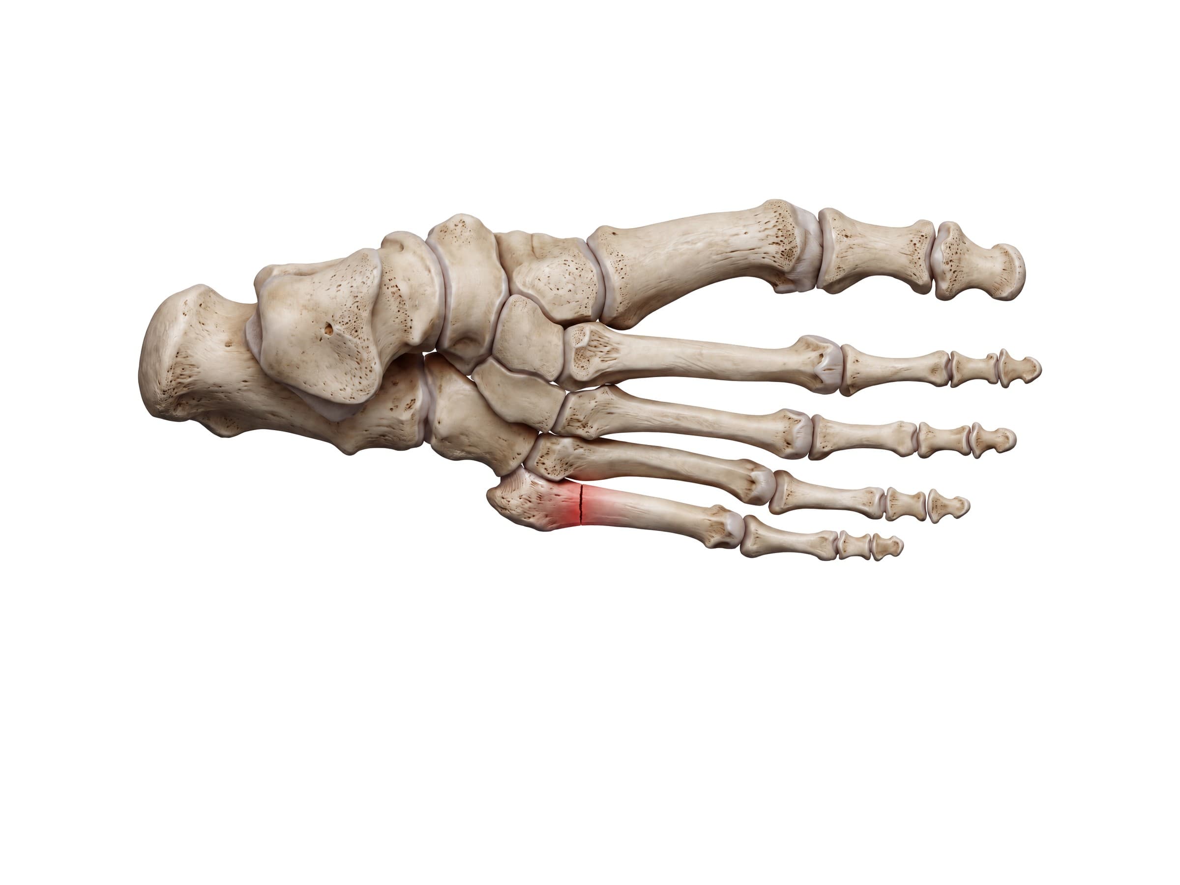
- Jones fracture: a break of the fifth metatarsal about 1.5 to 3 cm from its base, just beyond the tuberosity, where the bone's blood supply is poor. It heals slowly and can fail to heal, so it is often treated in a non-weight-bearing cast or, in athletes, with a screw. It is different from an avulsion fracture of the tuberosity, in which the tip of the base is pulled off during an ankle roll.
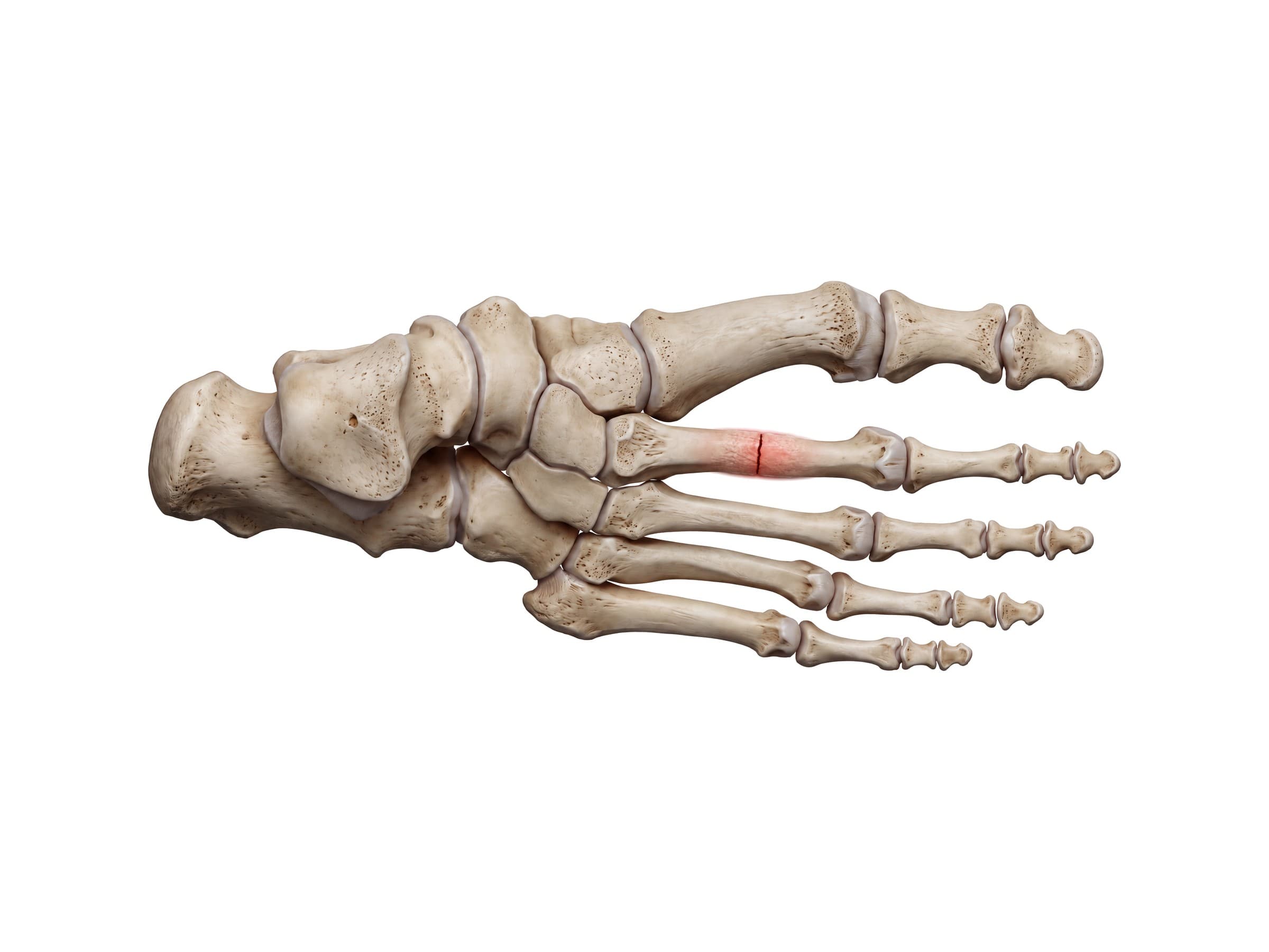
- Metatarsal stress fracture: a crack that builds up from repetitive loading, most often in the shaft of the second metatarsal (the "march fracture"). It is common in runners and military recruits. The first X-ray is often normal, and the fracture shows up weeks later as new bone forms, or straight away on MRI.
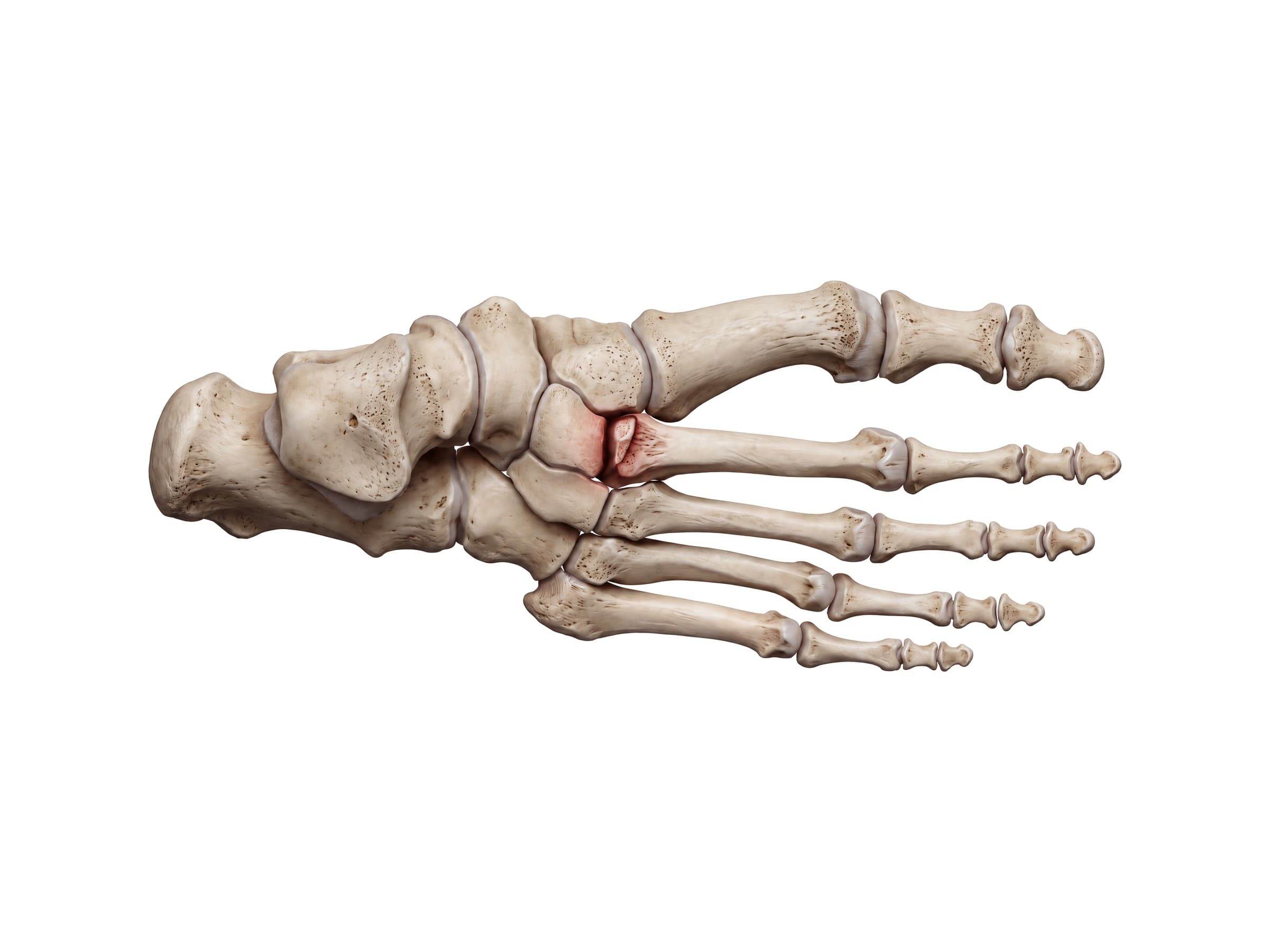
- Lisfranc injury: a sprain, fracture or dislocation at the tarsometatarsal joint, from a twist on a pointed foot, a fall from a height, or a crush injury in a motor vehicle accident. Subtle injuries are often missed on the first X-ray. Signs include a gap between the bases of the first and second metatarsals and a small fleck of bone (the "fleck sign"); weight-bearing X-rays, CT or MRI confirm it. Unstable injuries need surgery.
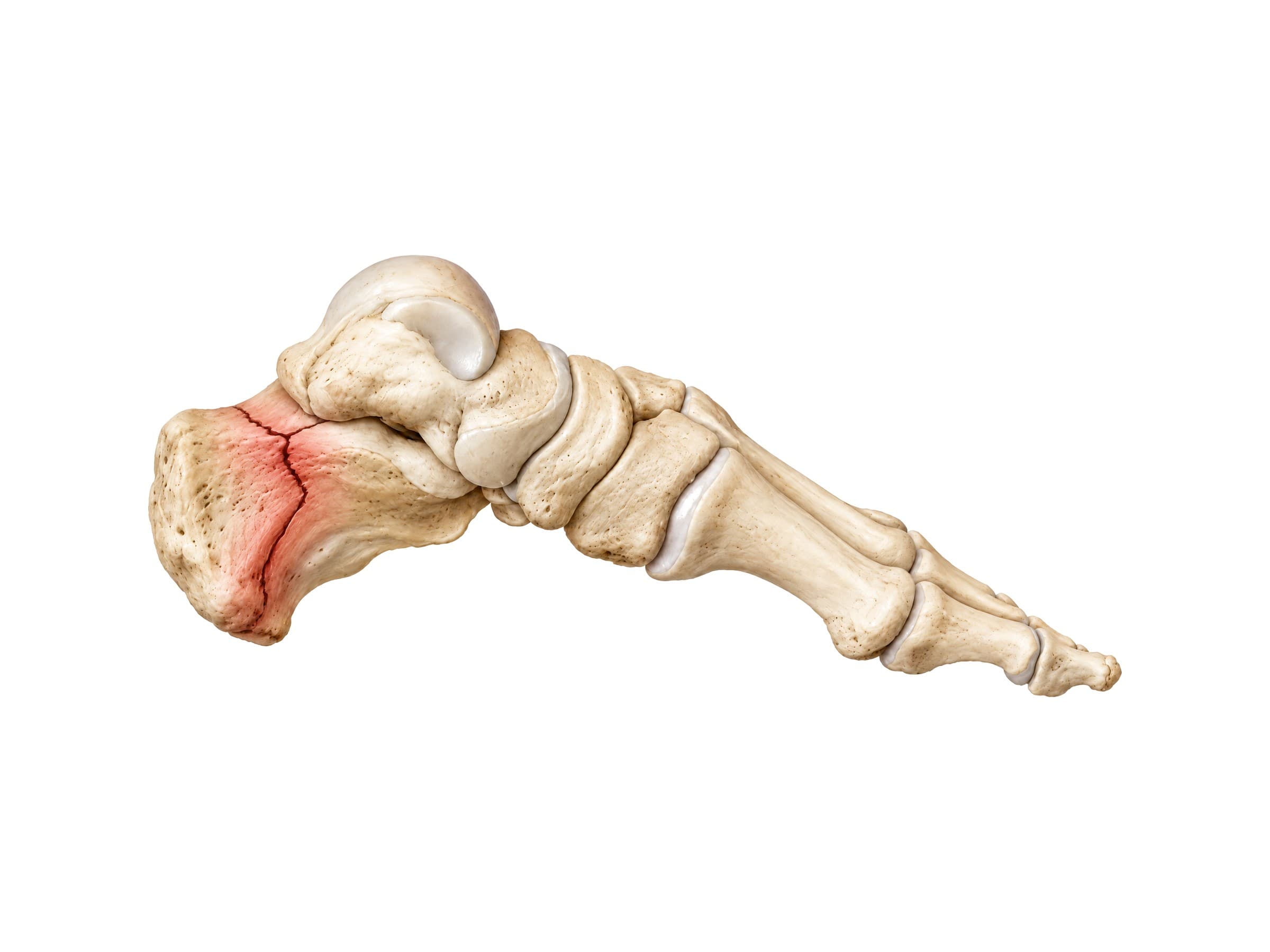
- Calcaneus fracture: usually from a fall from a height or a jump landing on the heel, when the talus is driven down into the calcaneus. Most extend into the subtalar joint. X-rays show a flattened heel (a reduced Böhler's angle), and CT maps the fracture lines for surgical planning. Because the force travels up the skeleton, spine fractures are checked for as well.
- Toe fracture: the most common foot fracture, usually from stubbing a toe or dropping something on the foot. Most heal with buddy taping, but fractures of the big toe that enter the joint may need fixation.
- Talar neck fracture: a high-energy injury, often from forcing the foot upward in a car crash, with a risk of avascular necrosis.
- Navicular stress fracture: pain on the top of the midfoot in sprinters and jumpers, frequently missed on X-ray and best seen on CT or MRI.
How the foot bones are shown in a personal injury case
Foot fractures are common in personal injury claims: crush injuries from a car's footwell in a frontal collision, falls from ladders and scaffolding, slips on stairs, and heavy objects dropped on the foot at work. A labeled illustration of the normal foot bones is the baseline exhibit. It shows the jury where the calcaneus, the Lisfranc joint or the fifth metatarsal sits in an uninjured foot before the injury-specific exhibit is introduced: the crushed heel, the displaced midfoot, the plate and screws. In Lisfranc and navicular cases, where the injury was missed on the first X-ray, an illustration of the bones and their alignment helps explain what the first reading should have shown and why the delay changed the outcome.
Natomy creates custom medical legal illustrations of foot injuries for demand packages and trial exhibits, and the role of these visuals at trial is covered in demonstrative evidence. For the joint above the foot, see ankle anatomy; for the rest of the lower limb, knee anatomy and hip anatomy; and for the upper limb, hand and wrist anatomy.
FAQ
How many bones are in the foot?+
Each foot has 26 bones: 7 tarsal bones in the back half of the foot, 5 metatarsals in the middle, and 14 phalanges in the toes (two in the big toe and three in each of the other four toes). Two small sesamoid bones under the big toe joint are usually not counted, and some people have extra accessory bones such as an os trigonum or an accessory navicular. Counting both feet, that is 52 bones, about a quarter of all the bones in the adult body.
What are the 7 tarsal bones called?+
The seven tarsal bones are the talus, calcaneus, navicular, cuboid, and the medial, intermediate and lateral cuneiforms. The talus and calcaneus form the hindfoot; the navicular, cuboid and three cuneiforms form the midfoot. A common mnemonic is "Tiger Cubs Need MILC": Talus, Calcaneus, Navicular, then Medial, Intermediate and Lateral cuneiforms, and Cuboid.
What is the bone on the side of my foot called?+
The bony bump halfway along the outer edge of the foot is the tuberosity at the base of the fifth metatarsal, the long bone that leads to the little toe. The peroneus brevis tendon attaches to it, which is why it can be pulled off in an ankle sprain (an avulsion fracture). Just behind it sits the cuboid. On the inner side of the foot, the bump in front of the ankle is the tuberosity of the navicular.
What is the most common bone to fracture in your foot?+
Toe and metatarsal fractures are the most common foot fractures. Among the metatarsals, the fifth metatarsal on the outer edge of the foot breaks most often, either as an avulsion of its base during an ankle roll or as a Jones fracture just beyond the base. Stress fractures, which build up from repetitive loading rather than a single injury, most often affect the second metatarsal.
Which tarsal bone is most commonly fractured?+
The calcaneus (heel bone) is the most commonly fractured tarsal bone, accounting for roughly 60 percent of tarsal fractures. Most happen when a person falls from a height or jumps and lands on the heel, driving the talus down into the calcaneus. About three quarters of calcaneus fractures extend into the subtalar joint, and because the same force travels up the leg, the spine should be checked as well.
What are the bones on the top of the foot?+
From the ankle toward the toes, the top of the foot is formed by the head and neck of the talus, the navicular and cuboid, the three cuneiform bones, the five metatarsals and the toe phalanges. Pain on the top of the foot often comes from a metatarsal stress fracture, a Lisfranc injury at the midfoot, or arthritis in the joints between the cuneiforms and metatarsals.
What are the sesamoid bones in the foot?+
The two sesamoid bones are pea-sized bones embedded in the tendons of the flexor hallucis brevis muscle, under the head of the first metatarsal at the base of the big toe. They act like small pulleys for the tendons and take load when you push off from the big toe. Inflammation of these bones (sesamoiditis) and sesamoid fractures cause pain under the ball of the foot, especially in runners and dancers.
What is the difference between the hindfoot, midfoot and forefoot?+
The hindfoot is the heel and back of the foot: the talus and calcaneus. The midfoot is the arched middle section: the navicular, cuboid and three cuneiforms. The forefoot is the front of the foot: the five metatarsals and fourteen phalanges. The hindfoot and midfoot meet at the Chopart (transverse tarsal) joint, and the midfoot and forefoot meet at the Lisfranc (tarsometatarsal) joint.
Need a custom illustration of this joint?
Generate a labeled or exhibit-ready figure for a specific patient, injury, or case in minutes.
Try Natomy →