Horner Syndrome: Causes, Signs & Pathway, Illustrated
Horner syndrome is a droopy eyelid, small pupil and less sweating on one side of the face from a damaged nerve pathway. See the pathway illustrated.
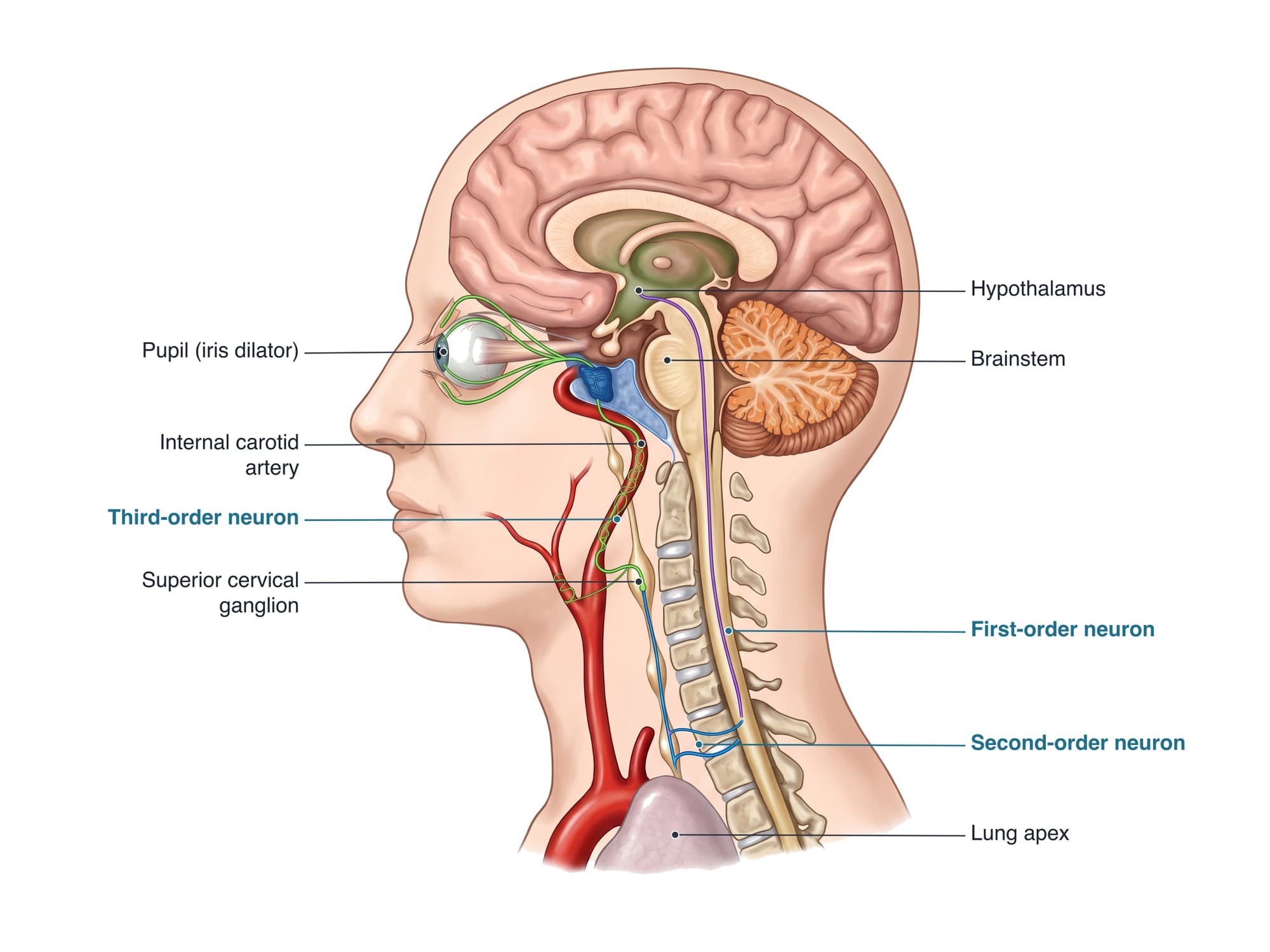
Horner syndrome is a group of signs on one side of the face, a slightly drooping upper eyelid (ptosis), a smaller pupil (miosis) and reduced sweating (anhidrosis), caused by damage to the sympathetic nerve pathway that runs from the brain down into the chest and back up the neck to the eye. The signs themselves are usually mild, but the cause can be serious, such as a carotid artery dissection, a stroke, or a tumor at the top of the lung. The illustration above shows the full pathway.
The anatomy
The sympathetic supply to the eye is a chain of three neurons:
- First-order neuron: starts in the hypothalamus and runs down through the brainstem and cervical spinal cord to the ciliospinal center at about C8 to T2.
- Second-order neuron: leaves the spinal cord at about T1, passes over the apex of the lung near the subclavian artery, and climbs the cervical sympathetic chain to the superior cervical ganglion high in the neck.
- Third-order neuron: travels on the wall of the internal carotid artery into the skull, through the cavernous sinus, and on to the eye. It supplies the iris dilator muscle, which widens the pupil, and the small Müller muscle, which helps hold up the upper eyelid. Sweat fibers to most of the face leave earlier, along the external carotid artery.
For the anatomy of the brain, see brain anatomy.
Types
| Lesion | Where | Examples of causes | Sweating |
|---|---|---|---|
| First-order (central) | Hypothalamus, brainstem, cervical spinal cord | Brainstem stroke (lateral medullary syndrome), multiple sclerosis, spinal cord injury, syringomyelia | Reduced over the face and often the body on that side |
| Second-order (preganglionic) | T1 root, lung apex, lower neck | Pancoast tumor at the top of the lung, chest or neck surgery, trauma, brachial plexus injury, central line placement | Reduced over the face |
| Third-order (postganglionic) | Along the internal carotid artery to the eye | Carotid artery dissection, cavernous sinus lesions, cluster headache | Little or none lost |
Causes
- Carotid artery dissection, spontaneous or after neck trauma, a common cause of a new painful Horner syndrome
- Stroke in the brainstem
- Tumors: a Pancoast tumor at the lung apex, thyroid or neck tumors, and in young children neuroblastoma
- Trauma to the neck, chest or spinal cord, including birth injury of the brachial plexus
- Surgery and procedures in the neck or chest
- Congenital Horner syndrome, sometimes with a lighter iris on the affected side
- In some people no cause is found
Symptoms
- A slightly drooping upper eyelid (ptosis), usually 1 to 2 mm
- A smaller pupil on the same side (miosis), most noticeable in dim light, where the affected pupil also dilates more slowly
- Reduced or absent sweating on that side of the face (anhidrosis)
- A slightly raised lower eyelid, which can make the eye look sunken
- Symptoms of the cause: neck, face or eye pain with a dissection, arm pain or weakness with a Pancoast tumor, or dizziness and trouble swallowing with a brainstem stroke
Diagnosis
The diagnosis starts with comparing the pupils in bright and dim light. Eye drops confirm it: apraclonidine drops make the affected pupil widen and the lid lift, and cocaine drops fail to dilate the affected pupil. Hydroxyamphetamine drops can help tell a third-order lesion from the others. Imaging then follows the pathway: MRI of the brain and cervical spine, MR or CT angiography of the neck to look for a carotid dissection, which shows as a narrowed artery with a blood clot within its wall, and imaging of the chest to look at the lung apex.
Treatment
Treatment depends on the cause: antiplatelet or anticoagulant medicine for a carotid dissection, stroke care, or surgery, radiation and chemotherapy for a tumor. The eye signs need no treatment of their own. A noticeable eyelid droop can be improved with eye drops in some people or with minor eyelid surgery.
Recovery
The signs may fade if the nerve recovers, for example as a carotid dissection heals over several months. When the nerve is permanently damaged, the droop and small pupil usually stay, but they are mostly cosmetic.
Complications
Complications come from the underlying cause rather than the syndrome: a stroke from a carotid dissection, the spread of a tumor, or spinal cord injury. A congenital Horner syndrome can leave the iris on that side lighter in color.
How Horner syndrome is shown in a personal injury case
Horner syndrome can follow car crashes, falls and sports injuries that cause a carotid artery dissection, as well as neck and chest surgery, central line placement and birth injuries to the brachial plexus. Because the signs are subtle, the case often turns on showing the jury where the nerve pathway runs and how the injury reached it, for example how a neck injury tore the carotid artery that carries the third-order neuron. A pathway illustration like the one above, matched to the imaging, makes that connection clear. For how these images are used at trial, see demonstrative evidence and medical legal illustration.
FAQ
Is Horner syndrome serious?+
The eye signs themselves are usually mild and do not affect vision, but the cause can be serious. A new Horner syndrome, especially with neck pain or headache, needs prompt evaluation because it can be the first sign of a carotid artery dissection, a stroke, or a tumor at the top of the lung.
What are the three main signs of Horner syndrome?+
A slightly drooping upper eyelid (ptosis), a smaller pupil (miosis), and reduced sweating on the same side of the face (anhidrosis). The difference in pupil size is most obvious in dim light.
Does Horner syndrome go away?+
It depends on the cause. When the underlying problem heals, as many carotid dissections do, the signs can improve over months. If the nerve is permanently damaged, the droop and small pupil usually remain but rarely cause problems.
Can a neck injury cause Horner syndrome?+
Yes. Trauma to the neck, including whiplash-type injuries, can tear the inner lining of the internal carotid artery (dissection), which stretches the sympathetic fibers running on its wall. Injuries to the lower neck and upper chest, brachial plexus injuries and neck surgery can also damage the pathway.
Does Horner syndrome affect vision?+
Usually not. The small pupil lets in a little less light, and the droop may slightly cover the top of the visual field, but sight itself is normal.
How is Horner syndrome treated?+
Treatment is aimed at the cause, such as blood thinners for a carotid dissection or treatment of a lung tumor. A bothersome eyelid droop can be improved with eye drops in some people or with minor eyelid surgery.
Why does the location of the damage matter?+
The pathway has three neurons, and each runs through different territory: the brainstem and spinal cord, the top of the chest, and the neck and skull along the carotid artery. Working out which neuron is damaged narrows the list of causes and decides which part of the body is imaged.
Need this illustration for a patient or a case?
Describe the injury or upload a scan and get a clear, labeled medical illustration for a consult, a chart or a case exhibit.
Try Natomy →