Subdural Hematoma: Types, Causes, Symptoms & Treatment
A subdural hematoma is blood trapped between the dura and the brain, often from a torn vein after a fall or crash. Learn the types and treatment.
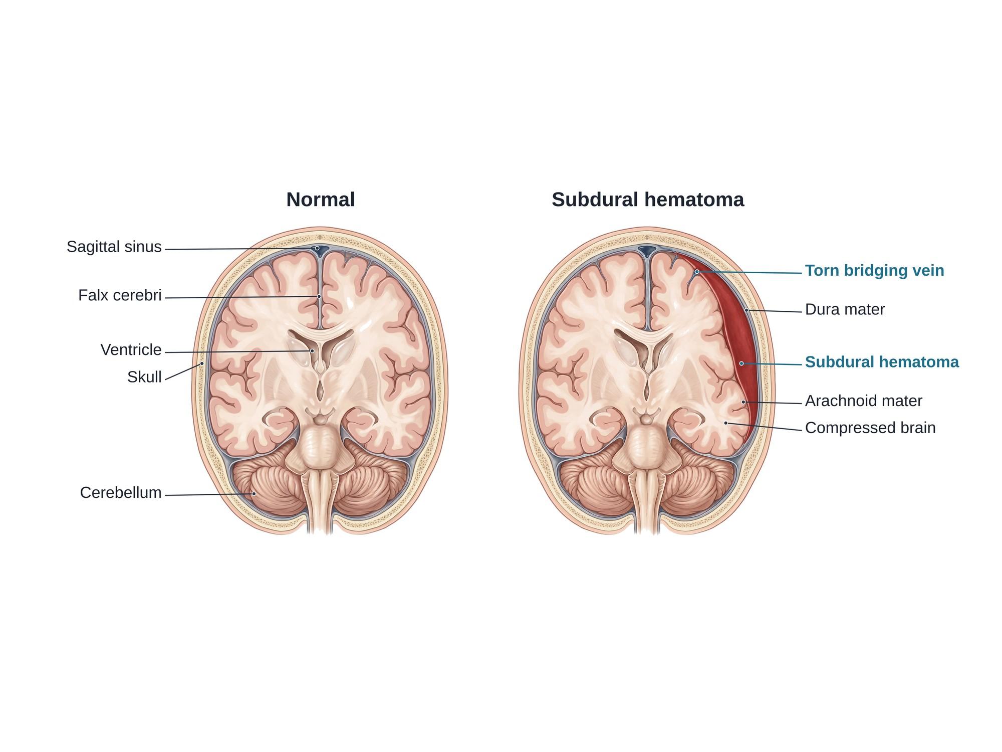
A subdural hematoma (SDH) is a collection of blood between the dura mater, the tough outer covering of the brain, and the arachnoid mater, the thin layer beneath it. It most often follows a traumatic brain injury from a fall, a car crash or an assault. The usual source is a torn bridging vein, a small vein that crosses from the brain surface to a large venous channel in the dura (see brain anatomy). This differs from an epidural hematoma, which forms between the skull and the dura, usually from a torn artery, and from a subarachnoid hemorrhage, where blood lies under the arachnoid. The blood spreads over the brain in a crescent shape and presses the brain inward. A large or growing hematoma is a medical emergency. The illustration above shows a normal brain beside one with an acute subdural hematoma.
Types
| Type | Age of the blood | Typical patient and course |
|---|---|---|
| Acute | Less than about 3 days | High-energy injury such as a crash or a hard fall; often with other brain injury; can be rapidly life-threatening |
| Subacute | About 3 days to 3 weeks | Symptoms build over days |
| Chronic | More than about 3 weeks | Usually an older adult, often after a minor fall; slow headaches, confusion or weakness |
| Acute-on-chronic | Mixed | Fresh bleeding into an older chronic collection |
Chronic subdural hematoma is becoming more common as people live longer. In a Finnish region, its yearly incidence doubled from 8 to 18 per 100,000 adults between 1990 and 2015. In people 80 and older it reached about 130 per 100,000 (Rauhala et al., 2020).
Causes
A sudden jolt makes the brain lag behind the skull and dura, which stretches the bridging veins until one can tear.
- Falls, the leading cause in older adults. A simple fall from standing can tear a bridging vein.
- Car, motorcycle and bicycle crashes, where rapid speeding up and slowing down shakes the brain.
- Assaults and sports injuries.
- Abusive head trauma in infants (shaken baby syndrome).
In the Finnish study, a head injury was documented in 59% of chronic cases. A ground-level fall caused 54% of cases in people 80 or older. By 2011 to 2015, 49% of patients were taking a blood thinner or an antiplatelet drug (Rauhala et al., 2020).
Risk factors include:
- Older age, because the brain shrinks and the bridging veins stretch across a wider gap
- Blood thinners (anticoagulants) and antiplatelet drugs such as aspirin
- Heavy alcohol use, which shrinks the brain and raises the risk of falls
- Bleeding disorders and low platelets
- Repeated falls, as with seizures or poor balance
Symptoms
Symptoms depend on how fast the blood collects.
Acute subdural hematoma:
- Headache, nausea and vomiting
- Confusion, drowsiness or loss of consciousness, sometimes after a short "lucid interval" when the person seems fine
- Weakness or numbness on one side of the body
- Trouble speaking
- One pupil larger than the other
- Seizures
Chronic subdural hematoma:
- Headaches that slowly get worse
- Unsteady walking and falls
- Memory loss, confusion or personality change
- Weakness of one arm or leg
- Drowsiness
In older adults, a chronic hematoma can look like dementia or a stroke. Any change in thinking or walking after a fall needs a scan.
Diagnosis
- CT scan of the head is the first test. A fresh subdural hematoma shows as a bright, crescent-shaped area along the inside of the skull. Older blood looks gray or dark. CT also shows how far the brain is pushed toward the other side (the midline shift).
- MRI can show small, older or mixed-age collections.
- Blood tests check clotting, platelets and the effect of any blood thinner.
- Neurological exam, including the Glasgow Coma Scale (GCS), a 3 to 15 score of eye opening, speech and movement.
Treatment
Emergency care comes first. Doctors protect the airway, control blood pressure, and reverse blood thinners.
Watching and waiting is common for thin hematomas that do not press much on the brain. Repeat CT scans make sure the blood is not growing.
Surgery removes the blood when it is large or causing harm. A key head injury guideline states: "An acute subdural hematoma (SDH) with a thickness greater than 10 mm or a midline shift greater than 5 mm on computed tomographic (CT) scan should be surgically evacuated, regardless of the patient's Glasgow Coma Scale (GCS) score" (Bullock et al., 2006).
- Craniotomy: a surgeon removes a piece of skull, opens the dura, washes out the clot and stops the bleeding vein. The bone is then put back.
- Decompressive craniectomy: the bone piece is left off so the swollen brain has room. See decompressive craniectomy.
- Burr hole drainage: for liquid chronic hematomas, the surgeon drills one or two small holes and drains the fluid. In a trial of 215 patients, leaving a drain in place cut recurrence to 9%, compared with 24% without a drain (Santarius et al., 2009).
- Middle meningeal artery embolization: a catheter is passed through the blood vessels to block the artery that feeds the hematoma lining. In a trial of 310 patients with chronic hematoma, treatment failed in 16% with embolization and 36% without it (Fiorella et al., 2025).
Recovery
- Patients with an acute hematoma often spend days in intensive care.
- Nurses and doctors check alertness, pupils and strength often in the first days.
- Seizure medicine may be given for a period after surgery.
- Many people need physical, occupational, speech or cognitive therapy.
- Older adults often need a rehabilitation stay before going home.
- Headaches, tiredness, poor focus and mood changes can last for months.
- Blood thinners are usually stopped for a time and restarted only when safe.
Complications
- Brain herniation, when pressure pushes part of the brain past a fold of dura; this can be fatal
- Recurrence of a chronic hematoma after drainage
- Seizures, early or late
- Infection after surgery
- Lasting brain injury: weakness, speech problems, memory loss and personality change
- Death
Outcomes depend on the patient's state on arrival. Among 1,427 adults with an acute traumatic subdural hematoma at a Level I trauma center, inpatient mortality was 16% (Ryan et al., 2012). For comatose patients (GCS 8 or less) treated with surgery since 2000, pooled mortality was 41% (van Essen et al., 2023).
How subdural hematoma is shown in a personal injury case
A subdural hematoma is one of the most serious findings on a head CT after a car crash, a fall on a wet floor or an assault. It shows that the injury reached the brain, not just the scalp. A torn bridging vein links the force of the event to the bleed. In older adults, a chronic hematoma may appear weeks after a fall, so the timeline and the treating records matter. Malpractice claims often ask whether a growing bleed was missed, whether blood thinners were reversed in time, or whether surgery was delayed. A medical illustration of a normal brain beside one with a subdural hematoma shows a jury where the blood collected, how it pressed on the brain, and why surgery was needed. For how these images are used at trial, see demonstrative evidence and medical legal illustration.
References
- Rauhala M, Luoto TM, Huhtala H, et al. The incidence of chronic subdural hematomas from 1990 to 2015 in a defined Finnish population. Journal of Neurosurgery. 2020;132(4):1147-1157.
- Bullock MR, Chesnut R, Ghajar J, et al. Surgical management of acute subdural hematomas. Neurosurgery. 2006;58(3 Suppl):S16-S24.
- Santarius T, Kirkpatrick PJ, Ganesan D, et al. Use of drains versus no drains after burr-hole evacuation of chronic subdural haematoma: a randomised controlled trial. Lancet. 2009;374(9695):1067-1073.
- Fiorella D, Monteith SJ, Hanel R, et al. Embolization of the Middle Meningeal Artery for Chronic Subdural Hematoma. New England Journal of Medicine. 2025;392(9):855-864.
- Ryan CG, Thompson RE, Temkin NR, et al. Acute traumatic subdural hematoma: current mortality and functional outcomes in adult patients at a Level I trauma center. Journal of Trauma and Acute Care Surgery. 2012;73(5):1348-1354.
- van Essen TA, Res L, Schoones J, et al. Mortality Reduction of Acute Surgery in Traumatic Acute Subdural Hematoma since the 19th Century: Systematic Review and Meta-Analysis with Dramatic Effect: Is Surgery the Obvious Parachute?. Journal of Neurotrauma. 2023;40(1-2):22-32.
Related pages
- Brain anatomyThe normal anatomy of the brain.
- Hemiplegic migraineHemiplegic migraine is a rare migraine with aura that causes weakness on one side of the body, often mistaken for a stroke.
- Horner syndromeHorner syndrome is a droopy eyelid, small pupil and less sweating on one side of the face. Learn the three-neuron pathway, causes and tests.
- Subarachnoid hemorrhageA subarachnoid hemorrhage is bleeding into the fluid-filled space around the brain, from a head injury or a burst aneurysm. See it beside normal anatomy.
FAQ
What is a subdural hemorrhage?+
A subdural hemorrhage is bleeding into the space between the dura, the tough outer covering of the brain, and the arachnoid, the thin layer under it. When the blood collects into a mass, it is called a subdural hematoma. The two terms are often used for the same thing.
What are the different types of subdural hematoma?+
Doctors classify a subdural hematoma by its age. An acute subdural hematoma is fresh blood, usually found within about 3 days of a head injury. A subacute one is about 3 days to 3 weeks old. A chronic subdural hematoma is older than about 3 weeks and is often a thin, dark fluid. Acute-on-chronic means fresh bleeding into an older collection.
How long does it take to recover from a brain bleed after a fall?+
It depends on the size of the bleed and the person's health. A small subdural hematoma that does not need surgery may clear over several weeks with rest and repeat scans. After surgery, most people stay in the hospital for days to a few weeks. Recovery of memory, balance and energy can take months, and older adults often need rehabilitation.
Can a minor fall cause a subdural hematoma?+
Yes, especially in older adults. As the brain shrinks with age, the bridging veins stretch and tear more easily. A simple trip and fall at home can be enough. People who take blood thinners are at higher risk, and many patients do not remember a head injury at all.
Does a subdural hematoma always need surgery?+
No. Small bleeds without symptoms or pressure on the brain are often watched with repeat CT scans while the body absorbs the blood. A thick hematoma, a large shift of the brain, or worsening symptoms usually needs surgery. Chronic hematomas are often drained through small burr holes.
What does a subdural hematoma feel like?+
An acute hematoma may cause a headache, confusion, drowsiness, vomiting or loss of consciousness soon after the injury. A chronic hematoma can come on slowly over weeks, with headaches, unsteady walking, weakness on one side, memory problems or personality changes. In older adults it can be mistaken for dementia or a stroke.
Can a subdural hematoma come back?+
Yes. Chronic subdural hematomas return after surgery in a meaningful share of patients, so doctors follow up with scans. A drain left in place after surgery lowers the risk. Embolization of the middle meningeal artery, the artery that feeds the hematoma lining, also lowers the chance of a repeat bleed.
What are the nursing care priorities for a patient with a subdural hematoma?+
Nurses check the level of consciousness, pupils and limb strength often, because a change can mean the bleed is growing. They keep the head of the bed raised, manage blood pressure and pain, watch for seizures, care for any surgical drain, and help prevent falls during recovery.
Need this illustration for a patient or a case?
Describe the injury or upload a scan and get a clear, labeled medical illustration for a consult, a chart or a case exhibit.
Try Natomy →