Decompressive Craniectomy: Procedure, Recovery & Risks
Decompressive craniectomy removes part of the skull so a swollen brain has room to expand. See the procedure shown beside the swollen brain before surgery.
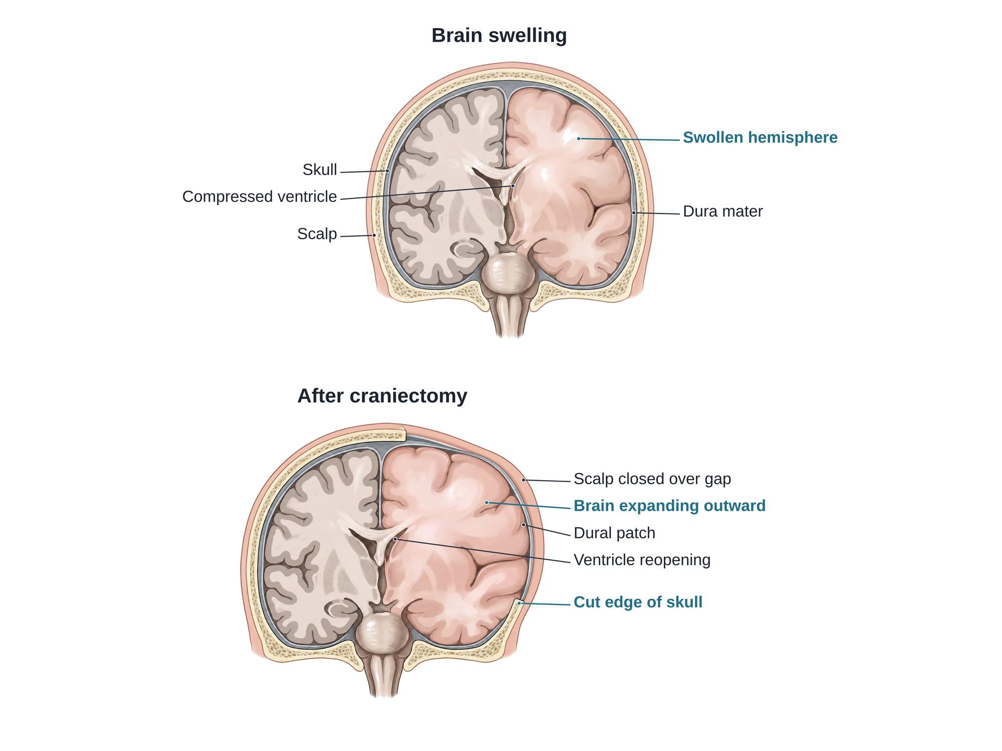
Decompressive craniectomy is an emergency brain operation in which a surgeon removes a large piece of the skull to relieve pressure from a swollen brain. The skull is a closed box. When the brain swells after a severe head injury or a large stroke, the pressure inside the skull (intracranial pressure) rises and squeezes the brain. Removing the bone, and opening the dura (the tough outer covering of the brain), lets the brain swell outward instead of being crushed. The bone is put back months later. The illustration above shows a swollen brain before surgery and the same brain after the bone is removed.
The anatomy
The brain sits inside the skull under three coverings called the meninges. The outer one, the dura mater, is a thick sheet lining the inside of the skull. A fold of dura, the falx cerebri, hangs down between the two halves of the brain. Fluid-filled spaces called the lateral ventricles sit deep inside each half. For the normal anatomy, see brain anatomy and skull anatomy.
When one half of the brain swells, it pushes the midline structures toward the other side. This is called midline shift. The ventricle on the swollen side gets squeezed flat. If the pressure keeps rising, brain tissue can be pushed under the falx or down through the opening at the base of the skull. This is called brain herniation, and it can be fatal.
Why it is done
Decompressive craniectomy is used when brain swelling threatens life and other treatment is not enough. Common reasons are:
- Severe traumatic brain injury (TBI) from a car crash, fall or assault, when pressure stays high despite medicine, sedation and fluid drainage
- A large stroke in the middle cerebral artery territory (malignant MCA infarction), which can swell over the first few days
- Bleeding in the brain, sometimes done at the same time as removing a blood clot such as an acute subdural hematoma
- Less often, severe brain infection or swelling from other causes
When the bone is removed during surgery to remove a clot, it is called a primary craniectomy. When it is done later because pressure keeps rising, it is called a secondary craniectomy.
Types
| Type | Bone removed | Typical use |
|---|---|---|
| Unilateral hemicraniectomy | A large flap over the front, side and back of one half of the head (frontotemporoparietal) | Swelling mostly on one side, such as a stroke or a subdural hematoma |
| Bifrontal craniectomy | Bone across the whole forehead region on both sides | Swelling of both frontal lobes after a diffuse brain injury |
The flap is usually large, often about the size of a hand. A small opening can let the brain push through its edges and get injured.
How the surgery is done
The operation is done under general anesthesia, usually as an emergency. It often takes 2 to 3 hours.
- Positioning. The head is turned and held still. The hair over the surgical area is shaved.
- Scalp incision. A large curved incision, often shaped like a question mark, is made from in front of the ear up over the top of the head. The scalp and the temporal muscle are folded back.
- Burr holes. Several small holes are drilled through the skull.
- Bone flap removal. A saw connects the burr holes, and the large piece of skull is lifted off. The bone is saved for later.
- Opening the dura. The dura is cut open so the brain can expand. Any blood clot is removed.
- Dural patch (duraplasty). A loose patch of graft material is sewn or laid over the brain to cover it while giving it room to bulge outward.
- Closing. The scalp is closed over the patch. A drain and a pressure monitor are often left in place.
Recovery
- Patients go to the intensive care unit, usually on a breathing machine at first. The pressure inside the head is watched closely.
- The ICU stay often lasts days to weeks, depending on the brain injury.
- Until the bone is replaced, the soft area of the head is protected with a helmet whenever the patient is out of bed.
- Cranioplasty, the operation to replace the bone or a custom implant, is usually done about 6 weeks to 6 months later.
- Most patients need months of rehabilitation. How much they recover depends mostly on the original brain injury, not the operation.
Risks
- Infection of the wound, the dural patch or the stored bone
- Bleeding, including new bleeding on either side of the brain
- Hydrocephalus, a build-up of brain fluid that may need a shunt
- Subdural hygroma, a fluid collection under the dura
- Seizures
- Brain injury at the bone edge, when swelling pushes the brain against the cut edge of the skull
- Sinking skin flap syndrome (syndrome of the trephined), where the scalp sinks in and causes headache, dizziness or new weakness until the bone is replaced
- Survival with severe disability, which families should understand before agreeing to surgery
How decompressive craniectomy is shown in a personal injury case
A decompressive craniectomy marks a severe traumatic brain injury. It usually follows a car crash, a fall from height, or an assault. It signals life-threatening swelling, a long hospital stay, a second operation for the cranioplasty, and often lifelong care needs. Imaging reports describe midline shift and effaced ventricles, but those words mean little to a jury. A medical illustration of the plaintiff's own swollen brain before surgery, and of the skull opened to let it expand, shows why the surgery was needed and what was removed. For how these images are used at trial, see demonstrative evidence and medical legal illustration.
Related pages
FAQ
What is the difference between a craniotomy and a craniectomy?+
In a craniotomy, the surgeon removes a piece of skull (the bone flap) to reach the brain and puts it back at the end of the operation. In a craniectomy, the bone flap is left out. A decompressive craniectomy leaves it out on purpose, so the swollen brain has room to expand. The bone is replaced weeks to months later in a second operation called a cranioplasty.
What is a burr hole?+
A burr hole is a small round hole drilled through the skull, usually about the size of a coin or smaller. It is used to drain a chronic subdural hematoma, to place a pressure monitor or drain, or as the starting point for cutting a larger bone flap. A decompressive craniectomy usually begins with several burr holes that are then joined to free the bone.
What is the survival rate after a decompressive craniectomy?+
It depends heavily on the cause, the patient's age and how severe the brain injury is. In severe traumatic brain injury with very high pressure, the operation lowers the risk of death compared with medicine alone. But many survivors live with moderate or severe disability. Doctors discuss these likely outcomes with the family before surgery.
How long is the skull left open after a craniectomy?+
The bone is usually replaced once the swelling has gone down and the patient is stable, most often about 6 weeks to 6 months later. Until then the brain is covered only by the dura and scalp, and the patient wears a protective helmet when out of bed.
Where is the bone flap kept?+
Most hospitals freeze the bone flap in a sterile bone bank. Some place it under the skin of the abdomen to keep it alive. If the bone cannot be used, the cranioplasty is done with a custom implant made of titanium, PEEK plastic or another material.
What are the long-term side effects of a craniectomy?+
Long-term effects come mostly from the brain injury itself: weakness on one side, speech and memory problems, personality changes and seizures. Problems from the operation include hydrocephalus (fluid build-up), infection, and a sunken skin flap that can cause headaches or new weakness until the bone is replaced.
What therapy is needed after a decompressive craniectomy?+
Most patients need intensive rehabilitation. This includes physical therapy for strength and walking, occupational therapy for daily tasks, and speech therapy for talking, swallowing and thinking skills. Rehab often starts in the hospital and continues for months.
Can you take a bath after brain surgery?+
Patients are usually told to keep the incision dry until it has healed and the stitches or staples are out, often about 1 to 2 weeks. Showers are usually allowed before baths. Soaking the head in a bath, pool or hot tub is avoided until the surgeon says the wound has fully healed.
Need a surgical illustration for consent or a case?
Describe the operation and get a clear, labeled medical illustration for patient consent, teaching or a case exhibit.
Try Natomy →