Colles Fracture: X-ray, Dinner Fork Deformity & Treatment
A Colles fracture is a break of the distal radius with the end tilted toward the back of the hand. See it on X-ray beside a labeled illustration.
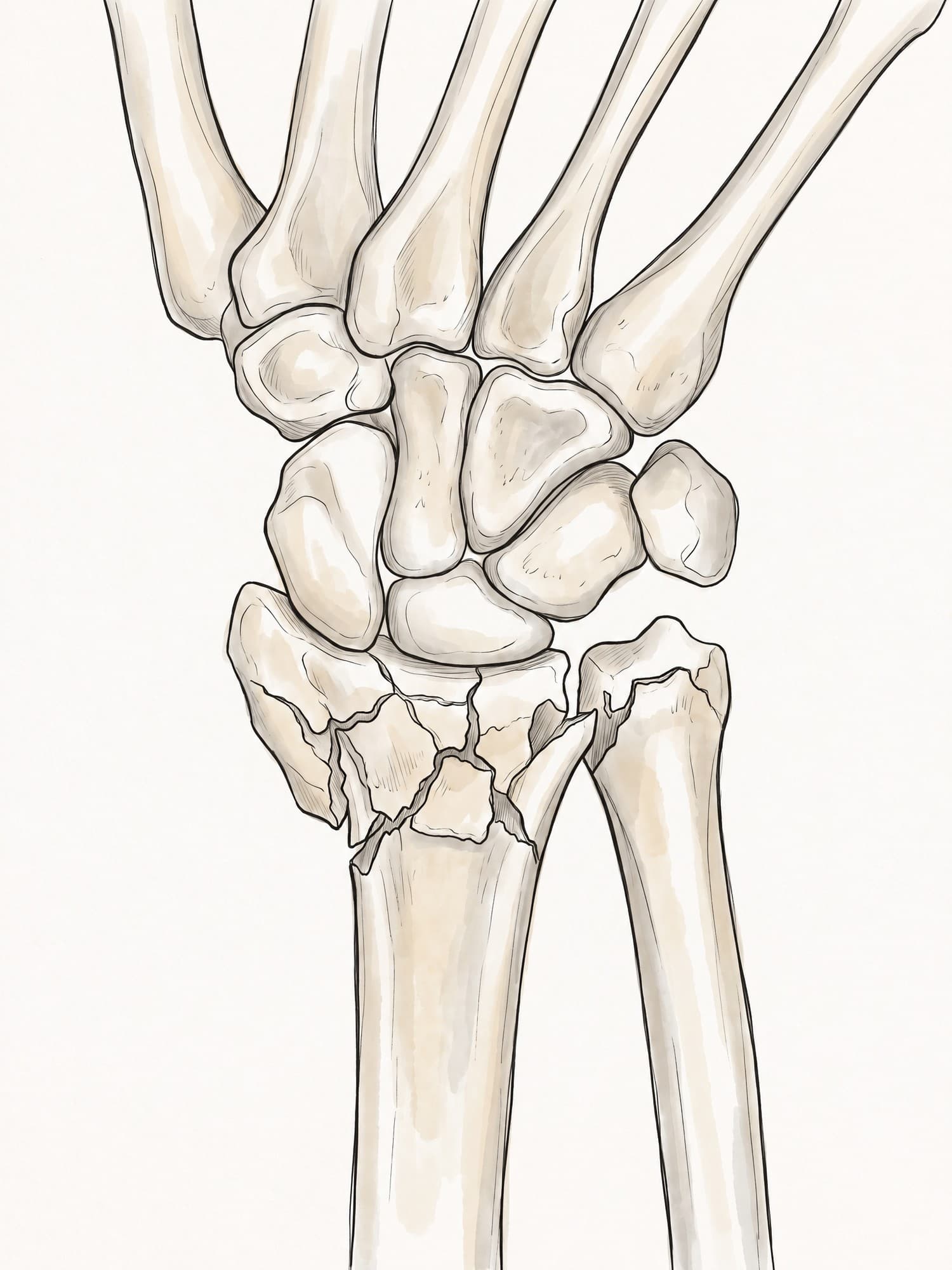
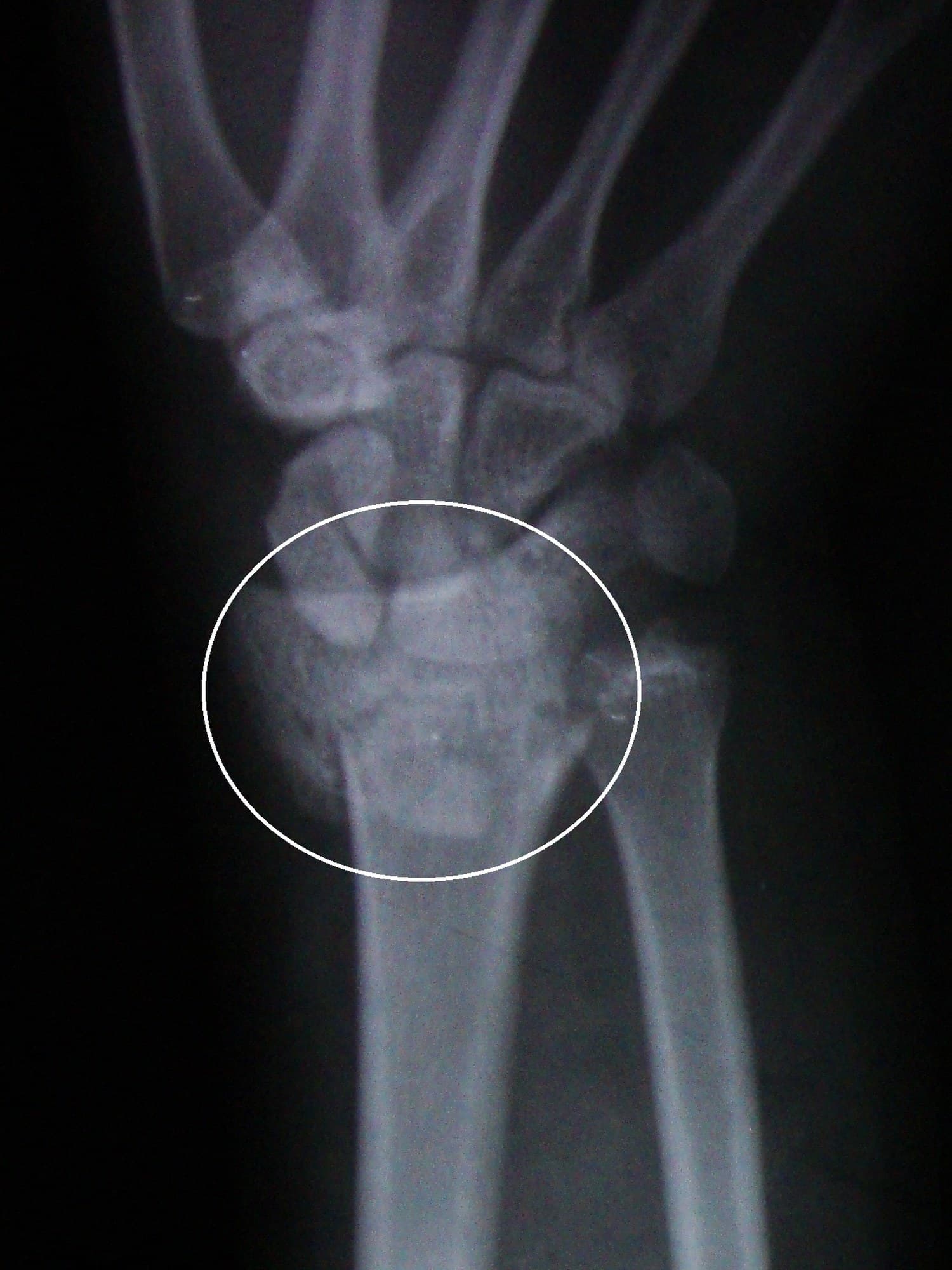
A Colles fracture is a break of the distal radius, the end of the forearm bone on the thumb side, about 2 to 3 cm above the wrist joint, in which the broken end shifts and tilts toward the back of the hand. It is the classic injury from a fall on an outstretched hand, and it gives the wrist a bent "dinner fork" shape when viewed from the side. The figure above shows a real wrist X-ray of a Colles fracture with a medical illustration drawn from it: drag the slider to compare them.
What is a Colles fracture?
The radius is the larger of the two forearm bones at the wrist, and its wide end forms most of the wrist joint with the scaphoid and lunate. In a Colles fracture this end breaks across, and the piece nearest the hand (the distal fragment) moves in three ways:
- Dorsal tilt: it tips toward the back of the hand instead of its normal slight tilt toward the palm.
- Dorsal shift: it slides toward the back of the wrist.
- Shortening: it is pushed up into the shaft, so the radius ends up shorter than the ulna.
The fracture was described by the Irish surgeon Abraham Colles in 1814, before X-rays existed, from the shape of the injured wrist alone. Strictly, the term means a fracture that stays outside the joint, but in practice it is used for most distal radius fractures displaced toward the back of the hand. The tip of the ulna, the ulnar styloid, breaks off at the same time in many cases, as it does in the X-ray above. For the normal bones and joints of the wrist, see hand and wrist anatomy.
Dinner fork deformity
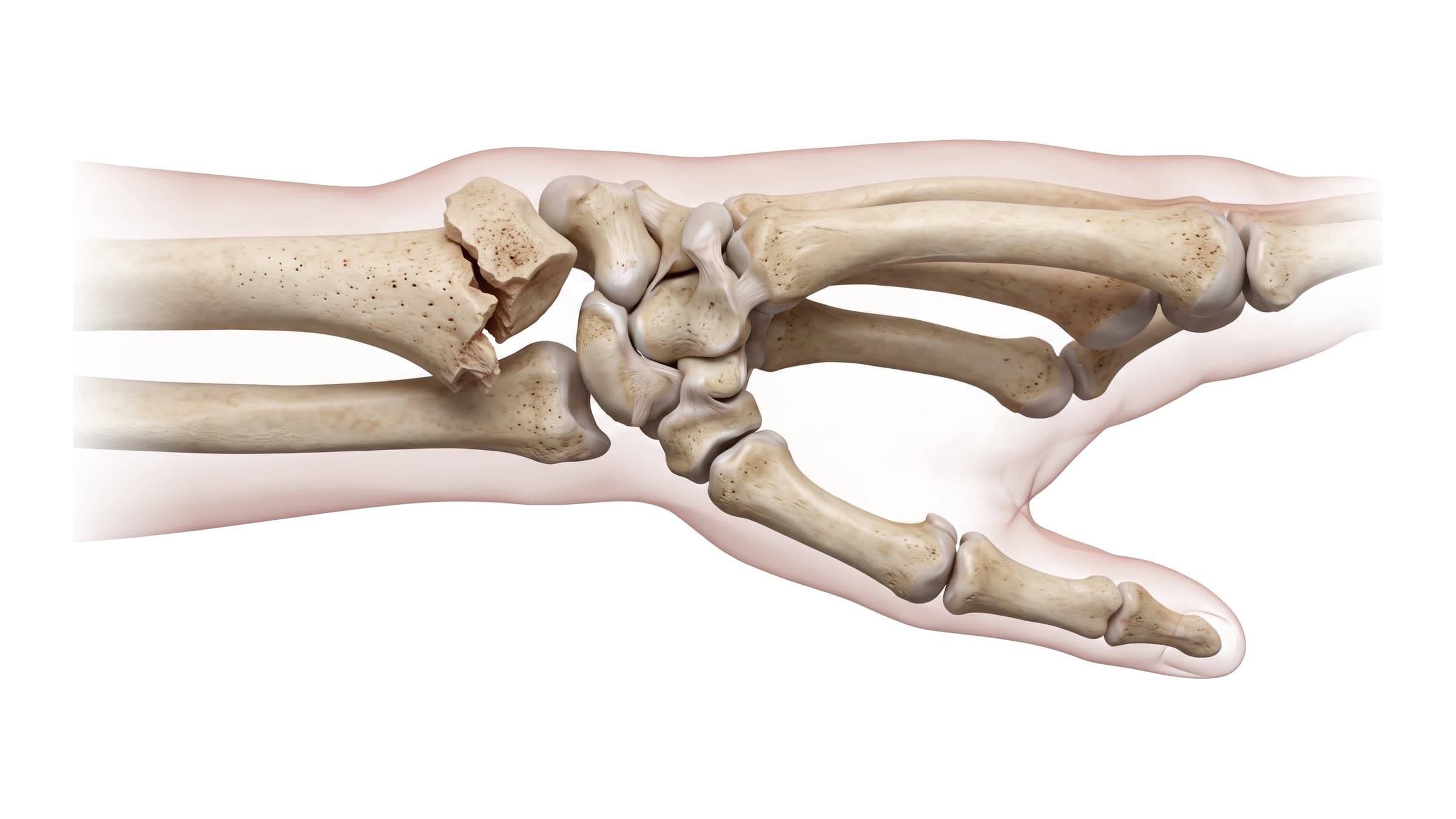
Seen from the side, a displaced Colles fracture bends the wrist into the shape of a dinner fork turned upside down: a step up at the back of the wrist where the fragment and the hand have shifted, and a dip just above it. It is the sign Colles described, and it is visible before any imaging. The deformity is less obvious when the fracture is not displaced, and swelling can hide it. The figure below shows the deformity from the side, with the back of the wrist at the top.
Colles fracture on X-ray
Two standard wrist views, front (PA) and side (lateral), show the fracture and how far it has moved. The measurements that guide treatment come from them:
| Measurement | View | Normal wrist | In a displaced Colles fracture |
|---|---|---|---|
| Palmar (volar) tilt | Lateral | About 11 degrees toward the palm | Tilted toward the back of the hand |
| Radial height | PA | About 11 to 12 mm | Reduced: the radius is shortened |
| Radial inclination | PA | About 22 degrees | Reduced |
| Joint surface | PA and lateral | Smooth | A step or gap if the fracture enters the joint |
On the PA view above, the end of the radius is broken into several pieces (comminuted) and the ulnar styloid is broken too. Comminution matters because a radius in many pieces is more likely to collapse and slip in a cast. CT is added when the fracture runs into the joint and the surgeon needs to see each fragment.
How a Colles fracture happens
Most Colles fractures come from a fall on an outstretched hand (often shortened to FOOSH) with the wrist bent back. The force travels up through the palm into the end of the radius, which fails on its palm side and collapses toward the back. Two groups are most affected:
- Older adults, especially women with osteoporosis, after a simple fall from standing. A wrist fracture from a low-energy fall is often the first sign of osteoporosis.
- Younger adults, after higher-energy injuries such as sports, bicycle or motorcycle crashes, and falls from a height. These fractures are more often comminuted and more often enter the joint.
Symptoms
- Pain and swelling at the wrist, worst when moving it or gripping
- The dinner fork shape from the side, when the fracture is displaced
- Bruising that spreads into the hand and forearm over the next days
- Numbness or tingling in the thumb, index and middle fingers if the median nerve is compressed
Related wrist fractures
The distal radius breaks in several named patterns. They are told apart on X-ray by which way the fragment moves and whether the joint surface is involved:
- Smith fracture: the reverse of a Colles fracture, with the fragment tilted toward the palm, usually from a fall onto a flexed wrist.
- Barton fracture: a piece of the joint rim breaks off and the carpal bones shift with it.
- Chauffeur's fracture: a break of the radial styloid, the bony point on the thumb side of the wrist.
- Buckle (torus) fracture: in children, the soft bone bulges on one side without breaking through.
Treatment
The aim is a radius of normal length, a near-normal tilt, and a smooth joint surface, held there until the bone heals.
- Closed reduction and casting: under a local anesthetic block, the fragment is pulled back into place and held in a splint, then a cast, for about 6 weeks. X-rays are repeated over the first weeks because some fractures slip in the cast.
- ORIF with a volar plate: open reduction and internal fixation, in which a surgeon realigns the fragments through an incision on the palm side of the wrist and fixes them with a plate and screws. It is used for unstable, comminuted or joint fractures and for fractures that slip after reduction.
- K-wires or external fixation: pins through the skin, or a frame outside the arm, to hold fragments that a plate cannot.
Commonly used targets for an acceptable result are radial shortening under about 3 mm, dorsal tilt under about 10 degrees, and a joint step under 2 mm, adjusted for the patient's age and activity.
Recovery time
The bone itself heals in about 6 to 8 weeks. Hand therapy then works on stiffness, grip and forearm rotation. Most people are back to everyday tasks within about 3 months, and strength and range of motion keep improving for 6 to 12 months. Recovery is slower after comminuted or joint fractures and in older patients.
Complications
- Malunion: the radius heals short or tilted, which can leave a visible deformity, weak grip and pain at the ulnar side of the wrist.
- Median nerve compression: acute carpal tunnel syndrome from swelling or the displaced fragment.
- Complex regional pain syndrome: severe pain, swelling, and skin and temperature changes out of proportion to the injury.
- Tendon rupture: the thumb's extensor pollicis longus tendon can fray against the fracture and rupture weeks later, even after a minor fracture.
- Post-traumatic arthritis: when the joint surface heals with a step.
- Stiffness: loss of wrist bending and forearm rotation.
How a Colles fracture is shown in a personal injury case
Wrist fractures from falls and vehicle crashes are common personal injury claims, and the X-ray is usually the key image. Most jurors cannot read one. An illustration drawn directly from the patient's own X-ray, like the one above, shows the same fracture in a form they can follow: which bone broke, which way it moved, and how many pieces it broke into. A typical set of exhibits pairs the X-ray and its illustration with a normal wrist for comparison, the fall that caused it, and the plate and screws placed at surgery. For how these images are used at trial, see demonstrative evidence, trial exhibits and medical legal illustration.
Related pages
FAQ
What is a Colles fracture?+
A Colles fracture is a break of the distal radius, about 2 to 3 cm above the wrist joint, in which the broken end of the radius shifts and tilts toward the back of the hand. It usually happens in a fall on an outstretched hand and is one of the most common fractures seen in emergency departments.
What is the dinner fork deformity?+
The dinner fork deformity is the shape of the wrist after a displaced Colles fracture, seen from the side. The broken end of the radius and the hand shift toward the back of the wrist, so the forearm, wrist and hand curve like the back of a fork.
What is the difference between a Colles fracture and a Smith fracture?+
Both are fractures of the distal radius. In a Colles fracture the broken end tilts toward the back of the hand (dorsal displacement), usually after a fall on an outstretched hand. In a Smith fracture, sometimes called a reverse Colles fracture, it tilts toward the palm, usually after a fall onto a flexed wrist.
Is a Colles fracture the same as a distal radius fracture?+
A Colles fracture is one type of distal radius fracture: the type in which the broken end is displaced toward the back of the hand. Other distal radius fractures include the Smith, Barton and chauffeur's (radial styloid) fractures, and fractures that are not displaced at all.
How long does a Colles fracture take to heal?+
The bone usually heals in about 6 to 8 weeks, which is roughly how long a cast or splint is worn. Stiffness and weakness often last longer: most people return to everyday tasks within about 3 months, and full strength can take 6 to 12 months, especially after surgery or in older patients.
Does a Colles fracture need surgery?+
Not always. Many Colles fractures are set back into position (closed reduction) and treated in a splint and cast. Surgery, most often a plate and screws on the palm side of the radius (ORIF), is considered when the fracture is unstable, slips after reduction, enters the joint with a step, or leaves the radius too short or too tilted.
What does a Colles fracture look like on an X-ray?+
On the side (lateral) view, the end of the radius is tilted toward the back of the wrist instead of slightly toward the palm. On the front (PA) view, the radius is often shortened and the fracture line runs across it just above the joint, sometimes with several fragments and a broken tip of the ulna (ulnar styloid).
Can you move your fingers with a Colles fracture?+
Usually yes, although it hurts. Being able to move the fingers does not rule out a fracture. Numbness or tingling in the thumb, index and middle fingers can mean pressure on the median nerve and needs prompt medical attention.
Need an illustration of a patient's own X-ray?
Upload an X-ray, CT or MRI and turn it into a clear, labeled medical illustration for a consult, a chart or a case exhibit.
Try Natomy →