Leg Nerves and Dermatomes: Lower Limb Nerves Labeled
Leg dermatome map and nerves of the lower limb labeled: L1 to S1 dermatomes, the femoral, sciatic, tibial and peroneal nerves, and nerves of the foot.
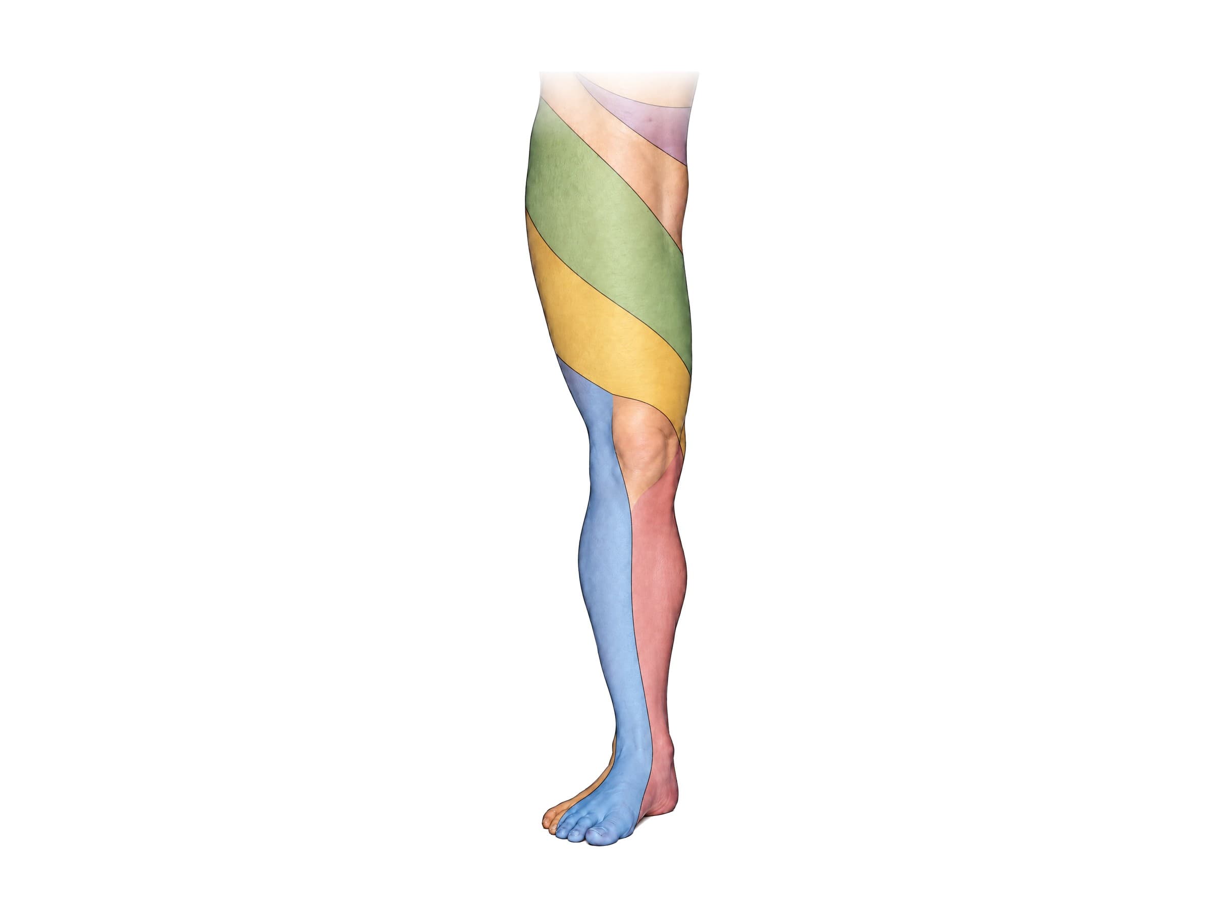
The nerves of the lower limb come from the lumbar and sacral spinal nerve roots (L1 to S3) and supply every muscle and patch of skin of the hip, thigh, leg and foot. Two maps describe how they reach the skin: the dermatome map, which shows the band of skin supplied by each spinal nerve root, and the cutaneous nerve map, which shows the area supplied by each named nerve, such as the saphenous or sural nerve. The main nerves are the femoral, obturator and lateral femoral cutaneous nerves from the lumbar plexus, and the sciatic nerve from the sacral plexus, which divides into the tibial and common peroneal (fibular) nerves. Matching a patient's numbness or weakness to these maps is how a clinician tells a pinched nerve root in the lower back from a nerve injury in the leg.
Leg dermatomes (L1 to S1)
The labeled view above shows the modern dermatome map on the front of the right leg. A dermatome is the strip of skin supplied by one spinal nerve root. Neighboring dermatomes overlap, so the borders are approximate, but the test points are consistent:
- L1: the groin and upper inner thigh, just below the crease of the hip.
- L2: an oblique band across the upper and middle front of the thigh. Test point: the middle of the front of the thigh.
- L3: an oblique band across the lower front of the thigh to the inner side of the knee. Test point: the inner knee.
- L4: the inner (medial) shin down to the inner ankle and the inner edge of the foot. Test point: the inner ankle (medial malleolus).
- L5: the outer (lateral) shin and the top of the foot, including the big toe and middle toes. Test point: the web space between the big toe and second toe.
- S1: the outer edge of the foot, the little toe, most of the sole and the heel. Test point: the outer side of the heel.
On the back of the leg, S1 continues up the outer calf, S2 forms a strip down the middle of the back of the thigh and calf, and S3 to S5 form rings around the buttocks, perineum and anus (the saddle area). Numbness in the saddle area is a warning sign of cauda equina syndrome; see lumbar spine anatomy.
The leg dermatomes follow the way the limb bud rotates in the embryo, which is why the bands spiral from the back of the hip around to the front of the thigh and down the inner leg, and why L5 ends on the top of the foot and S1 on the sole.
Myotomes and reflexes of the lower limb
Each root also powers particular movements (its myotome) and reflexes. A standard neurological exam tests:
- L2: hip flexion (lifting the thigh).
- L3: knee extension (straightening the knee).
- L4: ankle dorsiflexion (lifting the foot); the knee (patellar) reflex (L3, L4).
- L5: big toe extension (lifting the big toe).
- S1: ankle plantarflexion (standing on tiptoe); the ankle (Achilles) reflex.
Together with the dermatomes, these tests let a clinician name the compressed root: for example, numbness in the first web space with weak big toe extension and a normal ankle reflex points to L5, usually from an L4-L5 disc herniation.
Cutaneous nerves of the leg
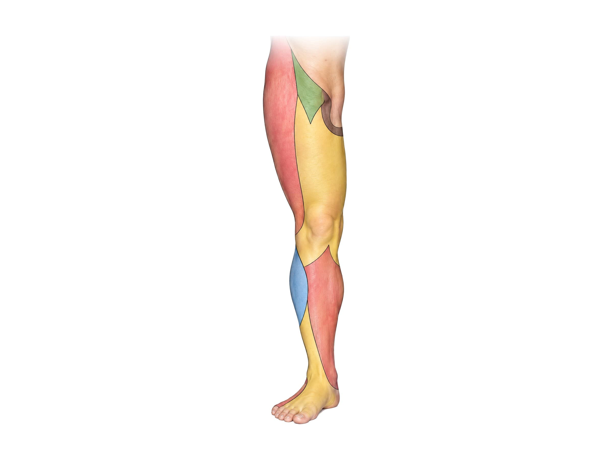
The cutaneous nerve map shows the skin supplied by each named peripheral nerve. On the front of the leg:
- Lateral femoral cutaneous nerve (L2, L3): the outer thigh. Compression under the inguinal ligament causes burning numbness of the outer thigh (meralgia paresthetica), common with tight belts, pregnancy and weight gain.
- Genitofemoral and ilioinguinal nerves (L1, L2): the groin and upper inner thigh.
- Anterior femoral cutaneous nerves (L2, L3): branches of the femoral nerve to the front and inner thigh.
- Saphenous nerve (L3, L4): the inner knee, inner shin and inner ankle; the long continuation of the femoral nerve.
- Lateral sural cutaneous nerve (from the common peroneal nerve): the upper outer calf.
- Superficial peroneal nerve (L4 to S1): the lower outer shin and most of the top of the foot. A small area between the big and second toes belongs to the deep peroneal nerve (L4, L5).
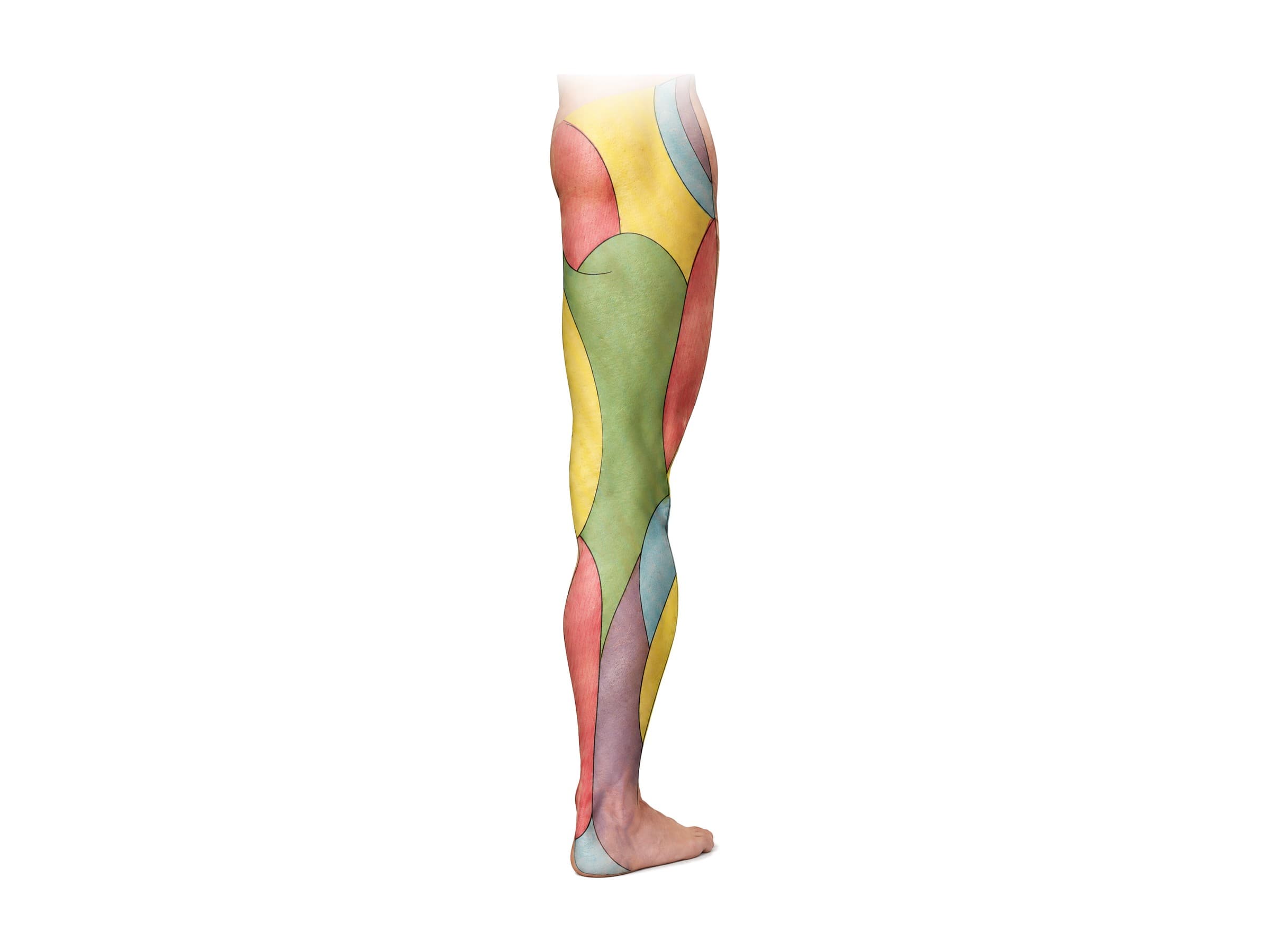
On the back of the leg:
- Cluneal nerves: the skin of the buttock.
- Posterior femoral cutaneous nerve (S1 to S3): the back of the thigh and the back of the knee.
- Sural nerve (S1, S2): the outer calf, outer ankle and outer edge of the foot. Surgeons often take it as a nerve graft because losing it leaves only a small numb patch.
- Tibial nerve (calcaneal branches) (S1, S2): the heel.
The difference between the two maps matters clinically. Numbness from an L5 root compression follows the L5 band down the outer shin and over the foot. Numbness from a common peroneal nerve injury at the knee covers a similar area but spares the sole and comes with weakness only of the muscles that nerve supplies.
Nerves of the lumbar plexus: the front of the thigh
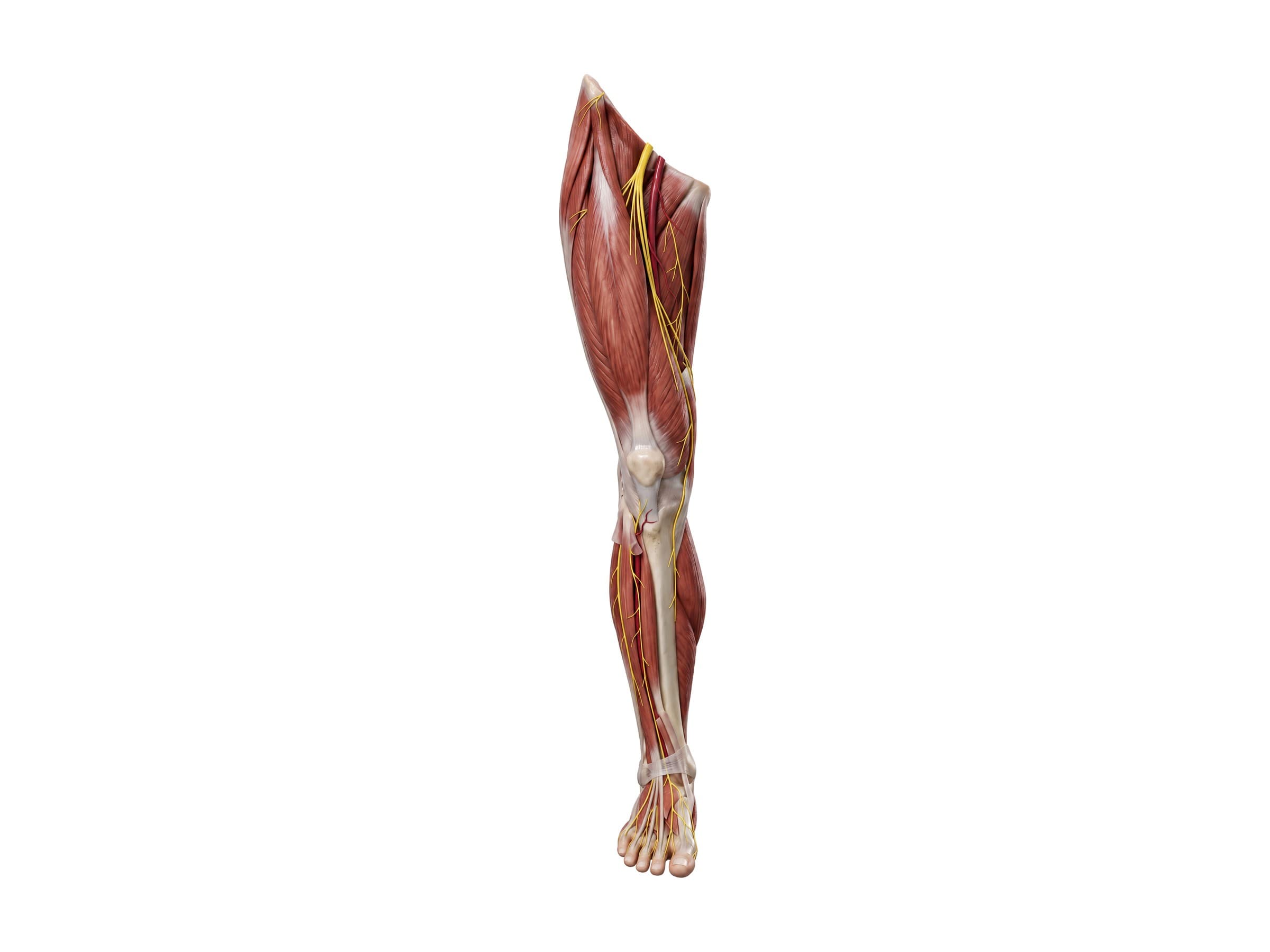
The lumbar plexus (L1 to L4) forms inside the psoas major muscle; see lumbar spine anatomy for the plexus itself. Its main branches to the leg are:
- Femoral nerve (L2 to L4): the largest branch. It passes under the inguinal ligament just outside the femoral artery, supplies the quadriceps (which straighten the knee) and the skin of the front of the thigh, and continues as the saphenous nerve down the inner leg. Femoral nerve injury causes a weak, buckling knee and an absent knee reflex.
- Obturator nerve (L2 to L4): runs through the obturator foramen of the pelvis to the inner thigh, supplying the adductor muscles that pull the legs together. It can be injured in pelvic fractures, hip surgery and childbirth.
- Lateral femoral cutaneous nerve (L2, L3): purely sensory, to the outer thigh.
Nerves of the sacral plexus: the sciatic nerve
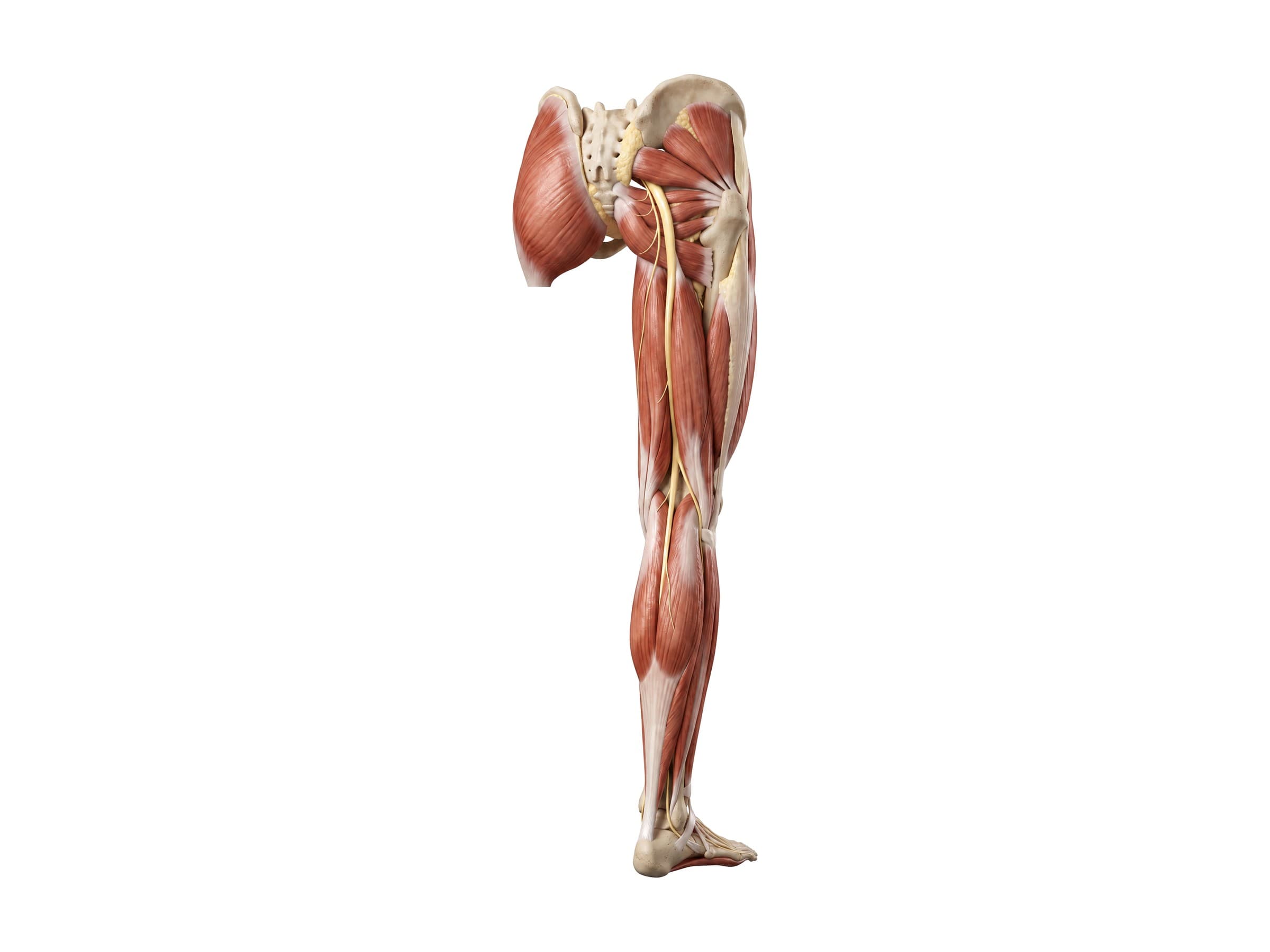
The sacral plexus (L4 to S3) forms on the back wall of the pelvis. Its main branch is the sciatic nerve, the largest nerve in the body, about as wide as a thumb where it leaves the pelvis through the greater sciatic notch below the piriformis muscle. It runs down the back of the thigh under the hamstrings, which it supplies, and divides above the knee, usually at the top of the popliteal fossa, into two nerves:
- Tibial nerve (L4 to S3): continues straight down through the back of the knee and the calf, supplies the calf muscles that point the foot and curl the toes, passes behind the inner ankle through the tarsal tunnel, and divides into the medial and lateral plantar nerves of the sole.
- Common peroneal (common fibular) nerve (L4 to S2): runs to the outer side of the knee and wraps around the neck of the fibula, just under the skin, where it divides into the superficial and deep peroneal nerves.
The sacral plexus also gives the superior and inferior gluteal nerves to the gluteal muscles, the posterior femoral cutaneous nerve, and the pudendal nerve to the perineum. "Sciatica" usually means pain from compression of the L4, L5 or S1 roots that form the sciatic nerve, not of the nerve itself.
Peroneal (fibular) nerves of the lower leg
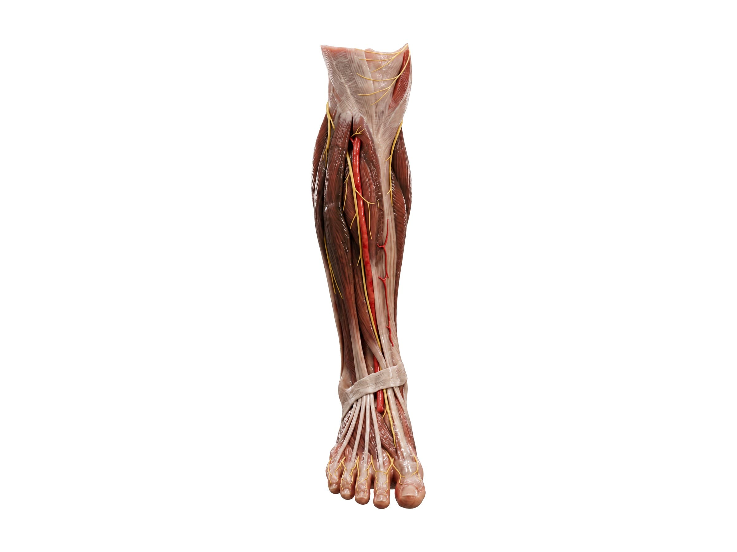
- Deep peroneal nerve: runs down the front of the shin with the anterior tibial artery, supplies the muscles that lift the foot and toes (tibialis anterior and the toe extensors), and ends as a small sensory branch to the web space between the big and second toes.
- Superficial peroneal nerve: runs down the outer side of the leg, supplies the peroneal muscles that turn the foot outward, and pierces the fascia above the ankle to supply most of the top of the foot.
Nerves of the foot
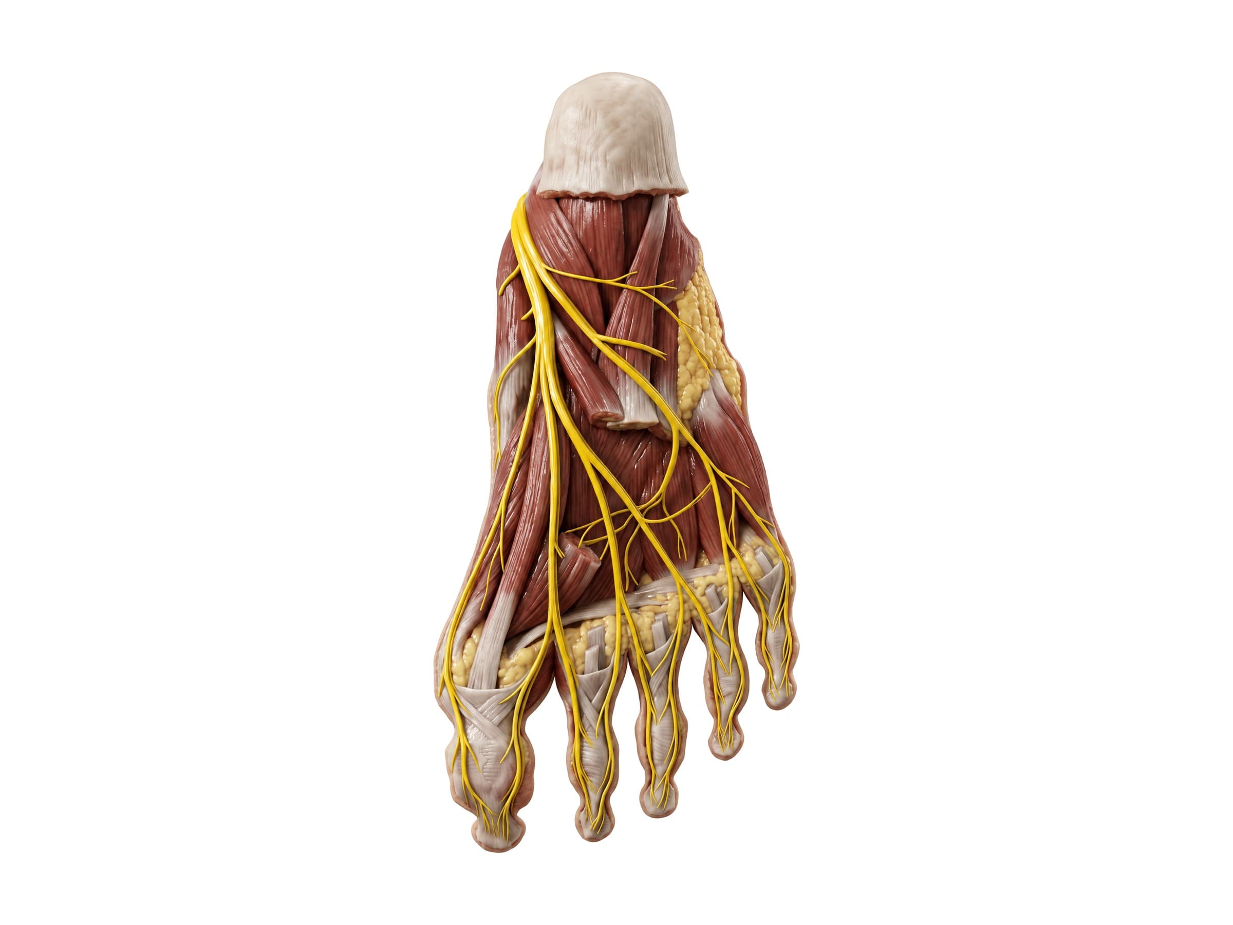
Behind the inner ankle the tibial nerve divides into the medial plantar nerve, which supplies the inner two-thirds of the sole and the big toe side (like the median nerve in the hand), and the lateral plantar nerve, which supplies the outer sole and the little toe side and most of the small muscles of the foot (like the ulnar nerve). Their plantar digital nerves run along the sides of each toe. A thickened digital nerve between the third and fourth toes is a Morton's neuroma, and compression of the tibial nerve behind the ankle is tarsal tunnel syndrome.
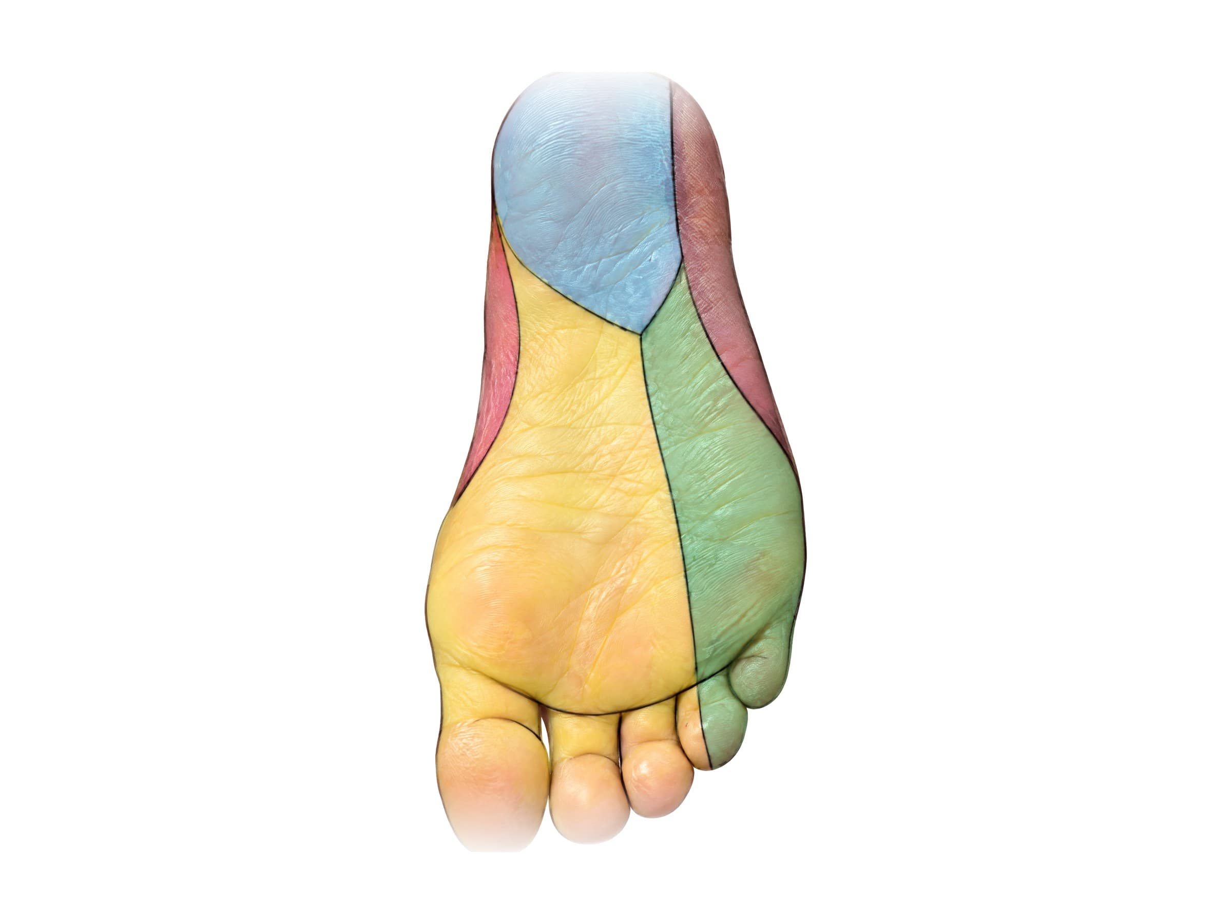
The skin of the sole is shared by five nerves: the tibial nerve's calcaneal branches over the heel, the medial plantar nerve over the inner sole, the lateral plantar nerve over the outer sole, the sural nerve along the outer edge, and the saphenous nerve along the inner edge. For the bones underneath, see foot bones.
Common nerve injuries of the leg and how they are diagnosed
Nerve problems are diagnosed by matching symptoms to the maps above, then confirmed with nerve conduction studies and electromyography (EMG), which show where along the nerve the signal is slowed or blocked. MRI shows compression of the nerve roots in the spine, and ultrasound or MRI can show a damaged nerve in the leg.
- Lumbar radiculopathy (sciatica): a disc herniation or stenosis compresses an L4, L5 or S1 root, causing pain, numbness and weakness in that root's dermatome and myotome. The most common cause of leg nerve symptoms after a back injury.
- Common peroneal nerve injury: the most common nerve injury in the leg, from a blow to the outer knee, a fracture of the fibular neck, a knee dislocation, a tight cast, prolonged crossing of the legs, or positioning during surgery. It causes foot drop: the foot slaps down and the toes catch, so the person lifts the knee high to walk (steppage gait), with numbness over the outer shin and top of the foot.
- Sciatic nerve injury: from posterior hip dislocation in a car crash (the knee hits the dashboard), acetabular fractures, hip replacement surgery, or a misplaced buttock injection. The peroneal part of the nerve is most vulnerable, so foot drop is the most common result.
- Femoral nerve injury: from pelvic fractures, bleeding in the psoas, or groin and hip surgery; causes knee buckling and numbness of the front of the thigh.
- Tibial nerve injury: from knee dislocations and fractures of the upper tibia; causes weakness pushing off and numbness of the sole.
- Compartment syndrome: swelling inside a closed muscle compartment of the leg after a fracture or crush injury cuts off blood flow to the nerves and muscles. Early numbness in the first web space (deep peroneal nerve) is a warning sign; untreated, it causes permanent nerve damage within hours.
- Peripheral neuropathy: damage to the longest nerves first, most often from diabetes, causing numbness and burning in both feet in a stocking pattern rather than in one dermatome or nerve territory.
How leg nerves are shown in a personal injury case
Nerve injuries are among the most consequential and least visible injuries in personal injury and medical malpractice claims. A plaintiff with foot drop or permanent numbness may look normal sitting in a courtroom, and the defense often argues that the symptoms are exaggerated or come from a pre-existing back problem rather than the incident. The dermatome and cutaneous nerve maps answer that argument directly: they show whether the pattern of numbness matches a nerve root in the spine (and therefore a disc injury) or a nerve in the leg (and therefore a direct injury at the knee or hip). Each injury follows a recognizable mechanism of injury: a peroneal nerve injury from a bumper striking the outer knee, a sciatic nerve injury from a dashboard driving the femur out of the hip socket, an L5 radiculopathy from a disc herniation after a rear-end collision. A labeled illustration of the normal nerves and dermatomes is the baseline exhibit, and an illustration of the injured nerve, with the matching patch of numb skin shaded, connects the EMG findings to what the plaintiff feels every day. In surgical malpractice claims, an illustration of where the nerve runs explains how a positioning injury, a misplaced injection or a retractor caused the damage.
Natomy creates custom medical legal illustrations of nerve injuries for demand packages and trial exhibits, and the rules for using these visuals at trial are covered in demonstrative evidence. For where the nerve roots start, see lumbar spine anatomy and thoracic spine anatomy. Related atlas pages: hip anatomy, knee anatomy, ankle anatomy and foot bones.
FAQ
What are dermatomes?+
A dermatome is the strip of skin supplied by the sensory fibers of a single spinal nerve root. Each root from C2 down to S5 has one, and together they form a map of bands around the body. Because a compressed nerve root causes numbness, tingling or pain in its own band, clinicians test sensation in each dermatome to work out which level of the spine is involved, often before an MRI confirms it.
What are the dermatomes of the leg?+
On the front of the leg, L1 covers the groin, L2 the upper front thigh and L3 the lower front thigh and inner knee, in oblique bands. L4 runs down the inner shin to the inner ankle. L5 covers the outer shin and the top of the foot, including the big toe. S1 covers the outer edge and sole of the foot, the little toe and the heel. On the back, S1 runs up the outer calf, S2 covers a strip down the back of the thigh and calf, and S3 to S5 form rings around the buttocks and perineum (the saddle area).
What is the L5 dermatome?+
The L5 dermatome covers the outer (lateral) side of the lower leg, the top of the foot and the big toe. A key test point is the web space between the big toe and the second toe. Numbness here, with weakness lifting the big toe or the front of the foot, points to an L5 nerve root problem, most often from a disc herniation at L4-L5.
What is the S1 dermatome?+
The S1 dermatome covers the outer edge of the foot, the little toe, most of the sole and heel, and a strip up the back of the calf. The test point is the outer side of the heel. Numbness here, with weakness standing on tiptoe and a reduced ankle reflex, points to an S1 nerve root problem, usually from a disc herniation at L5-S1.
What is the difference between a dermatome and a peripheral nerve distribution?+
A dermatome is the skin supplied by one spinal nerve root; a peripheral nerve territory is the skin supplied by one named nerve, such as the saphenous or sural nerve, which usually carries fibers from several roots. They overlap differently: numbness from an L5 root compression follows the L5 band, while numbness from a common peroneal nerve injury at the knee covers the outer shin and top of the foot but spares the sole. Comparing the pattern with both maps tells a clinician whether the problem is in the spine or in the leg.
What are the main nerves of the leg?+
The leg is supplied by two plexuses. From the lumbar plexus (L1 to L4), the femoral nerve supplies the front of the thigh and the quadriceps and continues as the saphenous nerve down the inner leg; the obturator nerve supplies the inner thigh; and the lateral femoral cutaneous nerve supplies the skin of the outer thigh. From the sacral plexus (L4 to S3), the sciatic nerve runs down the back of the thigh and divides above the knee into the tibial nerve (calf and sole) and the common peroneal (fibular) nerve (front and outer leg and top of the foot).
What does the common peroneal nerve do?+
The common peroneal (common fibular) nerve branches from the sciatic nerve above the knee and wraps around the neck of the fibula just below the outer knee, where it lies just under the skin. Its deep branch lifts the foot and toes, and its superficial branch turns the foot outward and supplies the skin of the outer shin and top of the foot. Because it is so exposed at the fibular neck, it is the most commonly injured nerve in the leg, and injury causes foot drop.
What are the symptoms of nerve problems in the legs?+
Nerve problems in the legs cause numbness, tingling or pins and needles, burning or electric pain, weakness, and sometimes muscle wasting. The pattern points to the cause: a band of symptoms down one leg suggests a pinched nerve root in the lower back (sciatica); symptoms in one nerve's territory, such as foot drop after a knee injury, suggest a peripheral nerve injury; and symmetrical numbness starting in both feet (stocking distribution) suggests peripheral neuropathy, most often from diabetes.
Need a custom illustration of this joint?
Generate a labeled or exhibit-ready figure for a specific patient, injury, or case in minutes.
Try Natomy →