Female Abdominal Anatomy: Organs, Pelvis & Uterus Labeled
Female abdominal anatomy labeled: abdominal organs by quadrant, uterus, ovaries, fallopian tubes, ureters, pelvic floor and a side section of the pelvis.
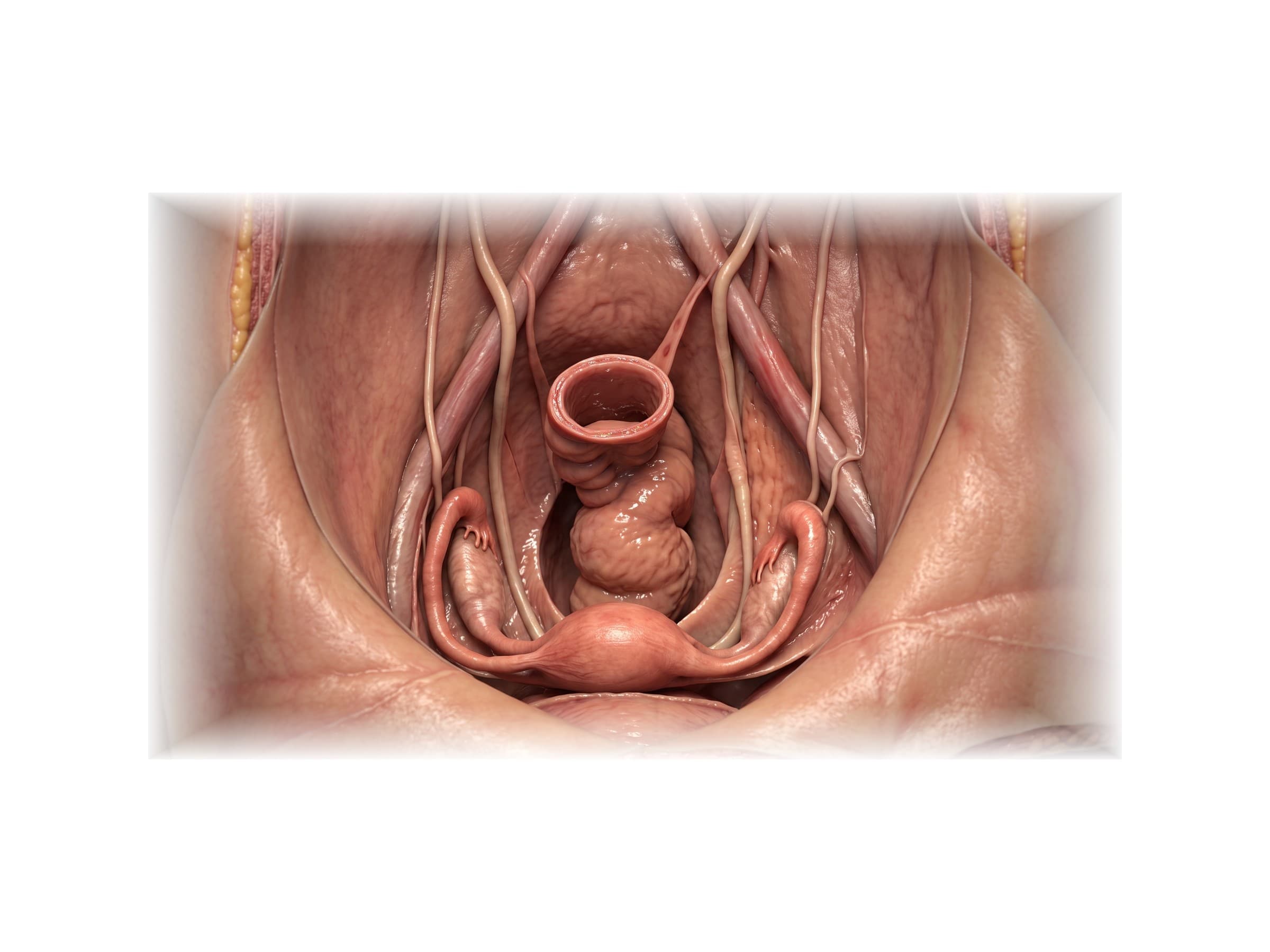
Female abdominal anatomy is the arrangement of the organs, muscles, vessels and bones between the diaphragm and the floor of the pelvis in a woman. The upper abdomen is the same in women and men: the liver, gallbladder, stomach, spleen, pancreas, kidneys and the small and large intestine, held in by the muscles of the abdominal wall and wrapped in a lining called the peritoneum. The difference is in the pelvis, where the uterus, ovaries and uterine (fallopian) tubes sit between the bladder in front and the rectum behind, the ureters pass close beside the cervix, and the bony pelvis is wider and shallower to allow childbirth. The figures on this page are labeled medical illustrations of each layer, from the abdominal organs in place down to the female pelvis, for reading imaging and operative reports, explaining a diagnosis or a surgery to a patient, and illustrating an injury or malpractice claim.
Abdominal regions and quadrants
Doctors describe the location of pain, a mass or a wound by dividing the abdomen into areas. Right and left always mean the patient's right and left.
- Four quadrants: a vertical and a horizontal line through the navel give the right upper (RUQ), left upper (LUQ), right lower (RLQ) and left lower (LLQ) quadrants. This is the system used in emergency medicine, imaging reports and the FAST ultrasound exam after trauma.
- Nine regions: two vertical lines through the middle of each collarbone and two horizontal lines (one under the ribs, one between the tops of the hip bones) give the right and left hypochondriac regions under the ribs, the epigastric region in the upper middle, the right and left lumbar (flank) regions, the umbilical region around the navel, the right and left iliac (inguinal) regions, and the hypogastric (suprapubic) region above the pubic bone.
In a woman, the hypogastric region and both lower quadrants sit over the pelvic organs, so pain there can come from the bowel, the bladder, the uterus or an ovary.
Organs of the female abdomen by quadrant
- Right upper quadrant: liver (most of it), gallbladder, duodenum, head of the pancreas, right kidney and adrenal gland, hepatic flexure of the colon.
- Left upper quadrant: stomach, spleen, tail of the pancreas, left kidney and adrenal gland, splenic flexure of the colon, part of the left lobe of the liver.
- Right lower quadrant: cecum and appendix, end of the ileum, ascending colon, right ureter, right ovary and uterine tube.
- Left lower quadrant: descending and sigmoid colon, left ureter, left ovary and uterine tube.
- Midline, low in the pelvis: the urinary bladder, the uterus and the rectum.
The kidneys, ureters, pancreas, most of the duodenum, the ascending and descending colon, the aorta and the inferior vena cava are retroperitoneal: they lie behind the peritoneum against the back wall of the abdomen, next to the lumbar spine and the psoas muscles.
Female pelvic organs
Looking down into the female pelvis from above and in front, as in the image at the top of this page, the organs sit in a row from front to back: the urinary bladder behind the pubic bone, the uterus in the middle, and the rectum against the sacrum. A uterine tube and ovary lie on each side of the uterus, against the side wall of the pelvis where the external iliac vessels and the ureter run.
The peritoneum drapes over the top of these organs like a sheet over furniture, forming two pockets:
- Vesicouterine pouch: the shallow fold between the bladder and the front of the uterus.
- Rectouterine pouch (pouch of Douglas): the deeper fold between the back of the uterus and the rectum. It is the lowest point of the abdominal cavity, so blood, pus or fluid collects there first. Free fluid in the pouch of Douglas on ultrasound can mean a ruptured ectopic pregnancy, a ruptured ovarian cyst or bleeding after abdominal trauma.
Female pelvis side view (midsagittal section)
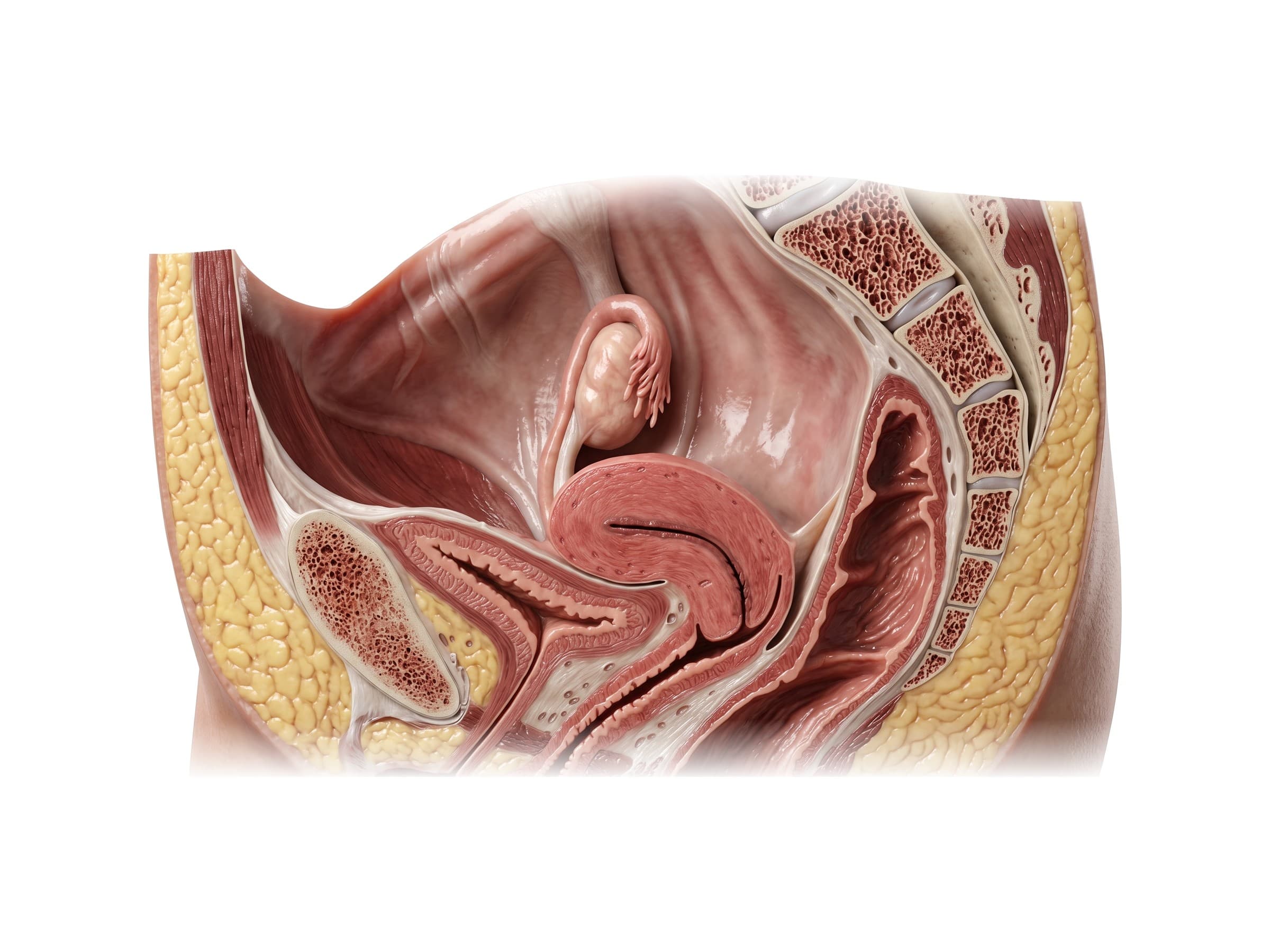
Cut down the midline, the female pelvis shows how closely the organs are stacked. The bladder sits directly behind the pubic symphysis. The uterus normally tips forward (anteverted) and bends forward on itself (anteflexed), so it rests on top of the bladder; this is why a full bladder pushes the uterus back and why pressure on the bladder is common in pregnancy. The cervix, the lower neck of the uterus, projects into the top of the vagina, which runs down and forward between the bladder and urethra in front and the rectum behind. The rectum follows the curve of the sacrum and coccyx.
Because the bladder, vagina and rectum share thin walls, an injury to one can involve another: a difficult delivery or pelvic surgery can leave a fistula, an abnormal opening between the bladder and vagina (vesicovaginal fistula) or the rectum and vagina (rectovaginal fistula).
Uterus, ovaries and fallopian tubes
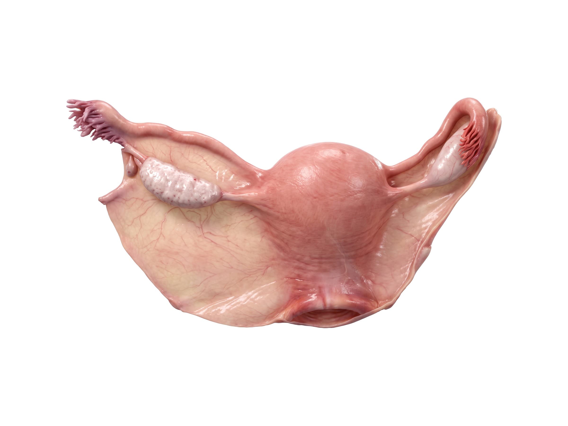
- Uterus: a pear-shaped muscular organ about 7 to 8 cm long in an adult who has not been pregnant. Its parts are the dome-shaped fundus at the top, the body, and the narrow cervix below. Its wall is mostly smooth muscle (myometrium), lined by the endometrium that is shed each month.
- Uterine (fallopian) tubes: about 10 cm long, running out from the upper corners of the uterus. Each ends in a funnel (infundibulum) fringed with finger-like fimbriae that sweep the egg in from the ovary. Fertilization usually happens in the wide middle part (ampulla), which is also the most common site of an ectopic pregnancy.
- Ovaries: almond-shaped, about 3 to 4 cm long, held by the ovarian ligament to the uterus and by the suspensory ligament to the pelvic wall. The suspensory ligament carries the ovarian artery and vein, and twisting of the ovary on these ligaments (torsion) cuts off its blood supply.
- Broad ligament: a wide double sheet of peritoneum draped over the uterus and tubes from side to side, like a sheet hung over a clothesline. It holds the uterus in the middle of the pelvis and carries the uterine vessels and the ureter near its base.
- Round ligament: a cord running from each upper corner of the uterus forward through the inguinal canal to the labia. It stretches in pregnancy and is a common cause of sharp groin pain (round ligament pain).
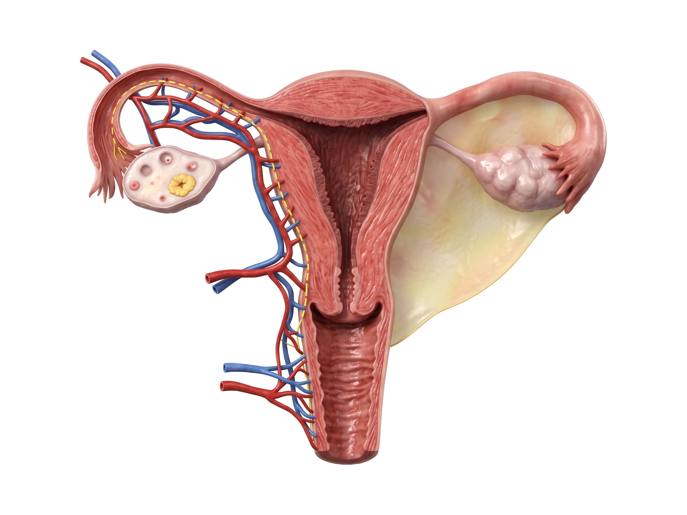
Cut open from the front, the wall of the uterus has three layers: the endometrium, the soft lining of the uterine cavity that thickens and sheds each month and where an embryo implants; the myometrium, the thick smooth muscle that contracts in labor and where fibroids grow; and the perimetrium, the thin peritoneal covering. The triangular uterine cavity narrows into the cervical canal, which opens into the vagina at the external os. These layers matter in OB/GYN claims: a uterine perforation during a D&C or IUD insertion passes through all three, a cesarean scar runs through the lower myometrium, and endometrial thickness on ultrasound is measured across the lining.
Blood supply and the ureters
The uterus is supplied mainly by the two uterine arteries, branches of the internal iliac arteries on the side walls of the pelvis. Each runs inward in the base of the broad ligament to the side of the cervix, then climbs along the side of the uterus and joins the ovarian artery, which comes straight off the aorta just below the kidneys and reaches the ovary through the suspensory ligament. Both pairs enlarge greatly in pregnancy, which is why bleeding after childbirth (postpartum hemorrhage) can be so fast.
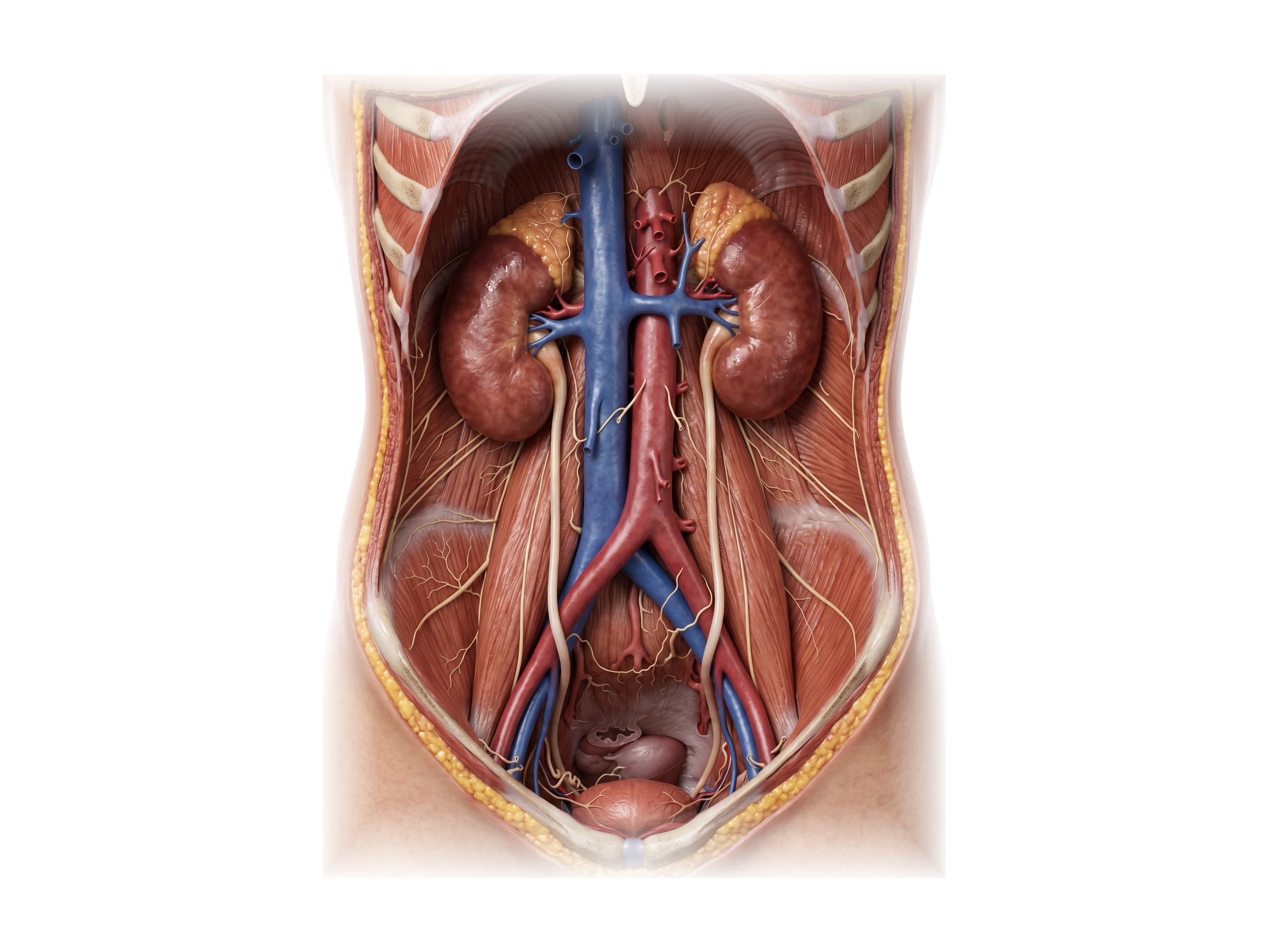
The ureters carry urine from the kidneys to the bladder. Each runs down the back wall of the abdomen on the psoas muscle, crosses the pelvic brim near the split of the common iliac artery, and runs down the side wall of the pelvis just below the ovary. It then turns forward and passes under the uterine artery about 1.5 to 2 cm to the side of the cervix before entering the bladder, a crossing surgeons remember as "water under the bridge".
This is the most common place for the ureter to be injured in gynecologic surgery: it can be clamped, tied, cut or burned while the surgeon controls the uterine artery during a hysterectomy. The ureter is also close to the ovarian vessels at the pelvic brim, where it can be caught when the ovary is removed.
The pelvic floor
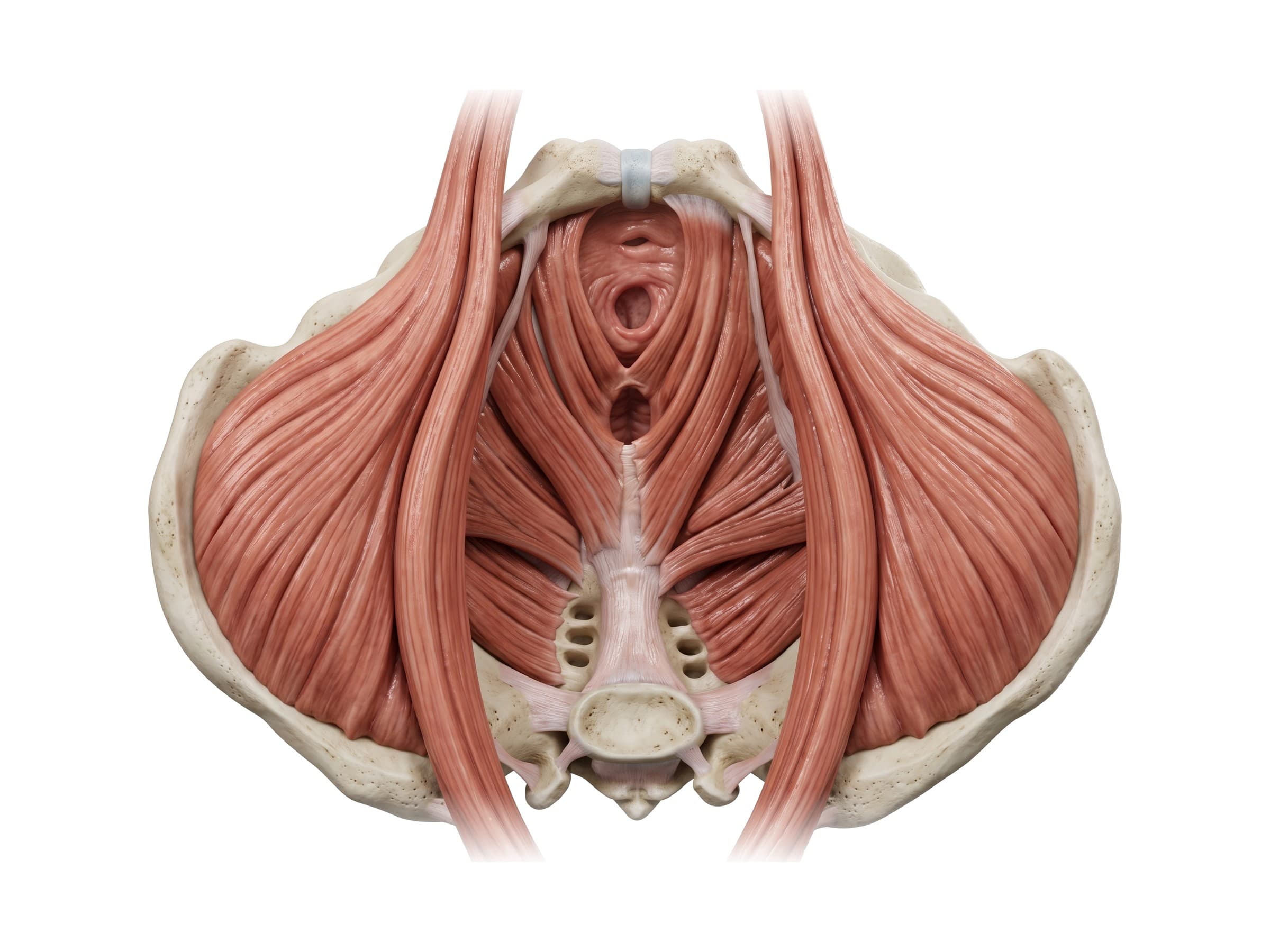
The bottom of the pelvis is closed by a hammock of muscle, the pelvic floor or pelvic diaphragm. Its main part is the levator ani, made of the pubococcygeus, puborectalis and iliococcygeus, which run from the back of the pubic bone and the side walls of the pelvis to the coccyx, with the small coccygeus behind them. In women three openings pass through it, the urethra, the vagina and the anal canal, which makes the female pelvic floor weaker than the male one. The puborectalis forms a sling around the rectum that helps keep stool in, and the perineal body, a knot of muscle and tendon between the vagina and anus, anchors the floor in the middle.
Below the levator ani lie the urogenital diaphragm (perineal membrane) and the external sphincters of the urethra and anus. The pudendal nerve supplies these muscles and the skin of the perineum; it can be stretched in a long or difficult vaginal delivery.
The female pelvis
The bony pelvis is a ring formed by the two hip bones (each made of the ilium, ischium and pubis) and the sacrum behind, joined in front at the pubic symphysis and behind at the sacroiliac joints. The bones and hip joints are shown on the hip anatomy page.
How the female pelvis differs from the male pelvis:
- Wider and shallower, with flared iliac wings.
- Pelvic inlet round or oval, instead of heart-shaped.
- Pubic arch (subpubic angle) wide, about 80 to 90 degrees, instead of about 60 degrees.
- Ischial spines and tuberosities farther apart, giving a roomier outlet.
- Sacrum shorter, wider and less curved.
- Obturator foramen more triangular, and the bones lighter and smoother.
The size and shape of the pelvic inlet and outlet decide whether a baby's head can pass in labor, and a narrow or misshapen pelvis (from a previous fracture, for example) can make vaginal delivery unsafe.
Common injuries and conditions
- Seatbelt injury: a lap belt worn too high, across the soft belly instead of the hip bones, can compress the abdomen in a crash and tear the small intestine or its mesentery, sometimes with a flexion fracture of the lumbar spine (a Chance fracture). A bruise across the lower abdomen, the "seatbelt sign", raises the chance of an internal injury. Bowel injuries can be missed on a first CT scan and show up a day or two later as peritonitis.
- Pelvic fracture: a break of the pelvic ring, from a fall in an older woman (often a stable pubic ramus fracture) or a high-energy car or motorcycle crash, where an open-book or vertical shear injury can tear the large veins at the back of the pelvis and cause life-threatening bleeding. Pelvic fractures in women can injure the bladder, urethra and vagina, and can narrow the birth canal, so a later delivery may need a cesarean section. Diagnosed with an AP pelvis X-ray and CT.
- Solid organ injury: blunt trauma to the abdomen most often tears the spleen (left upper quadrant, lower left ribs) or the liver (right upper quadrant). Bleeding shows up as free fluid on the FAST ultrasound and on CT.
- Ureteral injury during hysterectomy: the ureter can be clamped, cut or burned where it passes under the uterine artery beside the cervix, or at the pelvic brim near the ovarian vessels. If not recognized during the operation, it shows up days later as flank pain, fever, leaking urine (a ureterovaginal fistula) or a blocked, swollen kidney on CT urography.
- Bladder and bowel injury in pelvic surgery: the bladder sits in front of the cervix and can be cut during a cesarean section or hysterectomy, especially after a previous cesarean, and the bowel can be injured when a trocar or instrument enters the abdomen in laparoscopy.
- Cystocele and rectocele: when the pelvic floor and the wall of the vagina are weakened, usually by vaginal childbirth and aging, the bladder can bulge into the front wall of the vagina (cystocele) and the rectum into the back wall (rectocele). The uterus itself can drop (uterine prolapse). Symptoms include pressure, a bulge, urinary leakage and trouble emptying the bowel.
- Obstetric injuries: a severe perineal tear in childbirth (third- or fourth-degree tear) can reach the anal sphincter and cause long-term fecal incontinence. A uterine rupture, most often through an old cesarean scar during labor, is an emergency for both mother and baby. Pudendal nerve stretch in a prolonged second stage of labor can weaken the pelvic floor.
- Ectopic pregnancy and ovarian torsion: a pregnancy that implants in the uterine tube can rupture and bleed into the pouch of Douglas, and a twisted ovary loses its blood supply. Both cause sudden lower abdominal pain and are diagnosed with pelvic ultrasound.
How female abdominal anatomy is shown in a personal injury or medical malpractice case
Female abdominal and pelvic anatomy comes up in car crash claims (seatbelt injuries, pelvic fractures, splenic and liver lacerations), slip-and-fall cases involving pelvic fractures in older women, and medical malpractice claims involving gynecologic surgery and childbirth. A jury rarely knows where the ureter runs or how close the bladder sits to the cervix, so a labeled illustration of the normal anatomy comes first: it shows the uterus between the bladder and rectum, the ureter passing under the uterine artery beside the cervix, or the layers of the pelvic floor. The injury exhibit then builds on the same view: the clamp across the ureter at that crossing, the hole in the bladder, the fistula into the vagina, the torn mesentery under a seatbelt bruise, or the displaced pelvic ring. Showing the anatomy this way helps the medical expert witness explain why an injury was or was not avoidable, and why it caused the symptoms that followed. For OB injuries, a sagittal view of the pelvis makes it easy to show how a perineal tear reached the anal sphincter, or how a prolapse developed after delivery.
Natomy creates custom medical legal illustrations of abdominal and pelvic injuries for demand packages and trial exhibits. For how the force of a crash causes these injuries, see mechanism of injury, and for the rules on using illustrations in court, see demonstrative evidence. Surgeons can use the same figures in a surgical informed consent discussion. Related atlas pages: hip anatomy, lumbar spine anatomy, back muscles anatomy, leg nerves and dermatomes and heart anatomy.
FAQ
What organs are in a woman's lower abdomen?+
A woman's lower abdomen and pelvis hold the lower small intestine (ileum), the cecum and appendix on the right, the sigmoid colon on the left, the rectum, the urinary bladder behind the pubic bone, the uterus behind and above the bladder, the two ovaries and uterine (fallopian) tubes on either side of the uterus, and the lower ureters, which carry urine from the kidneys to the bladder. Pain low in the abdomen in a woman can therefore come from the bowel, the urinary tract or the reproductive organs.
What organs are on the left side of a woman's abdomen?+
The left upper abdomen holds the stomach, the spleen, the tail of the pancreas, the left kidney and the splenic flexure of the colon. The left lower abdomen holds the descending and sigmoid colon, loops of small intestine, the left ureter, and in women the left ovary and uterine tube. The left lower quadrant is a common site of diverticulitis, and in women also of ovarian cysts and ectopic pregnancy on that side.
What organs are on the right side of a woman's abdomen?+
The right upper abdomen holds most of the liver, the gallbladder, the duodenum, the head of the pancreas, the right kidney and the hepatic flexure of the colon. The right lower abdomen holds the cecum, the appendix, the end of the ileum, the ascending colon, the right ureter, and in women the right ovary and uterine tube. That is why right lower quadrant pain in a woman raises both appendicitis and ovarian problems such as torsion or a ruptured cyst.
What are the four quadrants of the abdomen?+
Two imaginary lines through the navel, one vertical and one horizontal, divide the abdomen into four quadrants: the right upper quadrant (RUQ), left upper quadrant (LUQ), right lower quadrant (RLQ) and left lower quadrant (LLQ). Right and left always refer to the patient's side, not the viewer's. Anatomists also use a finer grid of nine regions: the right and left hypochondriac, epigastric, right and left lumbar (flank), umbilical, right and left iliac (inguinal) and hypogastric (suprapubic) regions.
Where are the ovaries located?+
The ovaries sit on each side of the uterus against the side wall of the pelvis, in a shallow hollow called the ovarian fossa, just below the brim of the pelvis where the external iliac vessels and the ureter run. Each ovary is attached to the back of the broad ligament, joined to the uterus by the ovarian ligament and to the pelvic wall by the suspensory ligament, which carries its blood supply. On the skin, they lie roughly halfway between the front point of the hip bone and the pubic bone.
How is the female pelvis different from the male pelvis?+
The female pelvis is built for childbirth. Compared with the male pelvis it is wider and shallower, with a round or oval inlet instead of a heart-shaped one, a wider subpubic (pubic) arch of about 80 to 90 degrees instead of about 60, more widely spaced ischial spines and ischial tuberosities, a shorter and wider sacrum, and lighter, smoother bones with rounder, more triangular obturator foramina. These differences are used to estimate sex from a skeleton.
What is the rectouterine pouch (pouch of Douglas)?+
The rectouterine pouch, or pouch of Douglas, is the fold of peritoneum that dips down between the back of the uterus and the front of the rectum. In a standing or lying woman it is the lowest point of the abdominal cavity, so blood, pus or fluid collects there first. On ultrasound, free fluid in the pouch of Douglas can be a sign of a ruptured ectopic pregnancy, a ruptured ovarian cyst or bleeding from an abdominal injury, and it is a common site of endometriosis.
Why is the ureter at risk during a hysterectomy?+
Each ureter runs down the side wall of the pelvis and passes forward under the uterine artery about 1.5 to 2 cm to the side of the cervix, before entering the bladder. Surgeons remember it as 'water under the bridge'. When the uterine artery is clamped, cut or cauterized during a hysterectomy, the ureter can be clamped, tied, cut or burned with it. Ureteral injury happens in roughly 0.5 to 1 percent of hysterectomies and is a frequent subject of gynecologic malpractice claims, especially when it is not recognized during the operation.
Need a custom illustration of this joint?
Generate a labeled or exhibit-ready figure for a specific patient, injury, or case in minutes.
Try Natomy →