Osteophytes on the Spine: Bone Spurs, Causes & Treatment
Osteophytes are bone spurs that grow on the vertebrae as the spine wears with age. See them illustrated beside a normal lumbar spine.
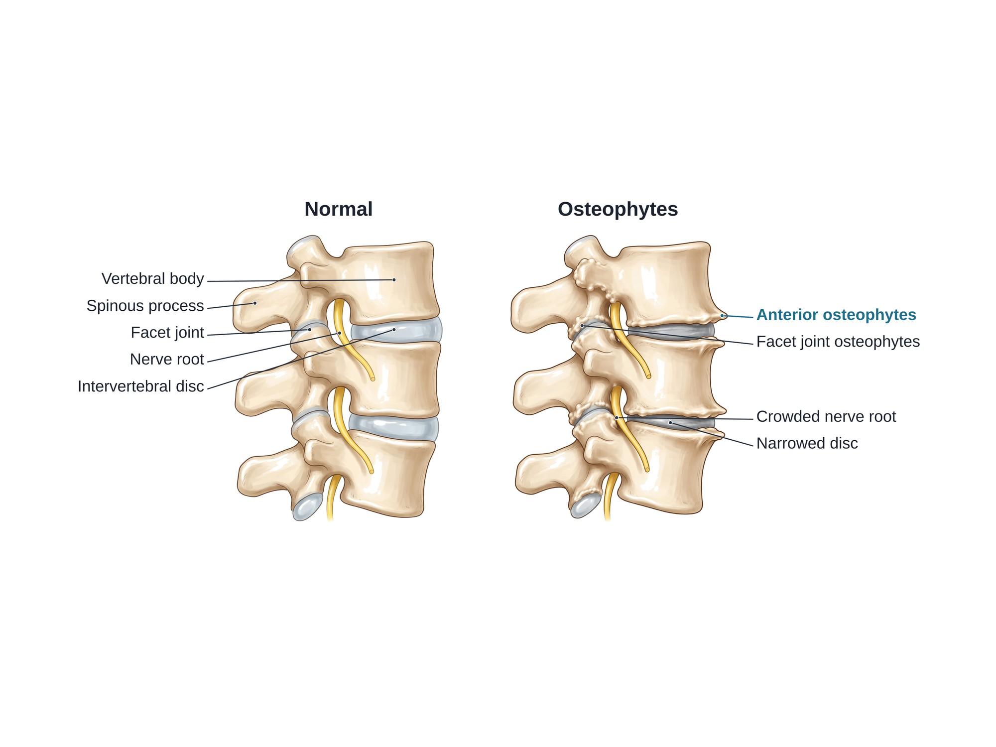
Spinal osteophytes are bone spurs, smooth bony growths that form on the edges of the vertebrae as the spine wears with age. They grow at the rims of the vertebral bodies next to the discs and around the facet joints at the back of the spine. Most cause no symptoms. When a spur narrows the space for a nerve root or the spinal cord, it can cause back or neck pain, and pain, numbness or weakness in a leg or arm. Spurs are most common in the lower back and the neck. The illustration above shows a normal lumbar segment beside one with osteophytes.
The anatomy
Each vertebra has a large, block-shaped vertebral body in front. A soft intervertebral disc sits between each pair of bodies and acts as a cushion. At the back, two small facet joints link each vertebra to the next. Between the vertebrae, on each side, is a small opening called the intervertebral foramen. A nerve root leaves the spinal canal through it. Bone spurs that grow into this opening can pinch the nerve. For the full anatomy, see lumbar spine anatomy.
Types
| Type | Where it grows | What it can do |
|---|---|---|
| Anterior osteophytes | Front rims of the vertebral bodies | Usually painless. In the neck, very large ones can press on the throat. |
| Posterior osteophytes | Back rims of the vertebral bodies, often with a bulging disc | Narrow the spinal canal or the foramen and press on nerves or the cord |
| Facet joint osteophytes | Around the facet joints | Part of facet arthropathy. Can narrow the foramen. |
| Uncovertebral osteophytes | Small joints at the sides of the neck vertebrae | A common cause of pinched nerves in the neck |
Two other bony growths look similar but are different. Syndesmophytes are thin bony bridges seen in ankylosing spondylitis. DISH (diffuse idiopathic skeletal hyperostosis) is thick, flowing bone along the front of many vertebrae.
Causes
Spurs are the body's response to stress on a joint. When a disc thins and the spine moves more than it should, the bone at the edges grows outward. This spreads the load and steadies the segment.
- Age-related wear: most adults over 60 have some spurs on imaging
- Disc degeneration: a thinner disc lets the vertebrae rub and shift
- Facet joint arthritis
- Old injury to a disc, a joint or a vertebra, which can speed up wear in that segment
- Repetitive heavy work, such as lifting and twisting
- Excess body weight
- Genetics and family history of arthritis
- Poor alignment, such as scoliosis or a slipped vertebra
Symptoms
Many people with spurs have no symptoms at all. When symptoms occur, they depend on where the spur is.
- Back or neck pain and stiffness, often worse after rest
- Pain spreading into the buttock or leg (sciatica) when a spur pinches a lumbar nerve root
- Pain, numbness or tingling down the arm when a spur pinches a neck nerve root
- Weakness in the muscles that nerve controls
- Leg pain or heaviness with walking, eased by sitting or bending forward (spinal stenosis)
- Clumsy hands or poor balance if a neck spur presses on the spinal cord (myelopathy, damage to the spinal cord)
- Trouble swallowing, in rare cases of very large front spurs in the neck
Diagnosis
The doctor asks about symptoms and checks strength, reflexes and feeling in the arms or legs. X-rays show spurs well as bony points at the edges of the vertebrae. They also show narrowed discs. CT shows the size and shape of each spur in fine detail. MRI shows the discs, nerves and spinal cord, and whether a spur is pressing on them. Nerve tests (EMG) can confirm a pinched nerve root. A spur only matters if it matches the patient's symptoms and exam.
Treatment
- Activity changes and staying active, rather than bed rest
- Physical therapy: core strength, posture and flexibility
- Weight control to reduce load on the spine
- Medicines: anti-inflammatory drugs and short courses of other pain medicine
- Epidural steroid injection to calm an irritated nerve root
- Facet joint injection or radiofrequency ablation when the facet joints are the pain source
- Surgery when a spur presses on a nerve or the cord and other care fails. A foraminotomy widens the nerve opening. A laminectomy enlarges the spinal canal. In the neck, ACDF surgery removes the disc and spurs from the front.
Recovery
Flares of pain from spinal arthritis often settle in a few weeks with activity changes, medicine and therapy. The spurs themselves stay, so symptoms can come back. Regular exercise is the best way to reduce flares. After surgery to remove a spur, leg or arm pain often improves quickly. Full recovery usually takes about 6 to 12 weeks.
Complications
- Foraminal stenosis, when spurs narrow the nerve opening
- Central spinal stenosis, narrowing of the main spinal canal
- Radiculopathy (a pinched nerve root), causing arm or leg pain, numbness or weakness
- Cervical myelopathy, pressure on the spinal cord in the neck
- Chronic back or neck pain
- Trouble swallowing from large front spurs in the neck (rare)
How spinal osteophytes are shown in a personal injury case
Osteophytes appear on many spine scans after car crashes and falls. Because bone spurs usually take months to years to form, spurs seen on a scan taken soon after an injury almost always existed before it. The defense often uses this to argue that the plaintiff's pain comes from pre-existing degeneration. The plaintiff's side may argue that the injury aggravated a spine that caused no trouble before. For example, a disc injury can push against a nerve that spurs had already crowded. Medical records and imaging from before the incident are key. Related degenerative findings, such as facet arthropathy, Schmorl nodes and Modic changes, raise the same question. A side-by-side illustration like the one above helps a jury see what a normal spine looks like and what spurs do. For how these images are used at trial, see demonstrative evidence and medical legal illustration.
Related pages
- Lumbar spine anatomyThe normal anatomy of the lumbar spine.
- Annular tearAn annular tear is a crack in the tough outer ring of a spinal disc, a common MRI finding in back pain cases. See it illustrated beside a normal disc.
- AnterolisthesisAnterolisthesis is a vertebra slipped forward over the one below, most often L4 on L5. See it illustrated beside normal anatomy.
- Facet arthropathyFacet arthropathy is arthritis of the small joints at the back of the spine, most often at L4-L5 and L5-S1. See it illustrated beside normal facet joints.
- Foraminal stenosisForaminal stenosis is narrowing of the opening where a nerve root leaves the spine, often at L4-L5 or L5-S1. See it illustrated beside normal anatomy.
- Lumbar disc herniationA herniated disc is disc gel pushed through a tear in its outer ring, often pressing on a nerve root. See protrusion, extrusion and sequestration illustrated.
- Modic changesModic changes are endplate and bone marrow changes next to a worn disc, seen on MRI as type 1, 2 or 3. See each type beside a normal spine.
- Schmorl nodeA Schmorl node is disc material pushed up or down through the vertebral endplate into the bone. See it beside a normal disc, with causes and treatment.
- SpondylolisthesisSpondylolisthesis is a vertebra that has slipped forward over the one below, often L5 on S1 after a pars fracture. See it illustrated beside normal anatomy.
FAQ
Are osteophytes the same as osteoarthritis?+
No, but they are closely linked. Osteoarthritis is the wear of a joint over time. Osteophytes are bone spurs that form as part of that wear. In the spine, spurs are one of the main signs of spondylosis, the general term for age-related spine wear.
What is a posterior disc osteophyte complex?+
It is a radiology term for a bulging disc combined with bone spurs at the back edge of the vertebral bodies. Together they push back into the spinal canal or the nerve openings. It is common in the neck and can press on the spinal cord or a nerve root.
Do bone spurs on the spine cause pain?+
Often they do not. Many people have spurs on a scan and no symptoms at all. Spurs cause trouble when they narrow the space for a nerve root or the spinal cord, or when they come with painful arthritis of the facet joints.
Can a car accident cause osteophytes?+
A crash does not create spurs overnight. Bone spurs usually take months to years to form. An injury can speed up wear in a damaged segment over time. It can also make spurs that caused no symptoms before start pressing on a nerve.
Do spinal bone spurs go away?+
No. Once formed, osteophytes do not shrink on their own. Symptoms, however, often settle with exercise, physical therapy and medicine. Surgery to remove a spur is needed only when it presses on a nerve or the spinal cord.
Can bone spurs on the spine be removed?+
Yes. A surgeon can shave spurs off during a foraminotomy, a laminectomy or an anterior neck surgery such as ACDF. This is done only when the spur is pinching a nerve or the cord and other treatment has failed.
What are the symptoms of bone spurs in the neck?+
Neck stiffness and pain are common. A spur pressing on a nerve root can cause pain, numbness or weakness down the arm. A spur pressing on the spinal cord can cause clumsy hands, balance problems or weakness in the legs. Rarely, large front spurs make swallowing hard.
Need this illustration for a patient or a case?
Describe the injury or upload a scan and get a clear, labeled medical illustration for a consult, a chart or a case exhibit.
Try Natomy →