Modic Changes: Types 1, 2 and 3, Causes & Treatment
Modic changes are endplate and bone marrow changes next to a worn disc, seen on MRI as type 1, 2 or 3. See each type beside a normal spine.
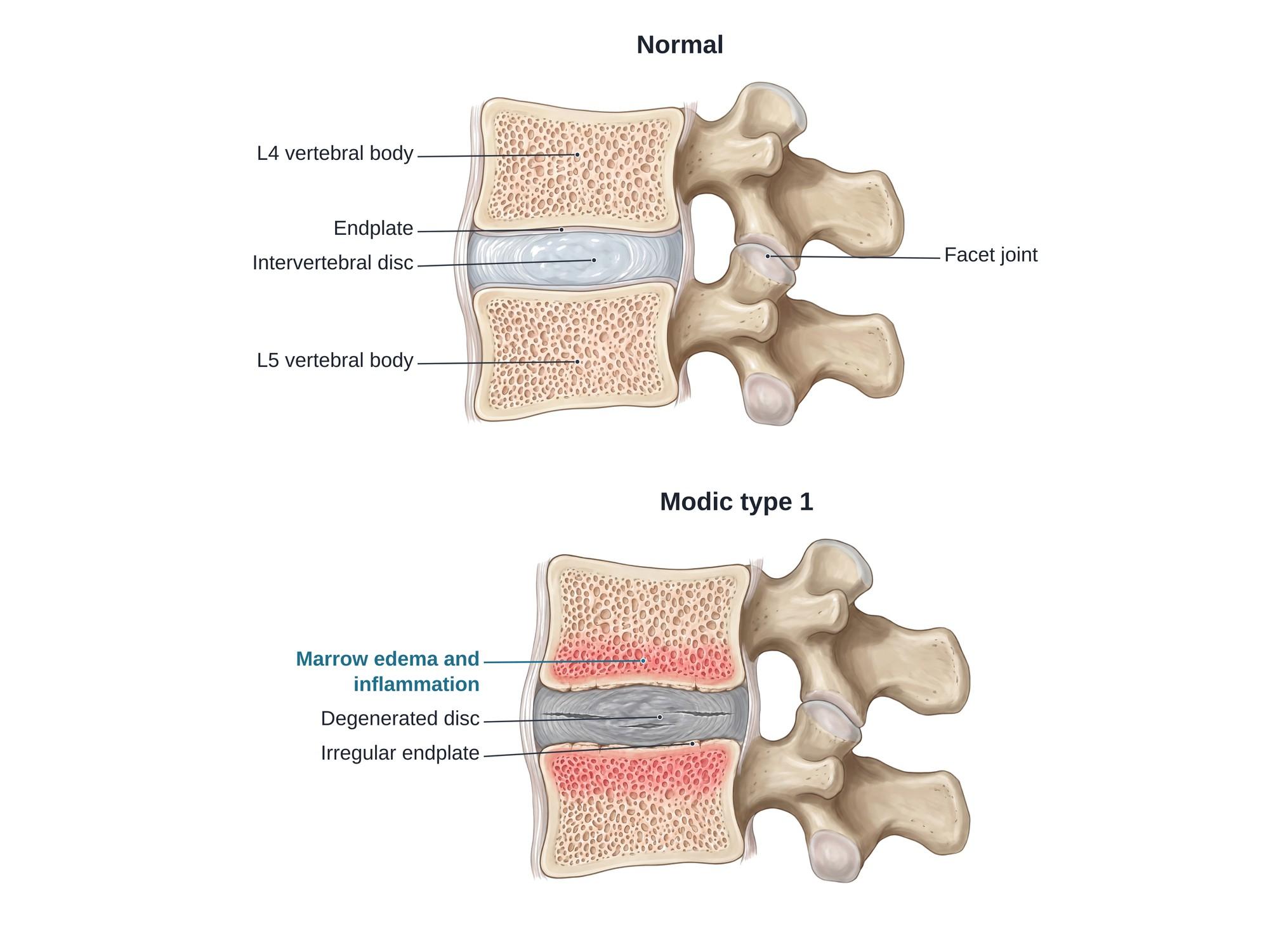
Modic changes are changes in the bone marrow next to the vertebral endplates, the thin layers of cartilage and bone that cap each vertebral body above and below a disc. They are seen on MRI and come in three types. Type 1 is swelling and inflammation (bone marrow edema). Type 2 is fat replacing the normal marrow. Type 3 is hardened, dense bone (sclerosis). They almost always sit next to a worn, degenerated disc, most often at L4-L5 or L5-S1. Type 1 is the type most closely linked to low back pain. The illustration above shows a normal L4-L5 segment beside one with Modic type 1 changes.
The anatomy
Each lumbar disc sits between two vertebral bodies. The disc has a soft center, the nucleus pulposus, and a tough outer ring, the annulus fibrosus. A thin endplate lines the top and bottom of each vertebral body where it meets the disc. The disc has almost no blood supply of its own, so it gets its nutrients by diffusion through the endplates. Just under the endplate lies spongy bone filled with marrow. The basivertebral nerve runs into the middle of the vertebral body and carries pain signals from the endplate. Modic changes happen in this layer of marrow, right next to the endplate. For the full anatomy, see lumbar spine anatomy.
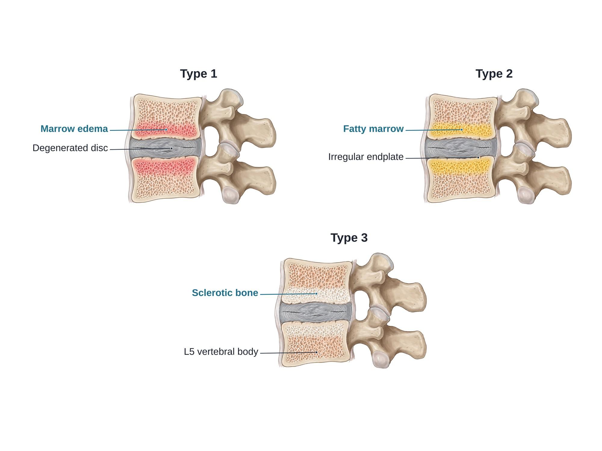
Types
Dr. Michael Modic described the types in 1988, based on how the marrow looks on two MRI sequences (T1 and T2).
| Type | What happens in the bone | MRI appearance | Usual course |
|---|---|---|---|
| Type 1 | Edema and inflammation, cracks in the endplate, new blood vessels | Dark on T1, bright on T2 | Often changes to type 2 over months to years |
| Type 2 | Fat replaces the red marrow | Bright on T1, bright or medium on T2 | Stable for years |
| Type 3 | Dense, hardened (sclerotic) bone | Dark on T1 and T2 | Stable, least common |
Mixed types, such as type 1/2 or type 2/3, are common. They show one type turning into another.
Causes
- Disc degeneration: as a disc wears, it loses water and height, and more load passes through the endplate. This is the most common setting.
- Endplate damage: tiny cracks in the endplate let disc material and fluid reach the marrow. This sets off inflammation. A Schmorl node is one form of endplate breakthrough.
- Disc herniation: type 1 changes often appear at the same level after a lumbar disc herniation, especially after surgery at that level.
- Low-grade infection (debated): some research found bacteria such as Cutibacterium acnes in removed discs. Whether this causes Modic changes is still unclear.
- Risk factors: older age, heavy physical work, smoking, higher body weight and a family history of disc disease.
Symptoms
Modic changes have no symptoms of their own that set them apart. When they do cause pain, it is usually:
- A deep, aching pain in the middle of the low back
- Pain that is worse with sitting, bending forward and lifting
- Morning stiffness, and sometimes pain at night
- Flare-ups that last weeks, with quieter times between
Leg pain, numbness or weakness points to a pinched nerve instead, such as foraminal stenosis or a herniated disc.
Diagnosis
Modic changes are found on MRI; plain X-rays cannot show them. On T1 images, type 1 bone looks darker than normal marrow and type 2 looks brighter. On T2 and STIR images, type 1 looks bright because it holds fluid. Type 3 looks dark on all sequences, and CT shows it as dense, white bone next to the endplate. The radiologist reports the level, the type and how much of the vertebral body is involved.
Type 1 can look like discitis-osteomyelitis, an infection of the disc and bone. Infection usually brings fever, high blood markers of inflammation and a bright, fluid-filled disc. Modic type 1 usually does not. When it is unclear, doctors compare with older scans, order blood tests or repeat the MRI.
Treatment
- Physical therapy and exercise: core and back strengthening, walking, and learning to lift and bend with less strain
- Medicines: anti-inflammatory drugs and other pain medicines for flare-ups
- Activity changes: shorter periods of sitting and less heavy lifting during flares
- Basivertebral nerve ablation: a heat probe passed into the vertebral body through a small tube burns the basivertebral nerve. It is used for chronic low back pain with type 1 or type 2 changes at L3 to S1.
- Antibiotics: long courses were tried for type 1 changes, but a large trial found little benefit, so they are rarely used now
- Fusion surgery: joining the two vertebrae removes motion at a painful level. It is reserved for selected people whose pain persists after other care.
Recovery
Many people with type 1 changes improve over 1 to 2 years as the inflammation settles or turns into type 2. Flare-ups often ease within weeks with therapy and medicine. After basivertebral nerve ablation, most people go home the same day and return to light activity within days. Pain relief builds over the next few weeks.
Complications
- Chronic low back pain with repeated flare-ups
- Further disc collapse and loss of disc height at the same level
- Strain on the nearby facet joints and the growth of bone spurs
- Narrowing of the nerve openings as the disc space collapses
- After treatment: nerve irritation, infection or bleeding from a procedure, and the usual risks of fusion surgery
How Modic changes are shown in a personal injury case
Modic changes often appear on an MRI taken after a car crash, fall or work injury. The defense may argue that they prove old, pre-existing disc disease. The type matters here. Type 2 and type 3 changes take many months or years to form, so they usually came before a recent injury. Type 1 changes can develop within weeks to months after a new disc injury or endplate crack at that level. An earlier MRI showing no change at that level, followed by type 1 after the injury, can support a new or worsened condition. A side by side illustration like the one above helps a jury see where the endplate and marrow are, and what each type looks like. For how these images are used at trial, see demonstrative evidence and medical legal illustration.
Related pages
- Lumbar spine anatomyThe normal anatomy of the lumbar spine.
- Annular tearAn annular tear is a crack in the tough outer ring of a spinal disc, a common MRI finding in back pain cases. See it illustrated beside a normal disc.
- AnterolisthesisAnterolisthesis is a vertebra slipped forward over the one below, most often L4 on L5. See it illustrated beside normal anatomy.
- Facet arthropathyFacet arthropathy is arthritis of the small joints at the back of the spine, most often at L4-L5 and L5-S1. See it illustrated beside normal facet joints.
- Foraminal stenosisForaminal stenosis is narrowing of the opening where a nerve root leaves the spine, often at L4-L5 or L5-S1. See it illustrated beside normal anatomy.
- Lumbar disc herniationA herniated disc is disc gel pushed through a tear in its outer ring, often pressing on a nerve root. See protrusion, extrusion and sequestration illustrated.
- Schmorl nodeA Schmorl node is disc material pushed up or down through the vertebral endplate into the bone. See it beside a normal disc, with causes and treatment.
- Spinal osteophytesOsteophytes are bone spurs that grow on the vertebrae as the spine wears with age. See them illustrated beside a normal lumbar spine.
- SpondylolisthesisSpondylolisthesis is a vertebra that has slipped forward over the one below, often L5 on S1 after a pars fracture. See it illustrated beside normal anatomy.
FAQ
What are Modic changes in the vertebrae?+
Modic changes are changes in the bone marrow just above and below a spinal disc, next to the vertebral endplates. They show up on MRI and come in three types: swelling and inflammation (type 1), fat (type 2) and hardened bone (type 3). They usually sit next to a worn, degenerated disc.
Is Modic type 1 serious?+
Modic type 1 is not dangerous in itself, but it is the type most often linked to ongoing low back pain. It can look like a spine infection on MRI, so doctors check blood tests and symptoms such as fever. Many type 1 changes settle or turn into type 2 over months to a few years.
Is Modic type 2 dangerous?+
No. Type 2 means fat has replaced part of the marrow next to the endplate. It is common with age and disc wear, it is more stable than type 1, and it is often found in people with no back pain at all.
What is the treatment for Modic changes?+
Most people start with physical therapy, exercise, activity changes and pain medicine. For lasting pain with type 1 or type 2 changes at L3 to S1, basivertebral nerve ablation is an option. Fusion surgery is used in selected cases. Long courses of antibiotics have been studied, but a large trial found little benefit.
Can Modic changes go away?+
They can change. Type 1 often turns into type 2 over time, and some type 1 changes fade. Type 2 and type 3 tend to stay. A change in type on follow-up MRI is common and helps show how long a finding has been there.
Can an injury cause Modic changes?+
Modic changes are mostly linked to disc wear. A new disc herniation or an endplate injury can be followed by type 1 changes within months at that level. Type 2 and type 3 changes take much longer to form, so they usually point to an older, pre-existing process.
What does Modic back pain feel like?+
It is usually a deep ache in the middle of the low back rather than leg pain. It often gets worse with sitting, bending and lifting, and many people feel stiff in the morning. Doctors sometimes call this vertebrogenic pain, meaning pain that comes from the vertebral body itself.
What are the long-term effects of degenerative disc disease with Modic changes?+
Many people have long quiet stretches with flare-ups now and then. The disc can keep losing height over the years, and nearby facet joints and nerve openings can narrow. Most people never need surgery.
Need this illustration for a patient or a case?
Describe the injury or upload a scan and get a clear, labeled medical illustration for a consult, a chart or a case exhibit.
Try Natomy →