Spondylolisthesis: Types, Grades, Causes & Treatment
Spondylolisthesis is a vertebra that has slipped forward over the one below, often L5 on S1 after a pars fracture. See it illustrated beside normal anatomy.
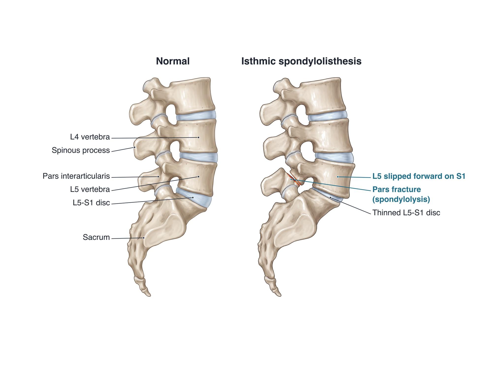
Spondylolisthesis is a slip of one vertebra over the vertebra below it, usually forward. It happens most often in the lower back, at L5 on S1 and L4 on L5. The two main types are isthmic, caused by a break in a thin bridge of bone called the pars interarticularis, and degenerative, caused by wear of the joints and disc with age. Many people have no symptoms. Others have back pain, tight hamstrings or leg pain from pinched nerves. The illustration above shows a normal lower lumbar spine beside isthmic spondylolisthesis of L5 on S1.
The anatomy
Each lumbar vertebra has a solid vertebral body in front. Behind the body, a ring of bone surrounds the spinal canal. Paired facet joints connect each vertebra to the next. They act like hooks that stop the vertebra from sliding forward. The pars interarticularis is the narrow neck of bone between the upper and lower facets. The intervertebral disc sits between the bodies and works as a cushion. If the pars breaks on both sides, the front of the vertebra loses its hook and can slide forward. For the full anatomy, see lumbar spine anatomy.
Types
| Type | Cause | Typical patient and level |
|---|---|---|
| Isthmic | Pars defect (spondylolysis), often a stress fracture | Starts in childhood or the teen years, usually L5-S1 |
| Degenerative | Worn facet joints and disc; pars intact | Adults over 50, more often women, usually L4-L5 |
| Dysplastic (congenital) | Malformed facets present from birth | Children and teens, L5-S1 |
| Traumatic | Acute fracture of the back of the vertebra (not the pars) | Any age, after high-energy injury |
| Pathologic | Bone weakened by tumor, infection or bone disease | Any age |
| Iatrogenic | Instability after earlier spine surgery | After decompression surgery |
In the isthmic type, the front of the vertebra slides forward but the back part stays behind. That is the gap shown in the illustration. In the degenerative type, the whole vertebra slides, so the spinal canal narrows more for the same amount of slip. That is why degenerative slips often cause spinal stenosis. See anterolisthesis for a degenerative slip of L4 on L5.
Grades
The Meyerding grade measures how far the upper vertebral body has moved forward. It is shown as a share of the depth of the body below.
| Grade | Slip |
|---|---|
| I | Up to 25% |
| II | 25% to 50% |
| III | 50% to 75% |
| IV | 75% to 100% |
| V (spondyloptosis) | More than 100%, fully off the vertebra below |
Grades I and II are low grade and are the most common. Grades III to V are high grade and are more likely to need surgery.
Causes
- Repeated back arching in sports such as gymnastics, football, diving, weightlifting and cricket bowling. This can cause a pars stress fracture in a growing spine.
- Age-related wear of the facet joints (facet arthropathy) and disc, which lets the vertebra drift forward.
- Genetics and anatomy: a family history of pars defects, or a steep sacral slope.
- Trauma: a high-energy fall or crash that fractures the back of the vertebra.
- Prior surgery that removed bone or joint for decompression.
Symptoms
- Low back pain, often worse with standing, walking or arching backward
- Tight hamstrings and a stiff, flattened back, especially in teenagers
- Pain, numbness or tingling down the buttock and leg (radiculopathy, meaning a pinched nerve root)
- Leg heaviness or cramping with walking that eases when sitting or bending forward (neurogenic claudication)
- In high-grade slips, a visible step in the lower back and a change in posture
- Rarely, weakness or loss of bladder or bowel control, which needs emergency care
Diagnosis
The doctor checks posture, back motion, hamstring tightness, strength, reflexes and sensation. A side view X-ray shows the slip. The doctor measures how far the back edge of the upper body sits in front of the one below, which gives the grade. Flexion and extension X-rays, taken bending forward and back, show whether the slip moves. CT shows the pars defect clearly. MRI shows the discs, the spinal canal and any pinched nerve roots. In young athletes with back pain and normal X-rays, CT or MRI can find an early pars stress reaction.
Treatment
- Activity changes: a break from sports and movements that arch the back
- Physical therapy: core and hip strengthening, hamstring stretching and flexion-based exercises
- Medicines: anti-inflammatory drugs and short courses of other pain medicine
- Bracing: sometimes used in teenagers with a fresh pars stress fracture, to help it heal
- Epidural steroid injection: for leg pain from a pinched nerve root
- Surgery: decompression to free the nerves, usually with spinal fusion using screws and rods, and often a spacer (cage) in the disc space. Surgery is considered for high-grade slips, a slip that keeps growing, nerve weakness, or pain that fails several months of treatment. In some young patients with a pars defect and no real slip, the pars can be repaired directly.
Recovery
Most people with a low-grade slip improve within a few weeks to a few months of therapy. Teen athletes often return to sport in about 3 to 6 months once pain-free. After fusion, patients usually walk within a day or two. Desk work often resumes in about 4 to 8 weeks. The fusion usually takes 6 to 12 months to become solid.
Complications
- The slip growing over time, more likely in children during growth spurts
- Spinal stenosis and foraminal stenosis, with chronic nerve root compression
- Ongoing back or leg pain
- A herniated disc at the slipped level or the level above
- After fusion: failure of the bones to fuse (nonunion), hardware problems, and extra wear at the next level (adjacent segment disease)
How spondylolisthesis is shown in a personal injury case
Spondylolisthesis often comes up after car crashes, falls and work injuries. The key question is whether the slip is new, or was there before and was made worse. Pars defects are found in a small share of adults who never knew they had one. Comparing earlier and later imaging, and flexion and extension X-rays that show new movement, helps answer that question. A side view illustration like the one above makes the pars break, the forward slip and the narrowed space for the nerves easy for a jury to follow. For how these images are used at trial, see demonstrative evidence and medical legal illustration.
Related pages
- Lumbar spine anatomyThe normal anatomy of the lumbar spine.
- AnterolisthesisAnterolisthesis is a vertebra slipped forward over the one below, most often L4 on L5. See it illustrated beside normal anatomy.
- Facet arthropathyFacet arthropathy is arthritis of the small joints at the back of the spine, most often at L4-L5 and L5-S1. See it illustrated beside normal facet joints.
- Foraminal stenosisForaminal stenosis is narrowing of the opening where a nerve root leaves the spine, often at L4-L5 or L5-S1. See it illustrated beside normal anatomy.
- Lumbar disc herniationA herniated disc is disc gel pushed through a tear in its outer ring, often pressing on a nerve root. See protrusion, extrusion and sequestration illustrated.
FAQ
How long does spondylolisthesis last?+
The slip itself usually stays, because the bone does not slide back on its own. The pain is different. Flare-ups often settle in a few weeks to a few months with physical therapy and activity changes. Many people then have long stretches with few symptoms.
What are the signs that spondylolisthesis is getting worse?+
Warning signs include leg pain that spreads further down the leg, new numbness or weakness, trouble walking more than a short distance, and changes in bladder or bowel control. The last one needs urgent care. A doctor may repeat X-rays to see if the slip has grown.
Can I live a normal life with spondylolisthesis?+
Most people can. Low-grade slips are common and often cause few or no symptoms. A strong core, healthy weight and smart lifting let most people work, exercise and play sports. Some high-impact sports may need to be limited while symptoms are active.
How do you fix spondylolisthesis?+
Most cases are treated without surgery: physical therapy, anti-inflammatory medicine, short activity changes and sometimes an epidural steroid injection. When that fails, or when nerves are badly pinched, surgeons remove pressure on the nerves and fuse the slipped level with screws and rods.
What makes spondylolisthesis worse?+
Repeated back arching (extension), heavy lifting, high-impact twisting and long periods of standing can aggravate it. In teenagers, sports like gymnastics, football line play and diving put the most stress on the pars.
What is the difference between spondylolysis and spondylolisthesis?+
Spondylolysis is a crack or defect in the pars interarticularis, with no slip. Spondylolisthesis means the vertebra has actually slipped. A pars defect on both sides can let the vertebra slide forward, which turns spondylolysis into isthmic spondylolisthesis.
Is spondylolisthesis the same as anterolisthesis?+
Anterolisthesis is a forward slip, which is by far the most common kind of spondylolisthesis. Spondylolisthesis is the broader term for any slip of one vertebra on another. A backward slip is called retrolisthesis.
Can a car accident cause spondylolisthesis?+
A high-energy crash or fall can fracture the back of the vertebra and cause a traumatic slip, though this is rare. More often, an injury makes an existing slip painful or unstable. Earlier imaging and flexion and extension X-rays help show what changed.
Need this illustration for a patient or a case?
Describe the injury or upload a scan and get a clear, labeled medical illustration for a consult, a chart or a case exhibit.
Try Natomy →