Foraminotomy: Procedure, Recovery & Risks
A foraminotomy widens the opening where a spinal nerve root leaves the spine to relieve a pinched nerve. See the foraminotomy procedure illustrated.
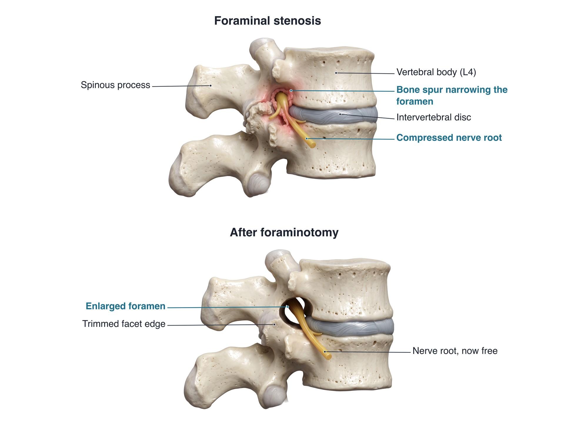
A foraminotomy is surgery that widens a neural foramen, the opening on each side of the spine where a spinal nerve root leaves the spinal canal. When this opening narrows, it squeezes the nerve root and causes pain, numbness or weakness down the leg or arm. The surgeon removes the bone spurs, thickened ligament or disc material that crowd the opening, so the nerve has room again. The illustration above shows a narrowed foramen pinching the nerve root, then the same foramen enlarged after foraminotomy.
Why it is done
The usual reason is foraminal stenosis, a narrowing of the foramen. It is most often caused by arthritis of the facet joints, the small joints at the back of the spine. The joints form bone spurs (osteophytes). The disc also loses height and bulges. Together they shrink the foramen and press on the nerve root. This is called radiculopathy, a pinched nerve root.
Symptoms that lead to surgery include:
- Pain that shoots down the leg (sciatica) or down the arm in cervical cases
- Numbness or tingling in the area served by that nerve
- Weakness in the muscles that nerve controls
- Symptoms that persist despite physical therapy, medicine and steroid injections, usually for 6 to 12 weeks or more
Foraminotomy is often combined with removal of disc material when a herniated disc also presses on the nerve. It can also be combined with a laminectomy when the central canal is narrow too. In the neck, a posterior foraminotomy is an alternative to ACDF surgery when a single nerve root is pinched, for example by a herniated disc in the neck, and the disc itself does not need to be removed.
How the surgery is done
The operation usually takes about 1 to 2 hours per level. The patient lies face down under general anesthesia.
- Incision. A small incision is made in the back, off the midline, over the affected level. X-ray confirms the correct level.
- Approach. The back muscles are gently spread apart, often through a tube (a tubular retractor). A microscope or endoscope gives a magnified view.
- Bone removal. A high-speed drill and small bone punches remove the bone spur and trim the edge of the facet joint behind the foramen. The surgeon keeps most of the facet joint so the spine stays stable.
- Freeing the nerve. Thickened ligament and any disc fragment pressing on the nerve root are removed. The surgeon follows the nerve out through the foramen to make sure it is free, as shown in the lower panel above.
- Closing. The muscles fall back into place. The skin is closed with stitches or skin glue.
Recovery
- Most patients go home the same day or the next day.
- Walking starts right away and is encouraged.
- Desk work can often resume in 1 to 3 weeks. Heavy work may take 6 to 12 weeks.
- Bending, lifting and twisting are limited for about 4 to 6 weeks.
- Physical therapy builds core and back strength.
- Leg pain often improves quickly. Numbness and weakness recover more slowly, sometimes over months.
Risks
- Dural tear, a tear in the sac around the nerves that leaks spinal fluid; usually repaired during surgery
- Nerve root injury, causing new numbness or weakness, uncommon
- Infection or bleeding at the incision
- Instability if too much facet joint is removed, which may later need a fusion
- Recurrence of narrowing over years
- Persistent pain when the nerve was damaged before surgery
How foraminotomy is shown in a personal injury case
Foraminotomy often follows a car crash or fall that turned an arthritic spine painful, or that caused a disc herniation into the foramen. The defense commonly argues that the bone spurs were pre-existing degeneration. The plaintiff's experts may argue the trauma made a silent condition symptomatic. An illustration of the plaintiff's own level, before and after surgery, shows the jury what was pinching the nerve and what the surgeon removed. It also helps explain the cost and the recovery time. For how these images are used at trial, see demonstrative evidence and medical legal illustration. For the normal anatomy, see lumbar spine anatomy.
Related pages
FAQ
Is a foraminotomy a major surgery?+
It is a real spine operation done under general anesthesia, but it is usually one of the smaller ones. Many foraminotomies are done through a small incision with a tube and a microscope or endoscope. Most patients go home the same day or the next day.
How long does it take to recover from a foraminotomy?+
Most people walk the same day and return to desk work in about 1 to 3 weeks. Lifting and bending are limited for about 4 to 6 weeks. Full recovery, including nerve healing, can take about 3 months. Numbness can take longer to fade than leg pain.
What is the difference between a foraminotomy and a laminectomy?+
A foraminotomy widens the side opening (the foramen) where one nerve root leaves the spine. A laminectomy removes the lamina, the back roof of the spinal canal, to relieve pressure on the central canal. The two are often done together when both areas are narrowed.
How painful is a foraminotomy?+
There is usually soreness at the incision and in the back muscles for a few days to a couple of weeks, controlled with pain medicine. Many patients notice their leg or arm pain is better right after surgery, because the nerve is no longer pinched.
What is the success rate of foraminotomy?+
For pain that runs down the leg or arm from a pinched nerve, most patients report good relief. Results are best when imaging clearly shows a narrowed foramen at the level that matches the symptoms. Back pain alone improves less reliably.
Can foraminal stenosis come back after surgery?+
Yes, over years. Arthritis can form new bone spurs, or the disc can collapse further and narrow the foramen again. Instability at the same level can also develop, which may need a fusion.
Is a cervical foraminotomy different?+
A cervical (neck) foraminotomy frees a nerve root that causes arm pain. It is usually done from the back of the neck through a small incision. It avoids fusion, so neck motion is kept. Recovery is similar to the lumbar operation.
Need a surgical illustration for consent or a case?
Describe the operation and get a clear, labeled medical illustration for patient consent, teaching or a case exhibit.
Try Natomy →