Annular Tear: Disc Fissure Symptoms, MRI & Treatment
An annular tear is a crack in the tough outer ring of a spinal disc, a common MRI finding in back pain cases. See it illustrated beside a normal disc.
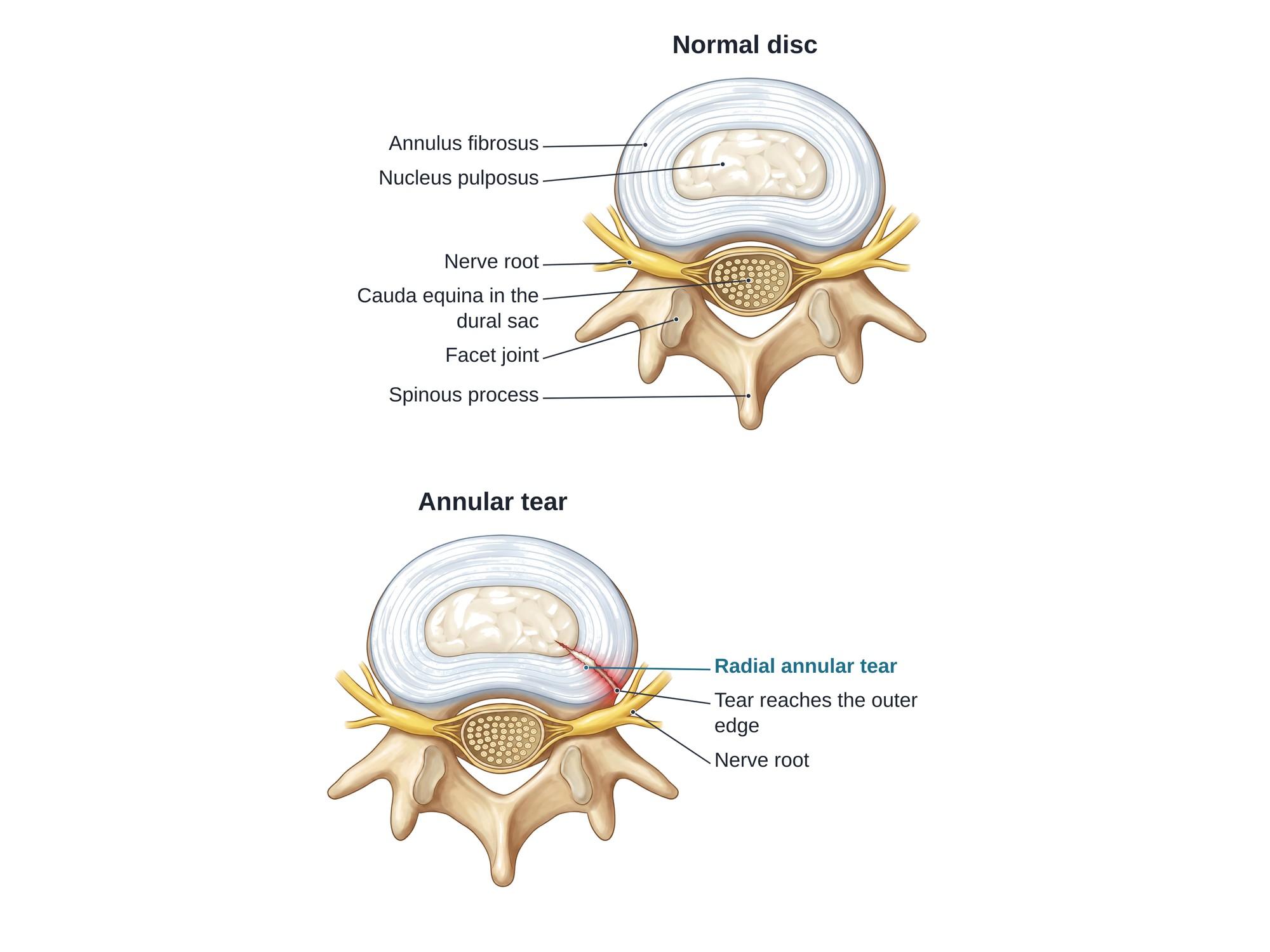
An annular tear is a crack or split in the annulus fibrosus, the tough outer ring of a spinal disc. It is also called an annular fissure. A tear can cause deep back pain, and fluid from the disc's soft center can leak out through it and irritate nearby nerves. Annular tears are among the most common findings on a lumbar MRI. They can come from a sudden injury or from wear over time. The illustration above shows a normal disc beside a disc with a radial annular tear, seen from above.
The anatomy
Each intervertebral disc sits between two vertebrae and acts as a cushion. See lumbar spine anatomy for the full layout.
- The nucleus pulposus is the soft, gel-like center of the disc.
- The annulus fibrosus is made of 15 to 25 tough rings (lamellae) of collagen that hold the nucleus in place, like the layers of an onion.
- Only the outer third of the annulus has nerve fibers. This is why a tear that reaches the outer rings can hurt.
- Behind the disc is the spinal canal. It holds the dural sac and the cauda equina, the bundle of nerve roots in the lower back.
- A nerve root leaves the canal on each side, just behind the back corner of the disc.
Most annular tears happen at the back or back corner of the disc, at L4-L5 or L5-S1. That is where the annulus is thinnest and closest to the nerves.
Types
| Type | Direction of the tear | What it means |
|---|---|---|
| Radial | From the nucleus outward through the rings | Most linked to pain; the path for a disc herniation |
| Concentric | Between rings, in a circle around the nucleus | Rings separate; often with wear |
| Transverse (rim lesion) | Near the outer edge where the disc attaches to bone | Often from injury or bone spur formation |
A tear that runs all the way to the outer edge is called full-thickness.
Disc protrusion and extrusion
An annular tear can be the first step toward a herniated disc.
- In a disc protrusion, the nucleus pushes into the tear and the outer rings bulge out, but the base of the bulge is wider than its tip. The outer annulus still holds.
- In a disc extrusion, nucleus material breaks through the tear and out of the disc. The piece outside is wider than the neck connecting it to the disc.
Both can press on a nerve root. See lumbar disc herniation for the full range from bulge to sequestration.
Causes
- Sudden injury: a car crash, a fall, a heavy or awkward lift, or a twist while bending.
- Disc degeneration: with age, discs lose water and the rings become weaker and crack more easily.
- Repeated strain: jobs or sports with heavy lifting, bending, twisting or vibration.
- Smoking, obesity and genetics, which speed up disc wear.
Symptoms
Many annular tears cause no symptoms. When they do:
- Deep, aching low back pain, often in the center of the back
- Pain that is worse with sitting, bending forward, lifting, coughing or sneezing
- Pain that eases when lying down or walking
- Pain into the buttock or leg (sciatica) if leaking disc material or a bulge irritates a nerve root
- Stiffness and muscle spasm
Diagnosis
MRI is the main test. On a T2-weighted MRI, a tear can show up as a small bright spot at the back of the disc, called a high-intensity zone (HIZ). This bright spot is often a sign of fluid or inflamed healing tissue in the tear.
- X-rays do not show the tear but can show disc space narrowing and bone spurs.
- Discography injects dye into the disc to show the tear and to see whether it reproduces the person's usual pain. It is used only in select cases.
- The exam looks for nerve signs, such as weakness, numbness or reduced reflexes.
Acute vs degenerative. An annular tear on MRI does not by itself show when it happened. Clues that a tear is new include a clear injury followed by new symptoms, no earlier back complaints, a bright HIZ, swelling, and nearby bone marrow changes. Clues that it is older include disc dehydration, disc height loss, bone spurs and Modic changes at the same level, along with earlier records of back pain. Tears are common in people with no symptoms, which is why the full picture matters.
Treatment
- Activity changes: short rest, then a gradual return to walking and daily tasks.
- Medicines: anti-inflammatory medicines, muscle relaxants for spasm, and other pain medicines.
- Physical therapy: core strength, posture and lifting technique.
- Epidural steroid injections, mainly for leg pain from nerve irritation.
- Other procedures for long-lasting disc pain, such as basivertebral nerve ablation in people with Modic changes. Some disc-sealing injections are still being studied.
- Surgery is uncommon. A microdiscectomy removes a herniated piece pressing on a nerve. Fusion or disc replacement is rarely used for pain from the disc itself.
Recovery
- Most people improve over 6 to 12 weeks, and many recover within 3 to 6 months.
- The tear heals slowly with scar tissue because the disc has little blood supply.
- Pain can flare with heavy lifting or long sitting during healing.
- Some people have long-lasting back pain from the disc, called discogenic pain.
Complications
- Disc herniation through the tear, which can press on a nerve root
- Chronic discogenic back pain
- Faster disc degeneration at that level
- Nerve root irritation (radiculitis) from leaking disc material, even without a herniation
- Rarely, a large herniation pressing on the cauda equina (cauda equina syndrome), which is a surgical emergency
How an annular tear is shown in a personal injury case
Annular tears are one of the most argued findings in back injury cases. The plaintiff's side often says a crash or fall tore the disc. The defense often says the tear is degenerative and was there before. A clear medical illustration of a normal disc beside a torn one shows a jury what the annulus is, how a tear runs through its rings, and why a tear next to the nerve root can cause pain without a herniation. It also helps explain the radiologist's report and the MRI findings that point to a new or old tear. For how these images are used at trial, see demonstrative evidence and medical legal illustration.
Related pages
- Lumbar spine anatomyThe normal anatomy of the lumbar spine.
- AnterolisthesisAnterolisthesis is a vertebra slipped forward over the one below, most often L4 on L5. See it illustrated beside normal anatomy.
- Facet arthropathyFacet arthropathy is arthritis of the small joints at the back of the spine, most often at L4-L5 and L5-S1. See it illustrated beside normal facet joints.
- Foraminal stenosisForaminal stenosis is narrowing of the opening where a nerve root leaves the spine, often at L4-L5 or L5-S1. See it illustrated beside normal anatomy.
- Lumbar disc herniationA herniated disc is disc gel pushed through a tear in its outer ring, often pressing on a nerve root. See protrusion, extrusion and sequestration illustrated.
- Modic changesModic changes are endplate and bone marrow changes next to a worn disc, seen on MRI as type 1, 2 or 3. See each type beside a normal spine.
- Schmorl nodeA Schmorl node is disc material pushed up or down through the vertebral endplate into the bone. See it beside a normal disc, with causes and treatment.
- Spinal osteophytesOsteophytes are bone spurs that grow on the vertebrae as the spine wears with age. See them illustrated beside a normal lumbar spine.
- SpondylolisthesisSpondylolisthesis is a vertebra that has slipped forward over the one below, often L5 on S1 after a pars fracture. See it illustrated beside normal anatomy.
FAQ
Why do annular tears hurt so much?+
The outer third of the annulus has nerve fibers, and a healing tear can grow new nerve endings deeper into the disc. Material from the nucleus can also leak out through the tear and irritate nearby nerves with inflammatory chemicals. This causes deep back pain and sometimes pain down the leg, even without a herniated disc.
How is an annular fissure treated?+
Most are treated without surgery: a short period of relative rest, anti-inflammatory or other pain medicines, physical therapy focused on core strength and posture, and a gradual return to activity. Epidural steroid injections may help when leg pain is present. Most people improve over weeks to months.
Does an annular fissure need surgery?+
Usually not. Surgery is considered only when severe pain lasts many months despite good treatment, or when the tear has led to a herniated disc pressing on a nerve. Options then include microdiscectomy for a herniation, or, rarely, fusion or disc replacement for pain from the disc itself.
What exercises should I do or avoid with an L5-S1 annular tear?+
Walking, gentle core strengthening and physical therapy exercises chosen for the person are often helpful. Many people are told to avoid heavy lifting, repeated bending forward, sit-ups and twisting under load early on, because these raise pressure in the disc. A physical therapist can guide what is safe.
What is the difference between an annular tear and a herniated disc?+
An annular tear is a crack in the outer ring of the disc. A herniated disc is when the soft center pushes out past the ring. A tear can happen without a herniation, and a herniation usually happens through a tear. Both can cause pain.
How long does an annular tear take to heal?+
Pain often improves within 6 to 12 weeks, and many people recover within a few months. The tear itself heals slowly because the disc has little blood supply. Scar tissue fills it over months to years, and the tear may still be seen on MRI after the pain is gone.
Can an annular tear be caused by a car accident?+
Yes. A sudden load on the spine, such as in a car crash, a fall or a heavy lift, can tear the annulus. Annular tears are also common with age and wear. Doctors look at symptoms, timing, past records and MRI features to judge whether a tear is new or older.
What is the best sleeping position for an annular tear?+
Many people find it easiest to sleep on their back with a pillow under the knees, or on their side with a pillow between the knees. These positions keep the spine neutral. Sleeping on the stomach often makes back pain worse.
Need this illustration for a patient or a case?
Describe the injury or upload a scan and get a clear, labeled medical illustration for a consult, a chart or a case exhibit.
Try Natomy →