Herniated Disc (Lumbar): Types & Symptoms, Illustrated
A herniated disc is disc gel pushed through a tear in its outer ring, often pressing on a nerve root. See protrusion, extrusion and sequestration illustrated.
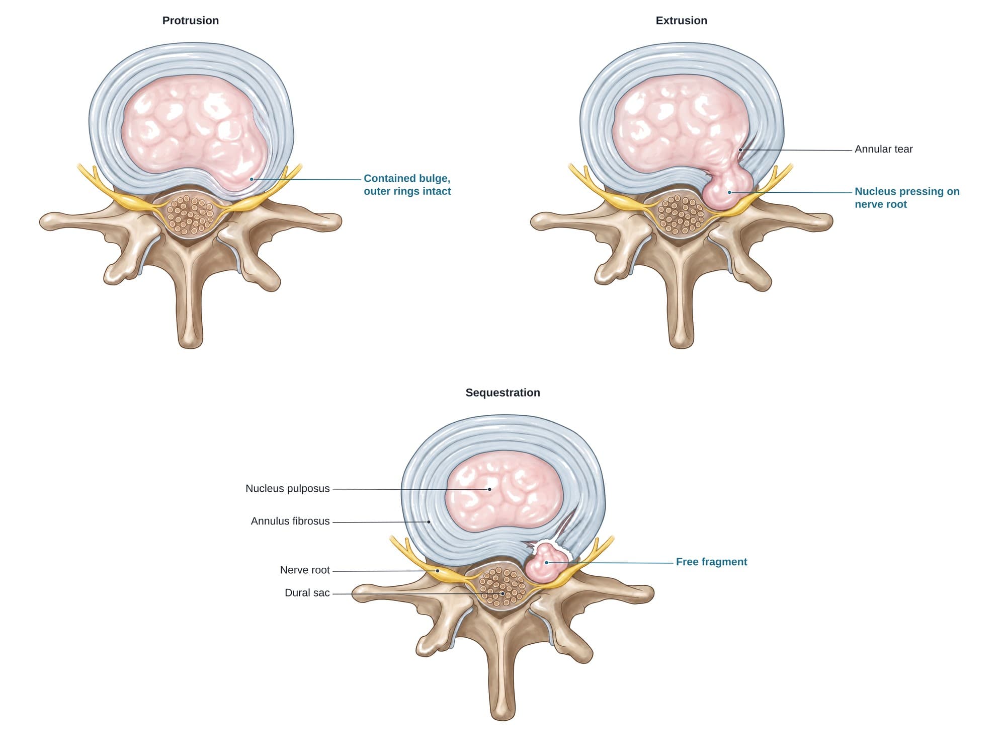
A lumbar disc herniation is a tear in the tough outer ring of a disc in the lower back that lets some of its soft center push out into the spinal canal. Most happen at L4-L5 or L5-S1. When the displaced disc material presses on a nerve root, it causes sciatica: pain, numbness or tingling down the leg. Herniations are described by shape, from a contained protrusion to an extrusion through the outer ring to a sequestration, where a piece breaks free. Most improve without surgery. The illustration above shows these three types from above.
The anatomy
Each intervertebral disc has a gel center, the nucleus pulposus, held in by layered rings of tough fiber, the annulus fibrosus. Directly behind the disc lies the spinal canal. In the lower back it holds the dural sac with the bundle of nerve roots called the cauda equina (the spinal cord itself ends higher up, around L1). A nerve root leaves the canal at each level through the intervertebral foramen. Because the back of the disc sits right against the canal and the roots, even a small herniation there can press on a nerve. For the full anatomy, see lumbar spine anatomy.
Types
| Type | What happens | Outer annulus |
|---|---|---|
| Bulge | The disc spreads evenly around much of its edge | Intact |
| Protrusion | Focal bulge, wider at its base than it sticks out; inner rings torn | Intact |
| Extrusion | Nucleus pushes through a full-thickness tear; the escaped part is wider than its neck | Torn |
| Sequestration | A piece of extruded nucleus breaks off and lies free in the canal, and can migrate up or down | Torn |
Herniations are also described by location: central, paracentral (just off center, the most common, usually pressing on the traversing root), foraminal and far lateral (pressing on the exiting root).
Causes
- Disc degeneration with age: the disc dries out and the annulus develops small tears
- Lifting, especially bending and twisting with a load
- Sudden trauma such as a car crash or a fall
- Repetitive strain from heavy physical work or prolonged vibration
- Risk factors: smoking, excess weight, family history, and a sedentary lifestyle
Symptoms
- Low back pain
- Sciatica: pain from the buttock down the leg, often below the knee
- Numbness or tingling in the leg or foot, following the pinched nerve root
- Weakness, such as trouble lifting the foot (L5) or rising on the toes (S1)
- Pain worse with sitting, bending, coughing or sneezing
- Cauda equina syndrome (rare, an emergency): numbness in the saddle area, bladder or bowel problems, and weakness in both legs
Diagnosis
The examination checks strength, reflexes and sensation in each leg. The straight leg raise test, lifting the straight leg while lying down, reproduces leg pain when a lower lumbar root is irritated. MRI is the main test: it shows the disc material outside its normal outline, its type and location, and how much it narrows the canal or presses on a root. X-rays do not show discs but rule out other causes. CT or CT myelography is used when MRI is not possible, and nerve studies (EMG) can confirm which root is affected.
Treatment
- Staying active within the limits of pain; strict bed rest is not recommended
- Physical therapy: core strengthening, nerve mobility and posture work
- Medicines: anti-inflammatories and, for short periods, other pain or nerve medicines
- Epidural steroid injection to calm an inflamed nerve root
- Microdiscectomy: removing the herniated fragment through a small opening to free the nerve, for persistent leg pain or worsening weakness
- Emergency surgery for cauda equina syndrome
Recovery
Most people improve within 6 to 12 weeks of non-surgical care. After a microdiscectomy, leg pain often eases quickly; people usually walk the same day and return to desk work in about 2 to 6 weeks, with heavier work later. Some numbness can take months to fade.
Complications
- Recurrent herniation at the same level
- Persistent numbness or weakness after severe or long-standing compression
- Chronic back pain and faster wear of the disc
- Cauda equina syndrome if a large central herniation is not treated in time
How a herniated disc is shown in a personal injury case
Herniated discs are among the most common injuries claimed after car crashes, falls and workplace lifting accidents. The key questions are whether the trauma caused the herniation or aggravated a degenerative disc, and whether the herniation explains the symptoms on the side and in the pattern the patient describes. An illustration that shows the disc type and the exact nerve root being compressed, drawn to match the MRI report, helps a jury follow the treating doctor's testimony. Disc injuries can also accompany a slipped vertebra; see anterolisthesis. For how these images are used at trial, see demonstrative evidence and medical legal illustration.
Related pages
FAQ
What is the difference between a bulging disc and a herniated disc?+
A bulging disc spreads out evenly around a wide part of its edge, like a hamburger too big for its bun, and the outer ring is intact. A herniated disc has a focal tear that lets the soft center push out in one spot. Herniations are more likely to press on a nerve root.
How long does a herniated disc take to heal?+
Most people improve a lot within about 6 to 12 weeks without surgery. Extruded and sequestered fragments often shrink over months as the body reabsorbs them. Some people have back or leg symptoms for longer.
Does a herniated disc need surgery?+
Usually not. Most are treated with activity changes, physical therapy, medicine and sometimes an epidural steroid injection. Surgery, usually a microdiscectomy, is considered for weakness that is getting worse, cauda equina syndrome, or leg pain that does not improve after about 6 weeks or more of treatment.
What does a herniated disc feel like?+
Low back pain is common, but the hallmark is sciatica: sharp, burning or electric pain that runs from the buttock down the back or side of the leg, often below the knee, with numbness or tingling. It is often worse with sitting, bending, coughing or sneezing.
Can a herniated disc get worse?+
A protrusion can progress to an extrusion with more strain, and symptoms can flare. Sudden numbness in the groin or saddle area, new bladder or bowel problems, or rapidly worsening leg weakness are emergencies.
Is a herniated disc permanent?+
The disc does not return to new, but the herniated part often shrinks and symptoms usually settle. Some people are left with intermittent back pain or numbness in part of the leg.
Can a car accident cause a herniated disc?+
Yes. A sudden load or twisting force can tear the annulus, especially in a disc that is already worn. In injury cases the question is usually whether the crash caused the herniation or made an existing, painless one symptomatic.
Which discs herniate most often?+
In the lower back, L4-L5 and L5-S1 account for the large majority. An L4-L5 herniation usually affects the L5 nerve root and an L5-S1 herniation the S1 root.
Need this illustration for a patient or a case?
Describe the injury or upload a scan and get a clear, labeled medical illustration for a consult, a chart or a case exhibit.
Try Natomy →