Facet Arthropathy: Stages, Symptoms & Treatment
Facet arthropathy is arthritis of the small joints at the back of the spine, most often at L4-L5 and L5-S1. See it illustrated beside normal facet joints.
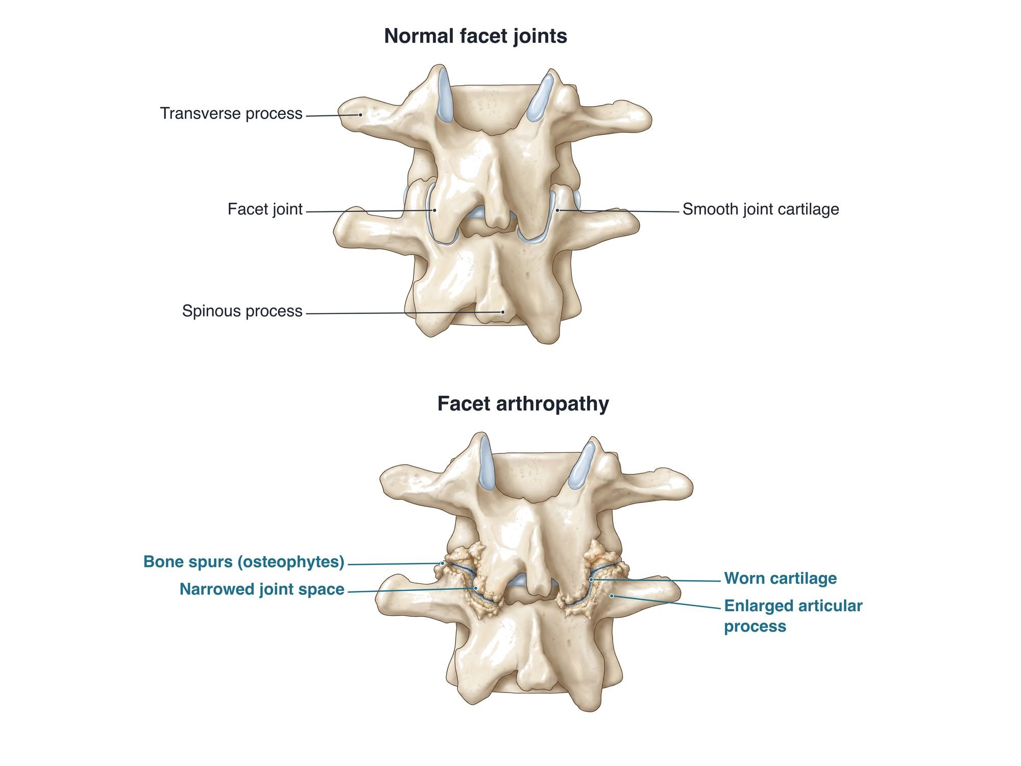
Facet arthropathy is arthritis of the facet joints, the small paired joints at the back of the spine that link each vertebra to the next. As the smooth cartilage in these joints wears down, the joint space narrows, the bone thickens, and bone spurs (osteophytes) form around the edges. This can cause low back or neck pain and stiffness. When the enlarged joints narrow the space for the nerves, it can also cause leg or arm pain. It is most common in the lower back at L4-L5 and L5-S1. The illustration above shows normal facet joints beside arthritic ones.
The anatomy
Each vertebra has two small joints at the back, one on each side, called facet joints or zygapophyseal joints. In each one, the inferior articular process of the vertebra above meets the superior articular process of the vertebra below. Like a knee or a hip, each facet joint is lined with smooth cartilage and enclosed in a joint capsule with fluid inside. The facet joints guide movement and stop the vertebrae from slipping and twisting too far. Each joint has its own small nerves, the medial branches, which carry pain signals from it. For the full anatomy, see lumbar spine anatomy.
Grades
Radiologists often grade facet arthropathy on CT or MRI:
| Grade | Joint space | Spurs and enlargement |
|---|---|---|
| 0 (normal) | Normal, 2 to 4 mm | None |
| 1 (mild) | Mildly narrowed | Small spurs, mild enlargement |
| 2 (moderate) | Narrowed | Moderate spurs and enlargement, small erosions |
| 3 (severe) | Very narrow or gone | Large spurs, marked enlargement, cysts |
The severity on a scan often does not match the amount of pain.
Causes
- Age-related wear: most adults over 60 have some facet arthritis on imaging
- Disc degeneration: when a disc loses height, more weight shifts onto the facet joints behind it
- Repetitive strain: years of heavy lifting, twisting or back arching
- Excess body weight, which adds load to the joints
- Injury: a facet joint sprain or fracture from a fall or crash, which can speed up wear
- Spinal alignment: scoliosis, or a vertebra that has slipped
- Inflammatory arthritis, such as rheumatoid arthritis or ankylosing spondylitis (less common)
Symptoms
- Dull, aching low back pain, often on one or both sides of the spine
- Pain spreading into the buttock or back of the thigh, usually not below the knee
- Pain that is worse leaning backward, twisting or standing for a long time
- Relief when bending forward or sitting
- Stiffness, especially in the morning or after rest
- In the neck: neck pain spreading to the shoulder blade or the back of the head
- Leg or arm pain, numbness or tingling when enlarged joints and spurs pinch a nerve root
Diagnosis
The doctor checks posture and movement, and presses over the joints to find tender spots. Pain when leaning back and twisting suggests the facet joints. X-rays show joint narrowing and spurs. CT shows the bone changes best. MRI shows fluid in the joint, cysts, inflammation and any pinched nerves. Because many people have facet arthritis on a scan without pain, the most reliable test is a diagnostic block. A numbing medicine is injected into the joint or onto its medial branch nerves. If the pain goes away for a few hours, the facet joint is the likely source.
Treatment
- Activity changes: staying active while avoiding movements that trigger pain
- Physical therapy: core strength, hip and hamstring flexibility, posture and flexion-based exercises
- Weight control to reduce load on the joints
- Medicines: anti-inflammatory drugs and short courses of other pain medicine
- Facet joint injection: a steroid placed into the joint for short-term relief
- Radiofrequency ablation: heat is used to turn off the medial branch nerves that carry pain from the joint. Relief often lasts about 6 to 12 months or longer and can be repeated.
- Surgery: rarely needed for facet pain alone. When enlarged joints narrow the nerve openings, a foraminotomy or laminectomy can free the nerves. Fusion is considered for instability, such as a degenerative spondylolisthesis.
Recovery
A flare of facet joint pain often settles within a few weeks with activity changes, medicine and therapy. Because the arthritis is long-term, flares can return. A regular exercise program is the best way to reduce them. After radiofrequency ablation, most people return to normal activity within a day or two, and relief builds over 2 to 3 weeks.
Complications
- Chronic low back or neck pain
- Foraminal stenosis, when spurs and enlarged joints narrow the nerve opening
- Central spinal stenosis, together with a thickened ligament and a bulging disc
- Facet joint cysts (synovial cysts) that can press on a nerve root
- Degenerative slipping of one vertebra on another (anterolisthesis)
How facet arthropathy is shown in a personal injury case
Facet arthropathy appears in many spine imaging reports after car crashes and falls. Because it develops over years, the defense often argues that a plaintiff's pain comes from this pre-existing arthritis rather than the injury. The plaintiff's side may argue that the injury made a joint painful that caused no problems before, an aggravation of a pre-existing condition. Earlier imaging, medical records from before the incident, and diagnostic blocks that confirm the pain source all matter. A side-by-side illustration like the one above helps a jury see the difference between a normal joint and an arthritic one. For how these images are used at trial, see demonstrative evidence and medical legal illustration.
Related pages
- Lumbar spine anatomyThe normal anatomy of the lumbar spine.
- AnterolisthesisAnterolisthesis is a vertebra slipped forward over the one below, most often L4 on L5. See it illustrated beside normal anatomy.
- Foraminal stenosisForaminal stenosis is narrowing of the opening where a nerve root leaves the spine, often at L4-L5 or L5-S1. See it illustrated beside normal anatomy.
- Lumbar disc herniationA herniated disc is disc gel pushed through a tear in its outer ring, often pressing on a nerve root. See protrusion, extrusion and sequestration illustrated.
- SpondylolisthesisSpondylolisthesis is a vertebra that has slipped forward over the one below, often L5 on S1 after a pars fracture. See it illustrated beside normal anatomy.
FAQ
What are the stages of facet arthropathy?+
Imaging scales usually describe four grades. Grade 0 is a normal joint. Grade 1 is mild narrowing or small spurs. Grade 2 is moderate narrowing, spurs and some enlargement of the joint. Grade 3 is severe narrowing with large spurs and marked enlargement. The grade does not always match the pain.
Is facet arthropathy serious?+
It is very common with age and is usually not dangerous. Many people with facet arthropathy on a scan have little or no pain. It becomes more significant when it causes daily back pain, or when enlarged joints narrow the space for the nerves and cause leg symptoms.
What not to do with facet arthropathy?+
Movements that load the facet joints often make pain worse. Examples are arching the back for long periods, heavy overhead lifting, and repeated twisting. Long periods of standing or lying flat on the stomach can also aggravate it. Staying active with gentle exercise is better than bed rest.
What are the symptoms of L5-S1 facet joint pain?+
A dull ache in the low back, often on one side, that can spread into the buttock and the back of the thigh. It usually does not go below the knee. Pain is often worse when leaning back, twisting or standing, and with stiffness in the morning.
Can facet arthropathy be cured?+
The joint changes do not reverse, but symptoms can usually be well controlled. Exercise, weight control, physical therapy and medicine help most people. For lasting pain, injections or radiofrequency ablation of the small nerves to the joint can give months of relief.
What is the difference between facet joint pain and sacroiliac joint pain?+
Facet joint pain usually sits on either side of the spine in the low back. Sacroiliac joint pain sits lower, over the dimple at the back of the pelvis, and often spreads to the buttock and groin. Exam tests and diagnostic injections help tell them apart.
Is facet arthropathy the same as spondylosis?+
They are related. Spondylosis is a broad term for age-related wear of the spine, including the discs and the facet joints. Facet arthropathy refers only to arthritis of the facet joints. The two often occur together.
Can a car accident cause facet arthropathy?+
The arthritis itself develops over years. But a crash can injure a facet joint capsule (a facet sprain) and cause pain, especially in the neck after whiplash. It can also make painful an arthritic joint that caused no trouble before.
Need this illustration for a patient or a case?
Describe the injury or upload a scan and get a clear, labeled medical illustration for a consult, a chart or a case exhibit.
Try Natomy →