Microdiscectomy: Surgery, Recovery Time & Risks
Microdiscectomy removes the piece of herniated disc pressing on a nerve root through a small window in the lamina. See it illustrated, with recovery time.
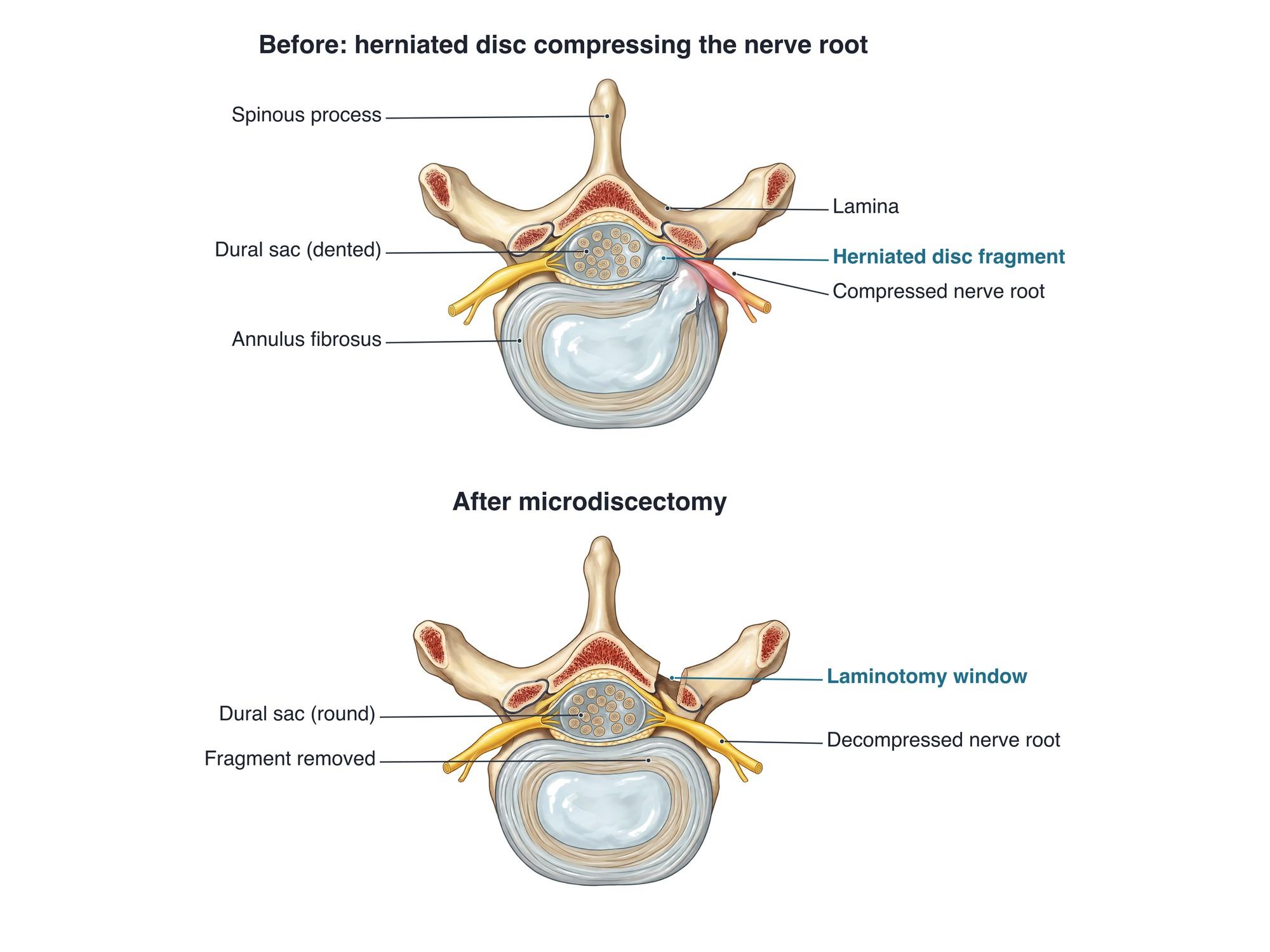
Microdiscectomy is surgery to remove the piece of a herniated disc that is pressing on a spinal nerve root. Through a small incision in the back, the surgeon uses a microscope to make a small window in the bone (a laminotomy). The surgeon gently moves the nerve root aside and removes the loose disc fragment. Most of the disc stays in place. It is the most common surgery for sciatica, usually at L4-L5 or L5-S1. The illustration above shows a herniated disc pinching a nerve root, and the same level after microdiscectomy.
Why it is done
Microdiscectomy is done to relieve radiculopathy, which means pain, numbness or weakness from a pinched nerve root. In the lower back, this is usually sciatica down one leg. It is considered when:
- Leg pain lasts beyond about 6 to 12 weeks despite therapy, medicine and injections
- There is weakness, such as foot drop, that is significant or getting worse
- Pain is severe enough that the patient cannot function
- MRI shows a herniation that matches the symptoms and exam, including a fragment inside the nerve opening (foraminal stenosis)
- Urgently for cauda equina syndrome: loss of bladder or bowel control, or numbness in the groin. This is an emergency.
It treats leg pain from nerve pressure. It is less reliable for back pain alone.
How the surgery is done
- The patient lies face down under general anesthesia. An X-ray confirms the correct level.
- The surgeon makes an incision of about 1 to 2 inches over the level, slightly to the side of the painful leg.
- The back muscles are gently pushed off the bone. Some surgeons use a tube (tubular retractor) to spread the muscle instead of cutting it.
- Under a microscope, the surgeon removes a small window of the lamina, the bony roof of the spinal canal, and the ligamentum flavum, a ligament under it. This is the laminotomy.
- The nerve root and dural sac (the sac holding the nerves) are gently moved toward the middle and protected.
- The herniated fragment is removed with small grasping tools. Any loose pieces inside the disc may also be taken out.
- The surgeon checks that the nerve root moves freely. The wound is closed with stitches or glue.
The whole operation usually takes about 1 hour.
Hardware
Microdiscectomy usually uses no hardware. There are no screws, rods or cages. The removed disc is not replaced. Some surgeons use a small device to close a large hole in the annulus in selected patients, but this is not routine. If the level is unstable, a different operation, such as a fusion, is needed instead.
Recovery
- Same day: walking starts within hours, and most people go home the same day or the next morning.
- Weeks 1 to 2: short, frequent walks. Avoid bending, lifting more than about 10 pounds, and twisting. Sitting may be limited to short periods.
- Weeks 2 to 6: most desk workers return to work. Physical therapy often starts around 2 to 6 weeks, focusing on core strength and posture.
- Weeks 6 to 12: lifting, sports and heavy work resume as cleared by the surgeon.
- Nerve recovery: leg pain often eases right away. Numbness and weakness can take weeks to months, and some may remain if the nerve was compressed for a long time.
Risks
- Recurrent disc herniation at the same level, the most common reason for repeat surgery
- Dural tear, a small leak of spinal fluid, usually repaired during surgery
- Nerve root injury, causing numbness or weakness (uncommon)
- Infection of the wound or, rarely, the disc space
- Persistent pain despite a technically good operation
- Bleeding and blood clots (rare)
- Surgery at the wrong level (rare, prevented with X-ray checks)
- Later back pain from disc degeneration at the same level
How microdiscectomy is shown in a personal injury case
Microdiscectomy is common in personal injury cases after car crashes, falls and lifting injuries that cause a disc herniation with sciatica. The surgery and its cost become part of the damages. Disputes often focus on whether the herniation was caused by the injury or was already there, and on future care, such as the risk of reherniation or a later fusion. Malpractice claims can involve a delayed diagnosis of cauda equina syndrome, wrong-level surgery or nerve injury. An illustration like the one above shows a jury what was pressing on the nerve and what the surgeon removed. For how these images are used at trial, see demonstrative evidence and medical legal illustration.
Related pages
FAQ
Is a microdiscectomy a serious surgery?+
It is real spine surgery done under general anesthesia, but it is one of the smallest. It usually takes about an hour through an incision of about 1 to 2 inches. Most people go home the same day. Serious complications are uncommon.
How long does it take to recover from a microdiscectomy?+
Most people walk the same day. Desk work often resumes in about 1 to 3 weeks, and physical jobs in about 6 to 12 weeks. Leg pain often improves right away. Numbness and weakness can take weeks to months to recover.
What is microdiscectomy recovery like week by week?+
In week 1, short walks and rest, with incision soreness. In weeks 2 to 4, longer walks and a return to desk work for many. In weeks 4 to 6, physical therapy builds core strength. From about 6 to 12 weeks, most people return to lifting, sports and heavy work as cleared by the surgeon.
What can you never do again after a microdiscectomy?+
Usually nothing is off limits for life. Most people return to work, exercise and sport. For the first 6 weeks or so, surgeons usually limit bending, lifting and twisting to let the annulus heal. Long-term, good lifting habits and core strength help protect the disc.
What is the success rate of microdiscectomy?+
For leg pain from a herniated disc that matches the imaging, most patients get good or excellent relief. Back pain improves less reliably than leg pain. The disc reherniates at the same level in a small share of patients, most often in the first year.
How long does it take to recover from an L5-S1 disc herniation without surgery?+
Many herniations improve on their own over 6 to 12 weeks as the body reabsorbs the disc material. Physical therapy, medicine and sometimes an injection help during that time. Surgery is considered if pain lasts beyond that, or sooner for weakness.
Is microdiscectomy the same as a laminectomy?+
No. A laminectomy removes most or all of the lamina to open the canal, usually for spinal stenosis. A microdiscectomy removes only a small window of lamina (a laminotomy) and then the herniated piece of disc.
Can the disc herniate again after microdiscectomy?+
Yes. A recurrent herniation at the same level happens in a minority of patients, usually within the first year. It often causes the same leg pain to return. It may be treated with therapy or a repeat operation.
Need a surgical illustration for consent or a case?
Describe the operation and get a clear, labeled medical illustration for patient consent, teaching or a case exhibit.
Try Natomy →