Stress Fracture in the Foot: Symptoms, MRI & Healing
A stress fracture is a small crack from repeated loading, often invisible on X-ray for weeks. See one on MRI beside a labeled illustration.
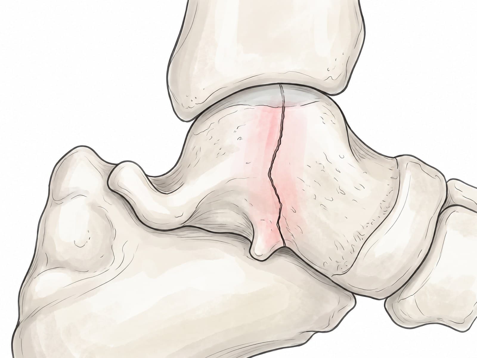
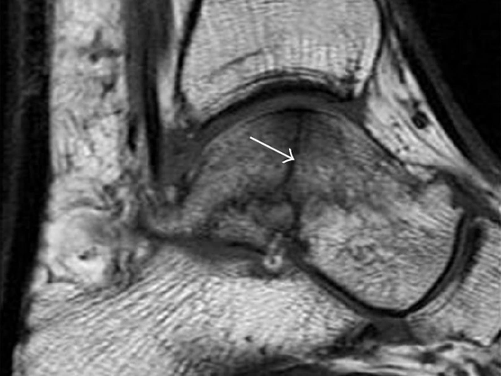
A stress fracture is a small crack in a bone caused by repeated loading rather than a single injury, as in running, marching or jumping. The bone breaks down faster than it can repair itself. Stress fractures are common in the foot, and they are often invisible on X-ray for the first few weeks, so MRI is usually what makes the diagnosis. The figure above shows a real MRI of a stress fracture with a medical illustration drawn from it: drag the slider to compare them.
Where stress fractures happen in the foot
- Second and third metatarsals: the most common site, the classic "march fracture" first described in soldiers, shown below.
- Navicular: a high-risk site in sprinters and jumpers that heals slowly.
- Base of the fifth metatarsal: overlaps with the Jones fracture zone.
- Calcaneus: the heel bone, in runners and military recruits.
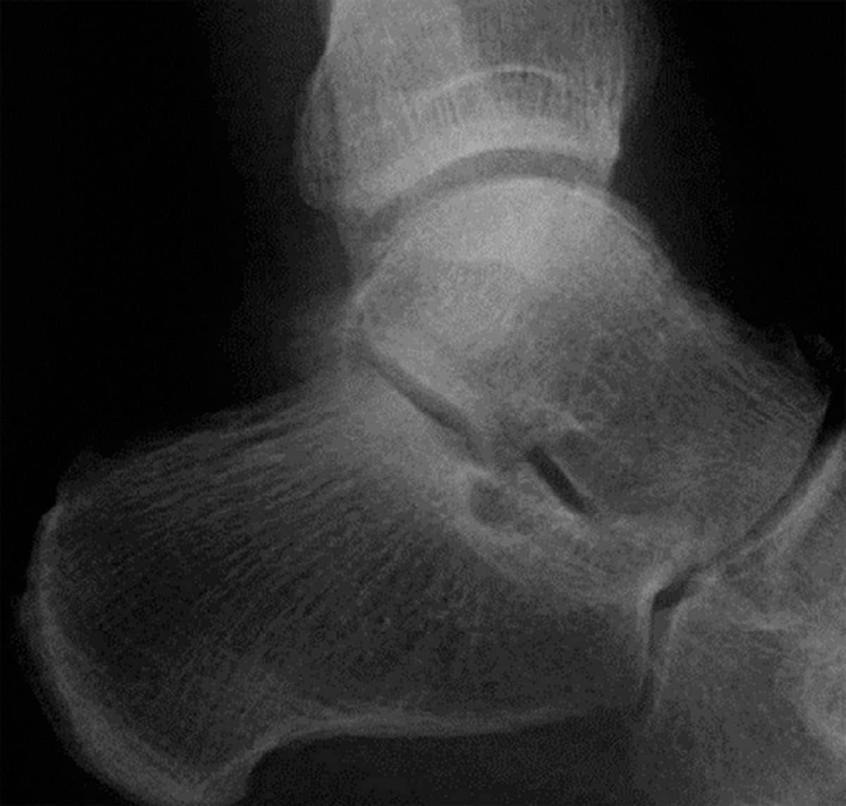
- Talus: less common, as in the case on this page, a 25-year-old basketball player with ankle and hindfoot pain and no injury.
For the normal bones of the foot, see foot bones anatomy.
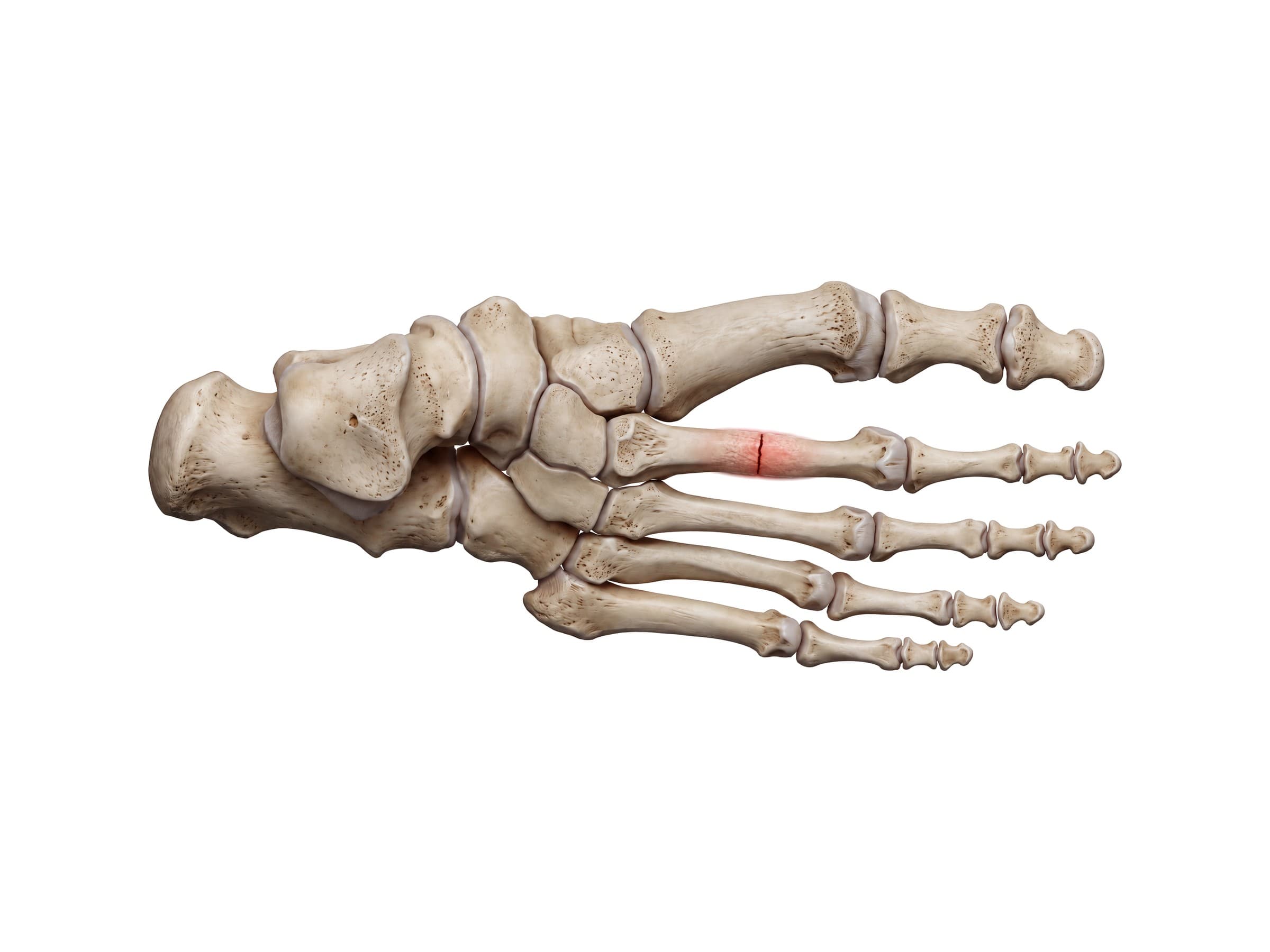
Why X-rays miss stress fractures
Early on, a stress fracture is a crack too fine for an X-ray to show. It becomes visible only after the bone starts to heal, as a faint line or a fluffy band of new bone, and some never show at all. In this case the X-ray taken a full month after the pain began still looked normal:
Stress fracture on MRI
MRI is the most sensitive test and the standard when a stress fracture is suspected and the X-ray is normal. It shows the problem within days:
| MRI finding | What it means |
|---|---|
| Bone marrow edema (dark on T1, bright on fluid-sensitive images) | The bone is overloaded: a stress reaction |
| A thin dark line within the edema | A fracture line has formed: a stress fracture |
| Swelling of the surrounding soft tissue | Often accompanies an active stress injury |
On the MRI above, a thin, irregular dark line runs down through the body of the talus, surrounded by an ill-defined dark area of bone marrow edema. Radiologists grade stress injuries from an early stress reaction to a complete fracture, which guides how long the athlete rests. CT is used for selected sites, such as the navicular, where it shows the fracture line in detail.
How a stress fracture happens
Bone constantly remodels in response to load. When training increases too quickly, the small cracks from repeated loading build up faster than the bone can repair them. Common triggers:
- A sudden increase in running distance, speed or hills
- A new sport or season, or military basic training
- Hard surfaces and worn or unsupportive shoes
- Low energy availability, low vitamin D or low bone density
Symptoms
- An ache in one spot that starts with activity and worsens over days to weeks
- Pinpoint tenderness on the bone, often on the top of the foot
- Mild swelling, and sometimes bruising
- Pain at rest or at night as it progresses
Treatment
- Relative rest: stopping running and impact activity, with low-impact exercise such as swimming or cycling.
- Stiff-soled shoe or walking boot: for most metatarsal stress fractures.
- Cast with no weight bearing: for high-risk sites such as the navicular, the base of the fifth metatarsal and the talus.
- Surgery: occasionally, for high-risk fractures that are displaced or do not heal.
- Addressing the cause: training load, footwear, nutrition, vitamin D and bone density.
Recovery time
Most metatarsal stress fractures heal in about 6 to 8 weeks. High-risk sites often take 8 to 12 weeks or longer. A return to running is gradual and starts only once walking and hopping are pain free.
Complications
- Progression to a complete fracture if training continues
- Delayed union or nonunion, especially at high-risk sites
- Recurrence if the underlying cause is not addressed
How a stress fracture is shown in a personal injury and malpractice case
Stress fractures come up in military and workplace claims and in missed-diagnosis cases, where a normal X-ray led to continued training and a complete fracture. The key evidence is usually the MRI, which jurors find even harder to read than an X-ray. An illustration drawn from the patient's own MRI, like the one above, shows exactly where the fracture runs and why the earlier X-ray looked normal. For how these images are used at trial, see demonstrative evidence and medical legal illustration.
Related pages
FAQ
What does a stress fracture in the foot feel like?+
A dull ache in one spot that starts during running, marching or long walks and gets worse each time, then eases with rest. Over time it may hurt even at rest or at night. There is usually a single tender point on the bone, often on the top of the foot, and sometimes mild swelling or bruising there.
Can a stress fracture be seen on an X-ray?+
Often not at first. In the first two to three weeks most stress fractures do not show on X-ray because the crack is too fine. They become visible later, if at all, as a faint line or a fluffy band of healing bone. MRI shows them within days, which is why it is the preferred test when a stress fracture is suspected.
Is it shin splints or a stress fracture?+
Shin splints cause a more diffuse ache along the inner edge of the shin that often warms up and eases during exercise. A stress fracture causes sharp, pinpoint tenderness on one spot of the bone that gets worse with activity and can hurt at rest. MRI can tell them apart and also grade an early stress reaction before it becomes a fracture.
Where do stress fractures in the foot happen?+
Most often in the shafts of the second and third metatarsals (the classic march fracture). Other sites include the navicular, the base of the fifth metatarsal, the calcaneus and, less often, the talus, like the case on this page.
How long does a stress fracture in the foot take to heal?+
Most metatarsal stress fractures heal in about 6 to 8 weeks with rest from impact activity and a stiff shoe or boot. High-risk sites, such as the navicular, the base of the fifth metatarsal and the talus, can take 8 to 12 weeks or more and may need a cast with no weight bearing or, occasionally, surgery.
What is a stress reaction?+
A stress reaction is the stage before a stress fracture: the bone is overloaded and swelling (bone marrow edema) shows on MRI, but no fracture line has formed yet. Caught at this stage, it usually heals faster with a period of reduced activity.
What causes stress fractures?+
Repeated loading faster than the bone can adapt: a sudden increase in running distance or intensity, a new sport, military training, hard surfaces or worn shoes. Low energy availability, low vitamin D, and low bone density raise the risk.
Need an illustration of a patient's own X-ray?
Upload an X-ray, CT or MRI and turn it into a clear, labeled medical illustration for a consult, a chart or a case exhibit.
Try Natomy →