Radial Head Fracture (Broken Elbow): X-ray & Recovery
A radial head fracture is a break at the top of the radius in the elbow, the most common adult elbow fracture. See one on X-ray beside an illustration.
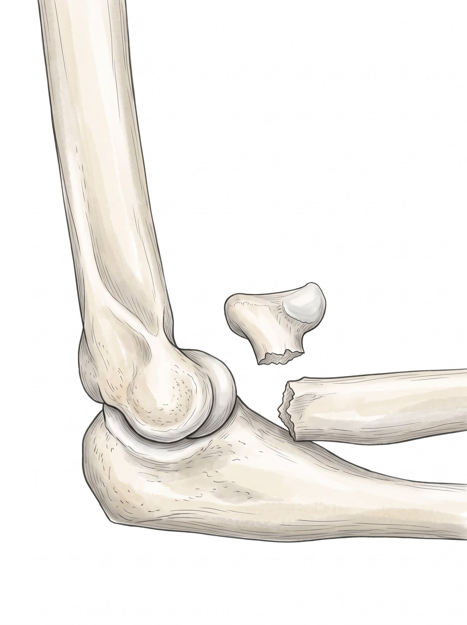
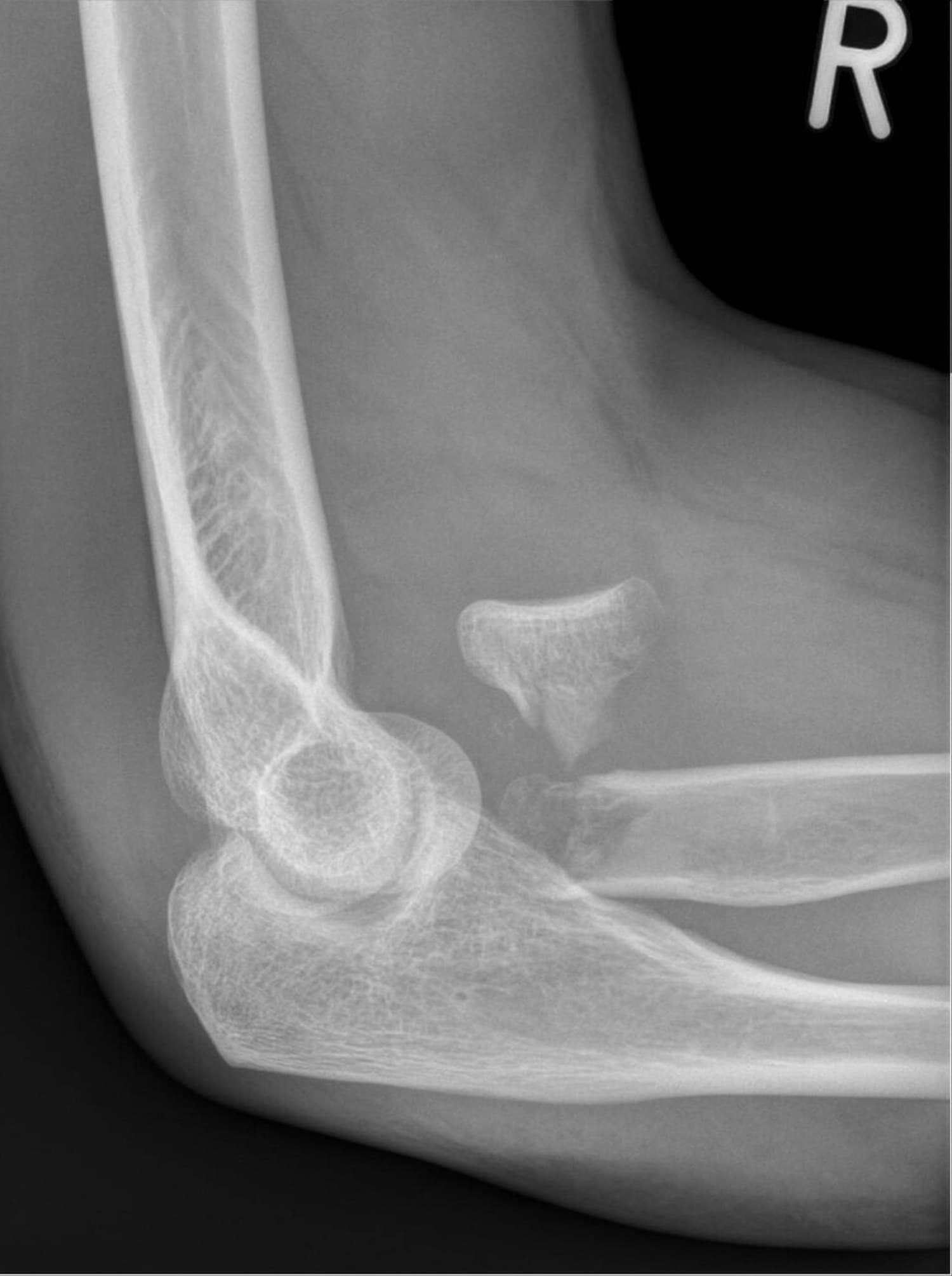
A radial head fracture is a break at the top of the radius, where it meets the humerus at the elbow. It is the most common elbow fracture in adults, usually from a fall on an outstretched hand. Most are small and heal with early movement, but displaced fractures can block forearm rotation and need surgery. The figure above shows a real X-ray of a displaced radial head fracture with a medical illustration drawn from it: drag the slider to compare them.
The radial head
The radius is the forearm bone on the thumb side. At the elbow it ends in the radial head, a disc that turns against the capitellum of the humerus and within the ring of the ulna as the palm turns up and down. Below the head is the narrower radial neck. The radial head also helps keep the elbow stable. For the full anatomy, see elbow anatomy.
Types of radial head fracture
The Mason classification:
| Type | Pattern | Usual treatment |
|---|---|---|
| I | Non-displaced or minimally displaced | Sling and early movement |
| II | Displaced partial fracture of the head | Movement, or fixation if rotation is blocked |
| III | Comminuted or completely displaced head or neck | Fixation or radial head replacement |
| IV | Any of the above with an elbow dislocation | Surgery to stabilize the elbow |
Radial head fracture on X-ray
Front, side and oblique views of the elbow are taken, sometimes with a dedicated radial head view. On the side view above, the radial head has broken off at the neck, rotated and moved forward and upward, so it no longer sits against the capitellum. This is a displaced Mason type III fracture.
| Finding | Why it matters |
|---|---|
| Displacement and comminution | Guide movement versus surgery |
| Fat pad sign | Points to a hidden fracture when the line is not visible |
| Radiocapitellar alignment | The radial head should line up with the capitellum in every view |
| Associated injuries | Coronoid fracture, dislocation, or wrist injury (Essex-Lopresti) |
How a radial head fracture happens
Almost always a fall on an outstretched hand with the elbow straight, driving the radial head into the capitellum. It happens in sports, cycling, falls on ice and stairs, and car crashes when the arm braces against the dashboard. Higher-energy injuries can add an elbow dislocation.
Symptoms
- Pain on the outer side of the elbow
- Swelling of the elbow
- Pain when turning the forearm palm up or down
- Inability to fully straighten the elbow
- Tenderness directly over the radial head
Treatment
- Sling and early movement: for Mason I and most Mason II fractures.
- Joint aspiration: draining blood from the joint can relieve pain and allow a rotation check.
- ORIF: small screws or a plate for displaced fractures that can be rebuilt.
- Radial head replacement: a metal implant for comminuted fractures that cannot be fixed, especially with instability.
- Physical therapy: to recover straightening and rotation.
Recovery time
Non-displaced fractures usually heal in 6 to 8 weeks, with most motion back within 2 to 3 months. After surgery, recovery often takes 3 to 6 months. A small loss of full straightening is common.
Complications
- Elbow stiffness, especially loss of straightening
- Post-traumatic arthritis
- Instability after complex injuries
- Heterotopic ossification (extra bone forming around the joint)
- Implant problems after radial head replacement
How a radial head fracture is shown in a personal injury case
Broken elbows follow falls on stairs and wet floors, cycling crashes, and car crashes when the arm braces against the dashboard. Hidden fractures that show only a fat pad sign are sometimes missed at first. An illustration drawn from the patient's own X-ray, like the one above, shows the radial head displaced from the joint and why turning the hand became difficult. For how these images are used at trial, see demonstrative evidence and medical legal illustration.
Related pages
FAQ
How long does a broken elbow take to heal?+
A non-displaced radial head fracture usually heals in about 6 to 8 weeks, and most people regain good motion within 2 to 3 months with early movement. Displaced fractures treated with surgery take longer, often 3 to 6 months for full recovery, and some loss of elbow straightening is common.
What does a broken elbow feel like?+
Pain on the outer side of the elbow after a fall on an outstretched hand, swelling, and pain when turning the palm up or down. Many people cannot fully straighten the elbow. A small fracture can still allow some movement, so it is easy to mistake for a sprain.
Does a radial head fracture need surgery?+
Most do not. Non-displaced and minimally displaced fractures are treated with a short period in a sling and early movement. Surgery is considered when the fragments block rotation, are badly displaced, or are broken into several pieces; options are screws or a plate, or replacing the radial head with an implant.
What is the Mason classification?+
A system for radial head fractures. Type I is non-displaced, type II is a displaced partial fracture, type III is comminuted or completely displaced like the one on this page, and type IV adds an elbow dislocation.
Why can a radial head fracture be hard to see on X-ray?+
Non-displaced fractures can be almost invisible. Doctors look for indirect signs such as the fat pad sign, where fluid in the joint lifts fat pads on the front and back of the elbow into view. An elbow with a positive fat pad sign after a fall is treated as fractured.
Can I move my elbow with a radial head fracture?+
Gentle movement is usually encouraged early, often within a week, for stable fractures, because the elbow stiffens quickly. Your doctor will check that the fracture does not block forearm rotation before starting.
What are the long-term effects of a broken elbow?+
Most people do well, but loss of the last degrees of straightening, aching with heavy use, and, after more severe fractures, post-traumatic arthritis or instability can persist.
Need an illustration of a patient's own X-ray?
Upload an X-ray, CT or MRI and turn it into a clear, labeled medical illustration for a consult, a chart or a case exhibit.
Try Natomy →