Intramedullary Nailing: Procedure, Recovery & Risks
Intramedullary nailing fixes a broken tibia or femur with a metal rod inside the bone. See the nail and locking screws beside the original fracture.
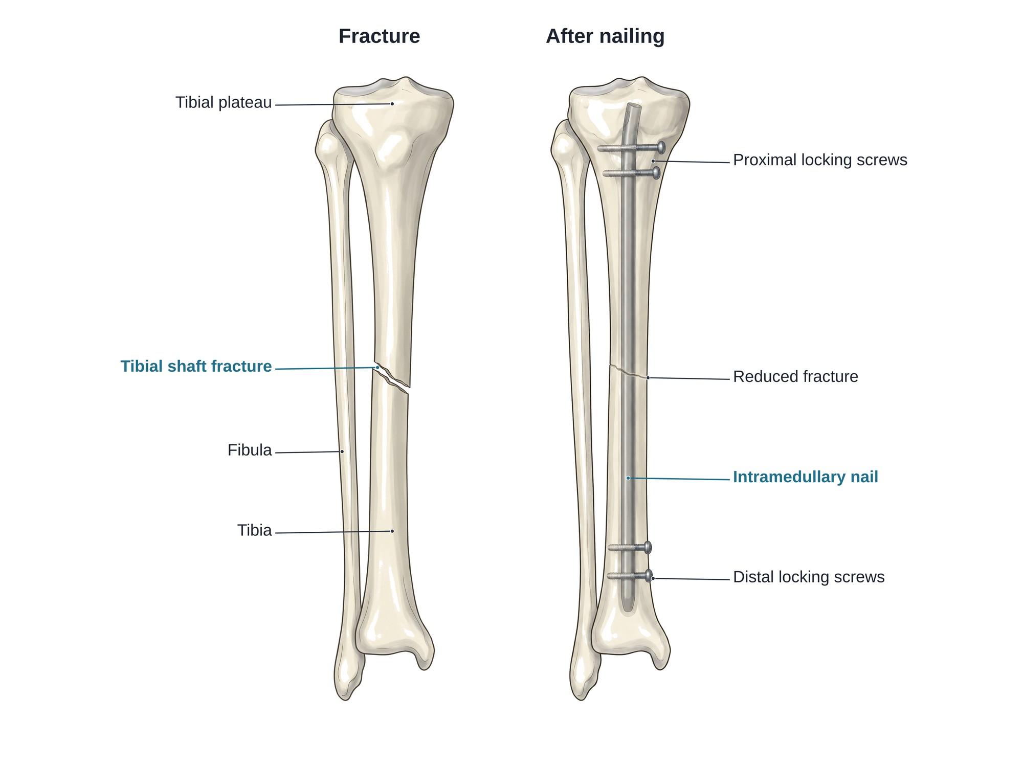
Intramedullary nailing is surgery to fix a broken long bone with a metal rod placed inside it. The rod is called an intramedullary nail (IM nail, or rod). It runs through the medullary canal, the hollow center of the bone where marrow lives. Screws through each end of the nail lock it in place. It is the standard treatment for most shaft fractures of the tibia (shin bone) and femur (thigh bone). The illustration above shows a tibial shaft fracture, then the same tibia held with a nail and locking screws.
Why it is done
Intramedullary nailing holds a broken bone in line while it heals. It is used most for fractures through the long middle part of a bone, called the shaft (diaphysis). Common uses are:
- Tibial shaft fractures, often from car crashes, motorcycle crashes and falls. See tibia and fibula fracture.
- Femoral shaft fractures, which usually come from high-energy trauma such as a car crash.
- Some fractures near the hip, using a short nail with a large screw into the femoral head. See hip fracture and hip fracture surgery.
- Some humerus (upper arm) fractures.
A nail is often chosen over a cast because shaft fractures of the tibia and femur are unstable. A cast cannot hold them well, and the patient would be off the leg for months. A nail lets most people move and bear weight sooner. It also needs only small incisions, which protects the skin and blood supply around the break. This matters for open fractures, where the bone has broken through the skin.
How the surgery is done
Tibial nailing usually takes about 1.5 to 2.5 hours. It is done under general or spinal anesthesia. Live X-ray (fluoroscopy) guides each step.
- Positioning. The patient lies on the back. The knee is bent, or kept nearly straight for a suprapatellar approach (going in above the kneecap).
- Reduction. The surgeon pulls and turns the leg to line up the broken ends. This is called closed reduction, because the fracture site is not opened. The fixed fracture in the right panel above shows the bone lined up.
- Entry point. A small incision is made at the front of the knee. An opening is made in the top of the tibia, just below and in front of the joint surface (tibial plateau).
- Guide wire and reaming. A long guide wire is passed down the canal, across the fracture, to the lower tibia. The canal is often widened with a drill called a reamer so a stronger nail fits.
- Nail insertion. The nail is passed over the guide wire and tapped down into the canal, across the fracture.
- Locking screws. Screws are placed through holes near both ends of the nail, crossing the bone from side to side. They stop the bone from shortening or rotating around the nail.
- Closing. The small incisions are closed with stitches or staples.
Femoral nailing follows the same steps. The nail goes in at the top of the femur, near the hip, or sometimes through the knee (retrograde nailing).
Hardware
- Intramedullary nail. A long rod of titanium or stainless steel. It is usually hollow (cannulated) so it can slide over the guide wire. Tibial nails have a slight bend near the top to match the bone.
- Proximal locking screws. Screws through the nail near the knee for a tibial nail.
- Distal locking screws. Screws through the nail above the ankle.
- End cap. Sometimes added to the top of the nail to make later removal easier.
When both ends are locked, the nail is called statically locked. Some surgeons use a slotted hole (dynamic locking) that lets the fracture ends press together as the patient walks.
Recovery
- Most patients go home in 1 to 4 days.
- Walking with crutches or a walker starts within a day or two. Many patients can put some or full weight on the leg early, as the surgeon allows.
- Physical therapy works on knee and ankle motion, strength and walking.
- The bone usually heals in about 3 to 6 months. X-rays are taken every few weeks to check healing.
- Return to desk work is often possible in a few weeks. Heavy labor and sports usually wait 4 to 6 months or longer.
- For a femur fracture fixed with a rod, recovery is similar. Thigh weakness and a limp can last several months.
Risks
- Knee pain at the front of the knee after tibial nailing. It is the most common complaint and can last a long time.
- Delayed union or nonunion, where the bone heals slowly or not at all. A second surgery may be needed.
- Malunion, where the bone heals crooked, shortened or rotated.
- Infection, more likely after open fractures.
- Compartment syndrome, a dangerous rise in pressure inside the leg muscles after injury or surgery. It needs urgent treatment.
- Fat embolism, where fat from the marrow travels to the lungs. Rare but serious.
- Painful or broken screws, which may need removal.
- Blood clots (deep vein thrombosis).
How intramedullary nailing is shown in a personal injury case
Tibial and femoral shaft fractures from car and motorcycle crashes are often fixed with a nail. The surgery changes the case. It adds an operation, a hospital stay, months of therapy, and hardware that usually stays for life. It can also mean a second surgery to remove painful screws or treat a nonunion. A side by side illustration of the plaintiff's own fracture and the nail that now holds it helps a jury see what the X-rays show. It also explains future knee pain, limp and work limits. Compare it with plate fixation in ORIF surgery and tibial plateau fracture surgery. For how these images are used at trial, see demonstrative evidence and medical legal illustration. For the normal anatomy around the entry point, see knee anatomy.
Related pages
- Knee anatomyThe normal anatomy of the knee.
- Below knee amputationA below knee amputation removes the lower leg through the tibia and fibula and pads the bone ends with calf muscle. See the cut level and the residual limb.
- Tibial plateau fracture surgeryTibial plateau fracture surgery lifts a sunken knee joint surface and holds it with a plate and screws. See the fracture and the repair side by side.
FAQ
How long does it take to recover from intramedullary nailing?+
Most tibia and femur fractures fixed with a nail heal in about 3 to 6 months. Many people walk with crutches within days, and some can put full weight on the leg early. Getting back to full strength and normal walking often takes 6 to 12 months.
Are intramedullary nails permanent?+
Usually, yes. The nail is designed to stay in the bone for life. It is removed only if it causes ongoing pain (often knee pain at the entry point), an infection, or another problem. Removal is a second, smaller surgery done after the bone has fully healed.
How long does an intramedullary nail last?+
The nail's job is to hold the bone while it heals. Once the fracture has healed, the bone carries the load again. A nail left in place can last a lifetime. A nail can break if the bone never heals (nonunion), because the metal then carries all the stress.
Is intramedullary nailing painful?+
The first days after surgery are painful, and pain medicine is given. Pain usually eases a lot over 2 to 6 weeks. After tibial nailing, knee pain at the front of the knee is common and can last months. Some people also feel the locking screws under the skin.
What is the recovery time for a broken femur with a rod?+
A femur (thigh bone) fixed with a rod usually heals in about 3 to 6 months. Walking with a walker or crutches starts within a few days. Most people need several months of physical therapy, and full recovery of strength can take up to a year.
Can you walk on a leg with an intramedullary nail?+
Often, yes. A nail shares the load with the bone, so many patients can put some or all of their weight on the leg soon after surgery. The surgeon decides based on the fracture pattern and how stable the fixation is.
What is the difference between an intramedullary nail and a plate?+
A nail sits inside the hollow center of the bone. A plate sits on the outside surface and is held with screws, as in open reduction and internal fixation (ORIF). For shaft fractures of the tibia and femur, a nail usually needs smaller incisions and lets patients bear weight sooner.
Will the metal set off airport metal detectors?+
It can. Modern scanners sometimes detect a tibial or femoral nail. A note from the surgeon is not required, but telling security staff about the implant can make screening faster.
Need a surgical illustration for consent or a case?
Describe the operation and get a clear, labeled medical illustration for patient consent, teaching or a case exhibit.
Try Natomy →