Hip Fracture: Femoral Neck X-ray, Surgery & Recovery
A hip fracture is a break at the top of the femur, most often the femoral neck after a fall. See one on X-ray beside a labeled illustration.
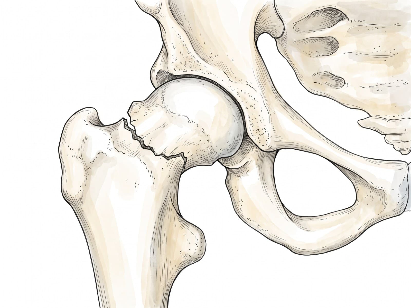
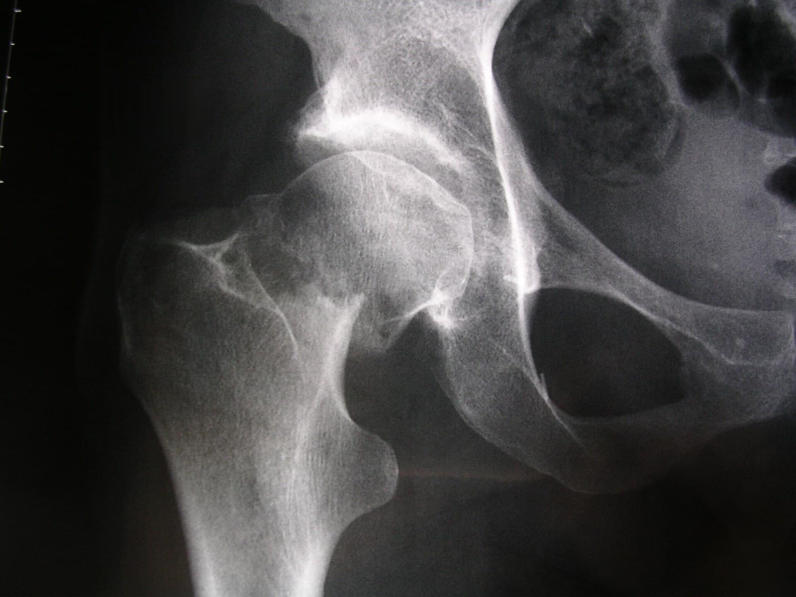
A hip fracture is a break at the top of the femur, the thighbone, close to the hip joint. The two main types are the femoral neck fracture, through the narrow neck just below the ball of the joint, and the intertrochanteric fracture, lower down between the two bony prominences of the upper femur. Most happen when an older adult falls from standing. The figure above shows a real X-ray of a displaced femoral neck fracture with a medical illustration drawn from it: drag the slider to compare them.
The upper femur
The top of the femur has four parts that matter for hip fractures:
- Femoral head: the ball that sits in the hip socket (acetabulum).
- Femoral neck: the narrow bridge between the head and the shaft, inside the joint capsule. Most of the blood supply to the femoral head runs along it.
- Greater trochanter: the bony point on the outside of the hip, where the gluteal muscles attach.
- Lesser trochanter: a smaller knob on the inner side, where the iliopsoas tendon attaches.
For the full anatomy of the hip, see hip anatomy.
Types of hip fracture
- Femoral neck (intracapsular) fracture: through the neck, inside the capsule. Divided by displacement (the Garden classification): non-displaced or impacted fractures (Garden I and II) and displaced fractures (Garden III and IV). The more displaced, the more likely the blood supply to the head is damaged.
- Intertrochanteric fracture: between the greater and lesser trochanters, outside the capsule. The blood supply is good, so the bone usually heals once fixed. When the bone breaks into several pieces it is a comminuted fracture.
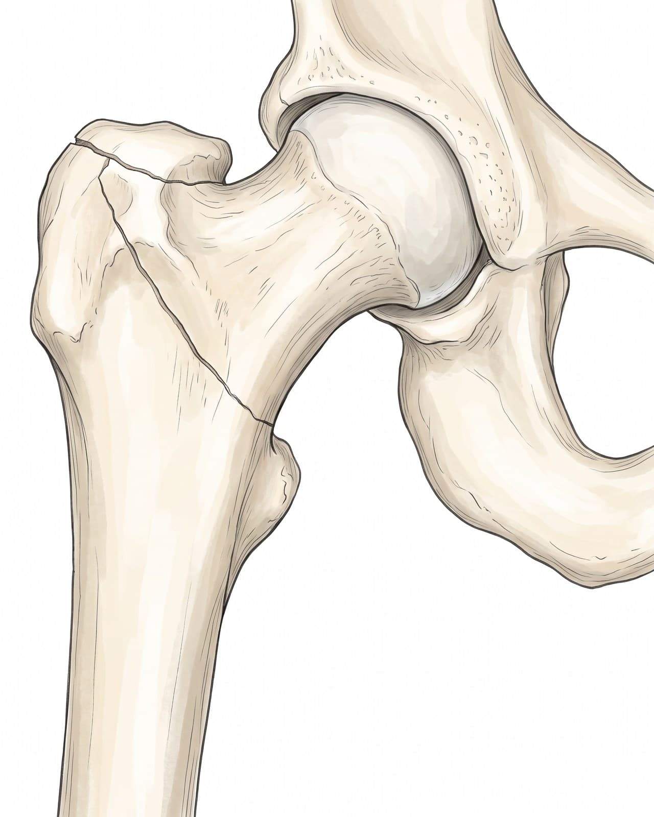
- Subtrochanteric fracture: just below the lesser trochanter, often from higher energy injuries or in bone weakened by disease.
Hip fracture on X-ray
A front (AP) view of the pelvis and a side view of the hip are taken. On the X-ray above, the femoral neck of the right hip is broken right at the base of the femoral head (a subcapital fracture). The head has tilted, and the neck and shaft have shifted and shortened, so the broken end overlaps the base of the head: a displaced femoral neck fracture.
| Finding | Why it matters |
|---|---|
| Location (neck, intertrochanteric or subtrochanteric) | Decides the type of surgery |
| Displacement (Garden grade for neck fractures) | Displaced neck fractures usually need a hip replacement |
| Shortening and outward rotation of the leg | Often visible on examination and on the X-ray |
| A normal X-ray with hip pain after a fall | MRI or CT to look for a hidden (occult) fracture |
Hairline and stress fractures of the hip
Not every hip fracture comes from a fall. A stress fracture of the femoral neck is a crack that builds up from repeated loading, in runners, military recruits and people who suddenly increase their activity, and an insufficiency fracture is the same thing in bone weakened by osteoporosis. These are often called hairline fractures because the crack is fine and the bone has not moved.
- Symptoms: a deep ache in the groin that comes on with activity, eases with rest, and gradually hurts with walking or at night.
- Diagnosis: X-rays are often normal at first; MRI shows the stress injury early. See stress fracture for how MRI finds them.
- Why they matter: a stress fracture on the tension (outer, upper) side of the femoral neck can progress to a complete, displaced fracture, so it is usually fixed with screws. One on the compression (inner, lower) side is often treated with rest and crutches.
How a hip fracture happens
More than 9 in 10 hip fractures in older adults come from a simple fall from standing, usually sideways onto the hip, in bone weakened by osteoporosis. Risk rises with age, female sex, low body weight, poor vision, medications that cause dizziness, and conditions that affect balance. In younger adults, hip fractures usually come from high-energy injuries such as car crashes and falls from a height.
Symptoms
- Pain in the groin or outer hip after a fall
- Inability to stand or put weight on the leg
- A leg that looks shorter and turned outward, with a displaced fracture
- Pain on moving the hip, sometimes felt in the knee
- Sometimes only mild pain, with an impacted or non-displaced fracture
Treatment
Surgery is usually done within a day or two of admission.
- Screw fixation (ORIF or percutaneous screws): for non-displaced or impacted femoral neck fractures, and for femoral neck fractures in younger adults, where the surgeon realigns the bone and fixes it to save the patient's own femoral head.
- Hemiarthroplasty or total hip replacement: for displaced femoral neck fractures in older adults, replacing the femoral head because its blood supply is often lost.
- Intramedullary nail or sliding hip screw: for intertrochanteric fractures.
- Early mobilization: standing and walking with help the day after surgery, with blood clot prevention and attention to bone health.
Recovery time
Most patients start walking with a walker within a day or two of surgery. Rehabilitation continues for about 3 months, and function often keeps improving for 6 to 12 months. Recovery depends heavily on health and mobility before the fracture, and many older adults need more support at home afterward.
Complications
- Avascular necrosis of the femoral head, after femoral neck fractures
- Nonunion, when a fixed neck fracture fails to heal
- Blood clots, pneumonia, pressure sores and delirium, especially with delayed surgery or prolonged bed rest
- Loss of independence and higher mortality in the year after the fracture in older adults
How a hip fracture is shown in a personal injury and nursing home case
Hip fractures are at the center of many fall cases: slips in stores and on stairs, falls in hospitals and nursing homes, and falls during transfers. Common questions include whether the fall was preventable, whether a fracture was missed on the first X-ray, and how long surgery was delayed. An illustration drawn from the patient's own X-ray, like the one above, shows which part of the femur broke, how far it moved, and why the head of the femur had to be replaced. For how these images are used at trial, see demonstrative evidence and medical legal illustration.
Related pages
FAQ
What does a fractured hip feel like?+
Usually sudden pain in the groin or outer hip after a fall, and difficulty or inability to stand or put weight on the leg. With a displaced fracture the leg often looks shorter and turned outward. Some people with a non-displaced fracture can still walk, with pain in the groin or knee.
Can you have a fractured hip and not know it?+
Yes. A non-displaced or impacted hip fracture can cause only mild pain, and the first X-ray can look normal. Older adults with hip pain after a fall and a normal X-ray are usually sent for MRI or CT, because an undiagnosed fracture can displace later and become much harder to treat.
Does a hip fracture always need surgery?+
Almost always. Surgery lets the patient get out of bed within a day or two, which lowers the risk of pneumonia, blood clots and pressure sores. Non-surgical treatment is reserved for people too unwell for an operation or for some stable impacted fractures, and it means weeks of limited mobility.
What surgery is done for a hip fracture?+
It depends on the type. A non-displaced femoral neck fracture is often fixed with screws. A displaced femoral neck fracture in an older adult is usually treated by replacing the femoral head, either a partial (hemiarthroplasty) or total hip replacement, because the blood supply to the head is often damaged. Intertrochanteric fractures are fixed with a nail inside the femur or a sliding hip screw and plate.
How long is recovery after hip fracture surgery?+
Most patients stand and start walking with a walker within a day or two. Rehabilitation continues for about 3 months, and many people take 6 to 12 months to reach their best level of function. A significant share of older adults do not return to their previous level of independence.
Can a hip fracture heal on its own?+
Rarely. A displaced femoral neck fracture usually cannot heal on its own, because the fragments are separated and the blood supply to the femoral head is often disrupted. Even stable fractures are usually fixed, because healing without surgery takes weeks of immobility that is dangerous for older adults.
How do you know if your hip is broken?+
The main signs are groin or outer hip pain after a fall, being unable to stand or walk on the leg, and a leg that looks shorter and turned outward. But a small or impacted fracture can let you walk with only mild pain, so the only way to know is an X-ray, and MRI or CT if the X-ray is normal and the pain continues.
What are the signs of a hip stress fracture?+
A deep ache in the groin or front of the hip that comes on with running or marching, gets worse with activity, eases with rest, and over time hurts with walking or at night. Pain at the end of hip movement, especially rotating the hip inward, is common. X-rays are often normal, so MRI is used to diagnose it.
What is the difference between a femoral neck fracture and an intertrochanteric fracture?+
Both are hip fractures of the upper femur. A femoral neck fracture breaks the narrow neck just below the femoral head, inside the hip joint capsule, where the blood supply to the head is at risk. An intertrochanteric fracture runs between the greater and lesser trochanters, outside the capsule, where the blood supply is good and the bone usually heals after fixation.
Need an illustration of a patient's own X-ray?
Upload an X-ray, CT or MRI and turn it into a clear, labeled medical illustration for a consult, a chart or a case exhibit.
Try Natomy →