Tibial Plateau Fracture Surgery: ORIF, Recovery & Risks
Tibial plateau fracture surgery lifts a sunken knee joint surface and holds it with a plate and screws. See the fracture and the repair side by side.
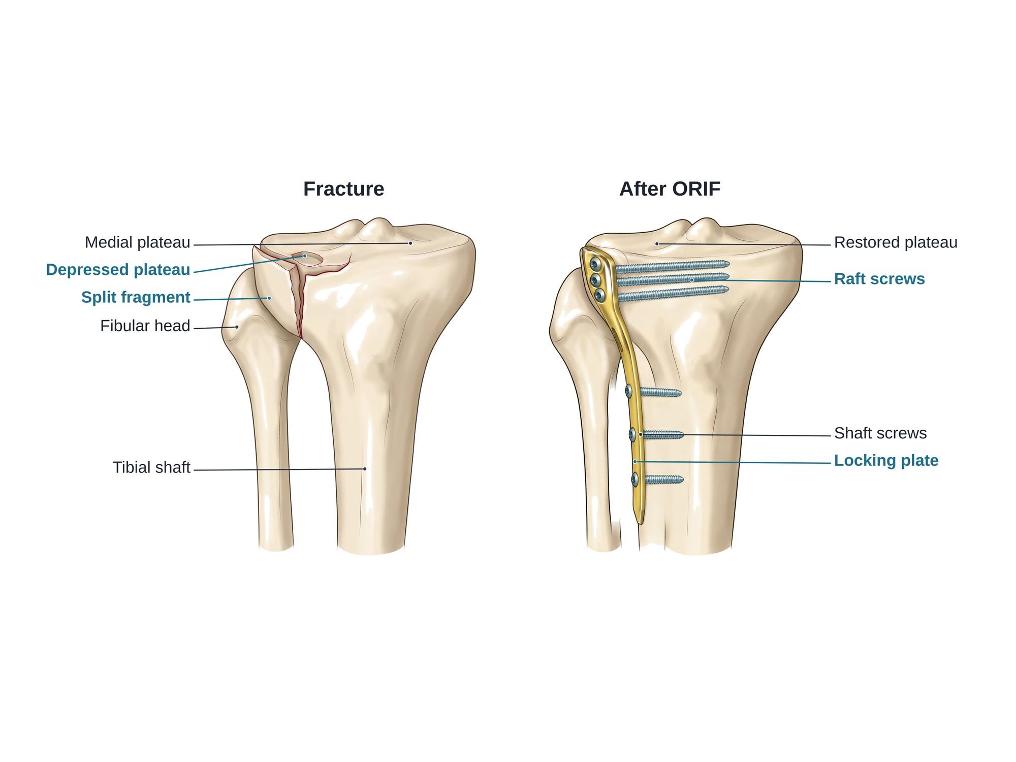
Tibial plateau fracture surgery repairs a break into the top of the tibia, the flat surface that forms the lower half of the knee joint. The usual operation is open reduction and internal fixation (ORIF). "Open reduction" means the surgeon opens the skin and puts the broken pieces back in place. "Internal fixation" means holding them there with a metal plate and screws. The main goal is a smooth, level joint surface, which protects the knee from arthritis. The illustration above shows a split-depression fracture of the outer plateau, then the same bone after repair.
Why it is done
A tibial plateau fracture enters the knee joint. If it heals with a step or a sunken area in the joint surface, the knee can become unstable, bent, and arthritic. Surgery is the usual tibial plateau fracture treatment when:
- Part of the joint surface is pushed down (depressed) by more than a few millimeters
- A fragment is split off and spread apart
- The knee is unstable or tilts inward or outward
- Both plateaus are broken (a bicondylar fracture, Schatzker types V and VI)
- The fracture is open (bone through the skin), or there is a nerve or artery injury
Small fractures that are not displaced can heal in a hinged brace without surgery.
Timing and swelling
These fractures often cause severe swelling. Operating through swollen, blistered skin raises the risk of wound problems and infection. In high-energy injuries, surgeons often place a temporary external fixator first. This is a frame of pins and bars outside the leg that holds it out to length. The definitive surgery follows 1 to 3 weeks later, once the skin wrinkles again. Doctors also watch closely for compartment syndrome, a dangerous rise of pressure in the leg muscles.
How the surgery is done
The operation usually takes 2 to 3 hours under general or spinal anesthesia. A CT scan beforehand maps every fragment.
- Incision. For an outer (lateral) plateau fracture, the incision runs along the front and outer side of the knee. Medial or bicondylar fractures may need a second incision on the inner or back side.
- Look at the joint. The surgeon lifts the meniscus or uses an arthroscope (a small camera) to see the joint surface. A torn meniscus is repaired.
- Open the split. The split fragment is opened like a book to reach the sunken area behind it.
- Lift the depressed surface. A bone tamp pushes the depressed cartilage and bone back up until it is level with the rest of the plateau.
- Fill the gap. The space left under the lifted surface is packed with bone graft or a bone substitute, so the surface does not sink again.
- Close the split and fix it. The split fragment is pressed back into place. A plate is laid against the outer tibia, with screws placed just under the joint surface and into the shaft below.
- Check and close. Live X-ray (fluoroscopy) confirms a level joint and good screw position. The wound is closed in layers.
Hardware
- Lateral locking plate. A pre-shaped plate that fits the outer side of the upper tibia. The screws lock into the plate, so the plate and screws act as one rigid frame. This is helpful in soft or broken-up bone.
- Raft screws. A row of screws placed parallel, just under the joint surface. They act like a raft that holds the lifted cartilage up.
- Shaft screws. Screws through the long part of the plate into the tibial shaft below the fracture.
- Buttress plates. Plates that press against a fragment to stop it sliding. A medial or back plate is often added for fractures of both plateaus.
- Lag screws alone. A simple split with good bone can sometimes be fixed with 2 or 3 screws and no plate, often through small incisions.
- External fixator or ring frame. Used as a first step in badly swollen legs, or as the final fixation for some severe or open fractures.
For the general principles of plates and screws, see ORIF surgery. Fractures lower down in the tibial shaft are usually fixed with a nail inside the bone instead, as shown in intramedullary nailing.
Recovery
Recovery is long, because the joint surface needs time to heal before it carries weight.
- First 2 weeks. Pain control, raising the leg, and wound care. Gentle knee bending often starts within days, sometimes with a motion machine.
- 6 weeks. The leg is kept off the ground or touches down lightly (toe-touch weight bearing). The goal is full knee straightening and bending past 90 degrees.
- 6 to 12 weeks. Weight bearing increases step by step once X-rays show healing.
- 3 to 6 months. Walking without crutches, rebuilding thigh and hip strength, and returning to most daily tasks. A limp and swelling are common at this stage.
- 6 months after the fracture. Many people are back to desk work, but stairs, kneeling and long walks may still hurt.
- 1 to 2 years. Strength and motion keep improving. Some stiffness, swelling and loss of high-impact activity often remain.
Return to driving depends on the side injured and on full control of the leg. Return to heavy labor or sports often takes 6 to 12 months.
Risks
Short-term risks include:
- Wound breakdown and infection, the main concern because the skin over the upper tibia is thin
- Compartment syndrome before or after surgery
- Deep vein thrombosis (a blood clot in a leg vein)
- Injury to the peroneal nerve, which runs around the fibular head and lifts the foot
- Loss of reduction, where the joint surface sinks again before it heals
Long-term effects of tibial plateau fractures include:
- Post-traumatic arthritis, wear of the joint cartilage after the injury. It is more likely with severe fractures, a remaining step in the joint, or a malaligned leg. Some patients later need a knee replacement.
- Stiffness and loss of full bending or straightening
- Knee instability from ligament or meniscus injury that occurred with the fracture
- Painful hardware, sometimes needing removal
- Nonunion or malunion, where the bone fails to heal or heals in a bent position
How tibial plateau fracture surgery is shown in a personal injury case
Tibial plateau fractures often come from pedestrian strikes, where a bumper hits the outside of the knee, and from car crashes and falls. The surgery, months of limited walking, and the risk of future arthritis and knee replacement drive much of the claim. A jury rarely understands what "split-depression fracture with articular step-off" means. An illustration of the plaintiff's own fracture beside the repair shows which part of the joint was crushed, why it had to be lifted and supported, and what hardware is now in the knee. That helps explain the length of recovery and the future medical costs in a life care plan. For how these images are used at trial, see demonstrative evidence and medical legal illustration. For the normal anatomy, see knee anatomy.
Related pages
- Knee anatomyThe normal anatomy of the knee.
- Below knee amputationA below knee amputation removes the lower leg through the tibia and fibula and pads the bone ends with calf muscle. See the cut level and the residual limb.
- Intramedullary nailingIntramedullary nailing fixes a broken tibia or femur with a metal rod inside the bone. See the nail and locking screws beside the original fracture.
FAQ
Does a tibial plateau fracture always need surgery?+
No. A fracture that is not displaced, with a smooth joint surface and a stable knee, can heal in a hinged knee brace with protected weight bearing. Surgery is usually advised when the joint surface is sunken or stepped, the fragments are spread apart, the knee is unstable, or both plateaus are broken.
How long does it take for a tibial plateau fracture to heal after surgery?+
The bone usually heals in about 3 months. Most people stay off the leg, or put only light weight on it, for about 6 to 12 weeks. Walking without crutches, rebuilding strength and getting back to normal daily life often takes 4 to 6 months. Some improvement can continue for a year or more.
What causes a tibial plateau fracture?+
A force that drives the femur down into the top of the tibia, often while the knee is bent sideways. Common causes are a car bumper striking the outside of the knee, a fall from a height, and skiing or sports injuries. In older adults with weak bones, a simple fall can be enough.
What is life like 2 years after a tibial plateau fracture?+
Many people are back to work and most activities by then. Stiffness, swelling after long days, aching with weather changes, and trouble with running or jumping are common. If the joint surface healed with a step, or the cartilage was badly bruised, arthritis can develop over the years.
Why is my knee still swollen months after tibial plateau surgery?+
Some swelling of the knee and lower leg is normal for 6 to 12 months after this surgery. It is usually worse at the end of the day and after activity. Raising the leg, compression and exercise help. New swelling with redness, fever, calf pain or wound drainage should be checked quickly, because it can mean infection or a blood clot.
When can I drive after tibial plateau fracture surgery?+
It depends on the side and on how well you can control the leg. After surgery on the left knee, people with an automatic car may drive once they stop taking opioid pain medicine. After surgery on the right knee, most need to wait until they can bear full weight and brake firmly, often around 8 to 12 weeks. The surgeon makes the final call.
Why do I still limp after a tibial plateau fracture?+
A limp is common for months. It usually comes from weak thigh and hip muscles, a stiff knee, or pain when the joint is loaded. Physical therapy usually improves it. A limp that does not improve can point to arthritis, a leg alignment problem, or a loose implant, and should be checked.
Will the plate and screws be removed?+
Usually not. The plate and screws are designed to stay in for life. They are taken out only if they rub on the tendons, cause pain, become infected, or need to come out for a later knee replacement.
Need a surgical illustration for consent or a case?
Describe the operation and get a clear, labeled medical illustration for patient consent, teaching or a case exhibit.
Try Natomy →