Hip Fracture Surgery: Types, Recovery & Risks
Hip fracture surgery fixes a broken hip with a sliding hip screw or replaces the femoral head. See both repairs and what hip fracture recovery involves.
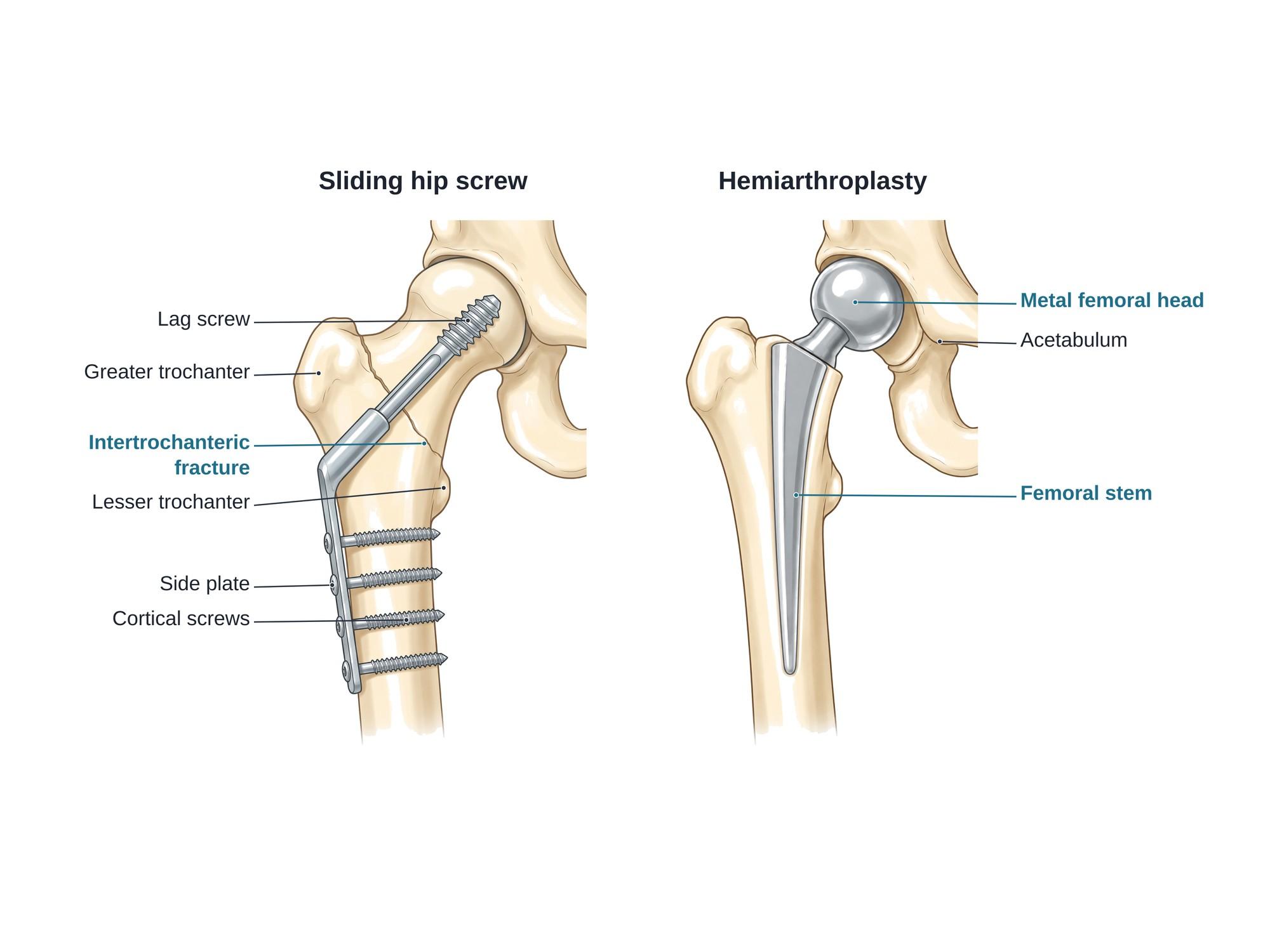
Hip fracture surgery is an operation to repair a broken hip, a break at the top of the femur (thigh bone) near the hip joint. The surgeon either holds the broken bone together with metal implants, called internal fixation, or replaces the broken ball of the hip with an artificial one, called arthroplasty. Two common repairs are the sliding hip screw (also called a dynamic hip screw) and the hemiarthroplasty (a partial hip replacement). The illustration above shows both, side by side, on the same right hip.
The anatomy
The hip is a ball-and-socket joint. The ball is the femoral head, at the top of the femur. It fits into the acetabulum, the cup-shaped socket of the pelvis. The femoral head sits on a short, angled femoral neck. Below the neck are two bony bumps where muscles attach: the greater trochanter on the outer side and the lesser trochanter on the inner side. For the full anatomy, see hip anatomy.
Where the bone breaks matters. The blood supply to the femoral head runs along the femoral neck. A break high in the neck can cut off that blood supply, so the head may die even if the bone is fixed. A break lower down, between the trochanters, keeps the blood supply and heals well once it is held in place.
Why it is done
Almost every hip fracture is treated with surgery. The main goal is to get the patient out of bed and walking quickly. Lying in bed for weeks is dangerous, especially for older adults. It leads to pneumonia, blood clots, pressure sores, muscle loss and confusion.
Most surgeons operate within 24 to 48 hours, once heart, lung and blood problems are checked and treated. Surgery also controls pain far better than bed rest.
Types of hip fracture surgery
The fracture type, the patient's age and how active they were before the fall decide the repair.
| Fracture | Usual surgery | What it does |
|---|---|---|
| Intertrochanteric (between the trochanters) | Sliding hip screw and plate, or a nail inside the femur | Holds the fragments while the bone heals |
| Subtrochanteric (just below the trochanters) | Long nail inside the femur | Bridges the break from the inside |
| Femoral neck, not displaced | Screws or a sliding hip screw | Holds the neck in place |
| Femoral neck, displaced, older adult | Hemiarthroplasty or total hip replacement | Replaces the femoral head |
| Femoral neck, displaced, younger adult | Urgent reduction and screw fixation | Tries to save the patient's own femoral head |
A nail inside the femur (a cephalomedullary nail) is now used for many intertrochanteric fractures. It works on the same idea as the sliding hip screw, but the support sits inside the bone, as in intramedullary nailing. Fixing a fracture with plates and screws after realigning it is described more generally under ORIF surgery.
How the surgery is done
Both operations are done under spinal or general anesthesia. Live X-ray (fluoroscopy) guides the implants.
Sliding hip screw, shown on the left above:
- Reduction. The patient lies on a special traction table. The leg is pulled and turned until the fracture lines up on X-ray.
- Incision. A cut of a few inches is made on the outer side of the upper thigh.
- Guide wire. A wire is drilled up through the femoral neck into the center of the femoral head.
- Lag screw. A large screw with coarse threads, the lag screw, is placed over the wire. Its tip stops just short of the joint surface.
- Side plate. A metal plate with a tube (the barrel) slides over the lag screw. The plate is fixed to the outer side of the femur with cortical screws, which pass through both walls of the bone.
- Closing. The wound is closed in layers.
The lag screw can slide inside the barrel. This lets the fracture settle and squeeze together when the patient stands, which helps it heal.
Hemiarthroplasty, shown on the right above:
- Approach. The surgeon opens the hip from the side or the back.
- Removal. The broken femoral head is removed. The femoral neck is cut cleanly above the lesser trochanter.
- Canal preparation. The hollow center of the femur (the medullary canal) is shaped with rasps.
- Stem. A metal femoral stem is placed in the canal, with or without bone cement.
- Head. A metal ball, sized to match the old femoral head, is fixed on the stem. It is set back into the patient's own acetabulum.
- Closing. The hip capsule and muscles are repaired, and the wound is closed.
Hardware
- Lag screw: a large screw that grips the femoral head.
- Side plate and barrel: hold the lag screw at the right angle and attach it to the shaft.
- Cortical screws: fix the side plate to the femur.
- Femoral stem: a metal post inside the thigh bone, cemented or press-fit.
- Prosthetic femoral head: a metal ball that replaces the broken head.
Fixation hardware usually stays in for life. It is removed only if it causes pain or another problem.
Hip fracture recovery
- Most patients sit up and stand with a walker within a day of surgery.
- After a hemiarthroplasty, full weight on the leg is usually allowed right away. After fixation, it usually is too, unless the surgeon says otherwise.
- The hospital stay is usually 3 to 7 days. Many patients then spend a few weeks in a rehab unit.
- Physical therapy builds strength and balance. Patients move from a walker to a cane over several weeks.
- Blood thinners are given for about 4 weeks to prevent blood clots.
- Bone healing takes about 3 months. Many people keep improving for 6 to 12 months.
- A bone density test and osteoporosis treatment help prevent the next fracture.
Hip fracture recovery is harder for older adults. Many need more help at home than before the fall, and some move to assisted living.
Risks
- Blood clots in the leg veins or lungs
- Pneumonia, confusion (delirium) and urinary infection, mostly in older patients
- Wound infection or deep infection around the implant
- Fixation failure: the lag screw cuts out of the femoral head, or the fracture does not heal (nonunion)
- Avascular necrosis, death of the femoral head from lost blood supply, after a femoral neck fracture is fixed
- Dislocation of the new ball out of the socket after hemiarthroplasty
- Fracture around the implant (periprosthetic fracture)
- Leg length difference or a limp
- Bleeding that needs a blood transfusion
Hip fractures carry a high risk of death in the following year for frail older adults. This risk comes mostly from the patient's other illnesses, not from the surgery itself.
How hip fracture surgery is shown in a personal injury case
Hip fractures often follow falls in stores, on icy walkways, in nursing homes and in car crashes. The surgery changes the value of a claim. It adds hospital and rehab costs, often a loss of independence, and sometimes a move into care. The defense may argue that osteoporosis, not the fall, caused the break.
An illustration of the plaintiff's own hip, showing the fracture and the hardware now inside it, helps a jury understand the injury. It explains the screws or metal ball seen on later imaging. It also shows why recovery takes months. For how these images are used at trial, see demonstrative evidence and medical legal illustration.
Related pages
FAQ
How long does it take to recover from hip fracture surgery?+
Most people stand and take a few steps with a walker within a day or two. The hospital stay is usually 3 to 7 days, often followed by a rehab unit. Bone healing takes about 3 months. Many people keep improving for 6 to 12 months, and some older adults do not return to their earlier level of independence.
What are the types of hip fracture surgery?+
There are two main groups. Fixation holds the broken bone together with metal: a sliding hip screw and plate, a nail inside the femur, or several screws. Replacement removes the broken femoral head: a hemiarthroplasty (partial hip replacement) or a total hip replacement. The type and location of the fracture decide which one is used.
Is hip fracture surgery safe for people over 80?+
Surgery carries real risk in very old patients, but leaving a hip fracture untreated is usually riskier. Without surgery a person may be stuck in bed for weeks, which leads to pneumonia, blood clots and pressure sores. Most surgeons operate within 24 to 48 hours once the patient is medically stable.
What causes death after a hip fracture?+
Deaths in the months after a hip fracture are usually caused by the patient's other illnesses, made worse by the injury and the time spent immobile. Common causes are pneumonia, heart problems, blood clots in the lungs (pulmonary embolism) and infection. Early surgery and early walking lower these risks.
Can you fracture your hip and not know it?+
Yes. An impacted or non-displaced hip fracture can cause only mild groin or knee pain, and some people can still walk. The first X-ray may look normal. If pain continues after a fall, MRI or CT is used to find the break before it shifts.
What is the difference between a hemiarthroplasty and a total hip replacement?+
A hemiarthroplasty replaces only the ball (the femoral head) and leaves the natural hip socket. A total hip replacement also lines the socket with a metal cup and liner. Hemiarthroplasty is common in older, less active patients. Total hip replacement is often chosen for active patients or when the socket already has arthritis.
How should a patient lie in bed after hip fracture surgery?+
Most patients lie on their back with a pillow between the knees. After a hip replacement, the care team may advise against crossing the legs, bending the hip past a right angle, or turning the foot inward for some weeks. These precautions lower the risk of the new hip dislocating.
What is a greater trochanteric fracture?+
It is a break of the greater trochanter, the bony bump on the outer side of the top of the femur. It often follows a direct fall on the side of the hip. An isolated greater trochanter fracture is usually treated without surgery, but MRI is often done to rule out a hidden intertrochanteric fracture.
Need a surgical illustration for consent or a case?
Describe the operation and get a clear, labeled medical illustration for patient consent, teaching or a case exhibit.
Try Natomy →