Degloving Injury: Types, Causes, Healing & Treatment
A degloving injury tears the skin and fat off the tissue beneath, open or closed (Morel-Lavallee). See it illustrated beside normal anatomy.
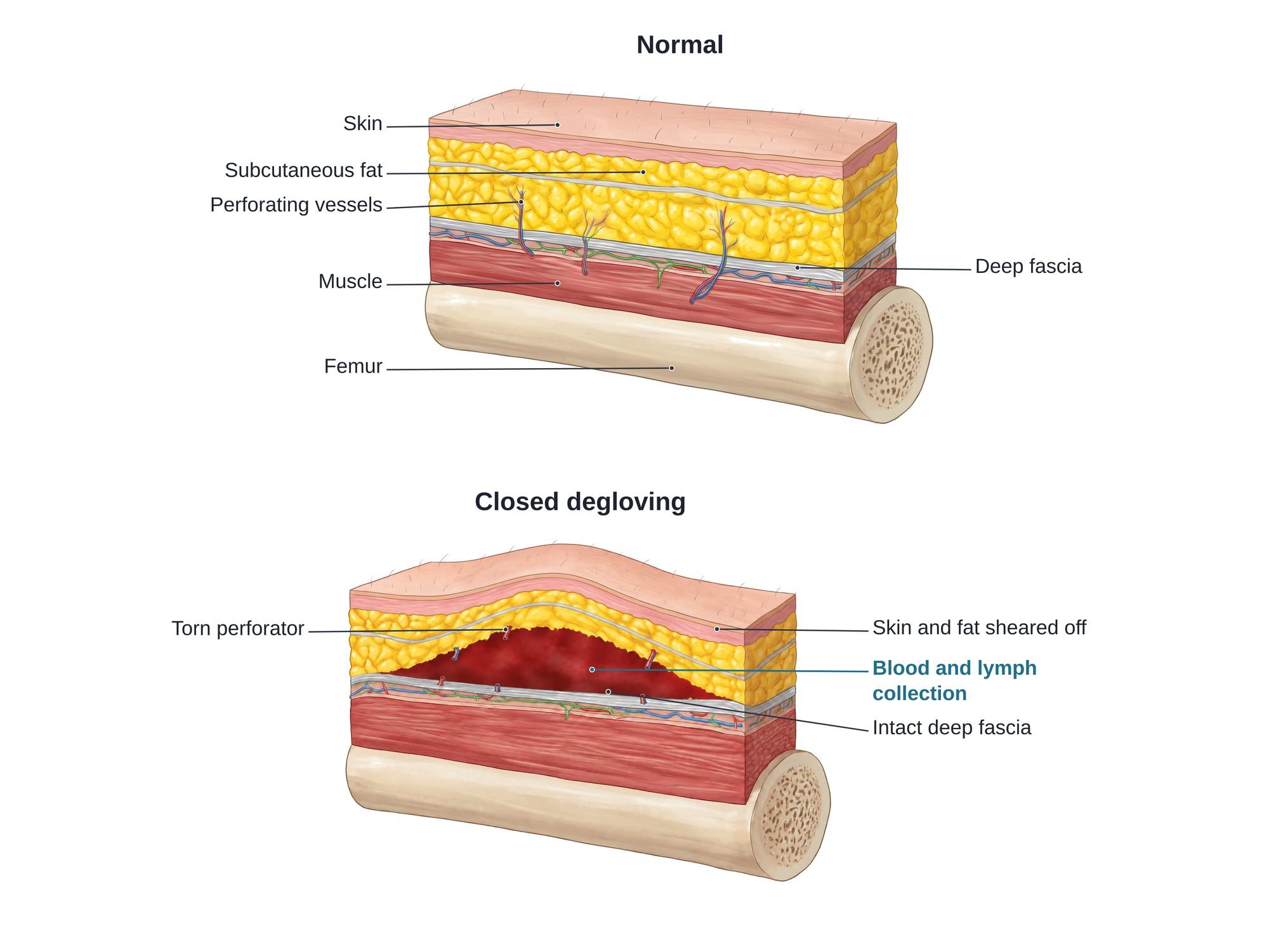
A degloving injury is a severe soft tissue injury where the skin and the fat under it are torn away from the deeper tissue, like a glove being pulled off a hand. A shearing force separates the layers at the deep fascia, the tough sheet that covers the muscles. In an open degloving injury the skin is torn and the deeper tissue is exposed. In a closed degloving injury, also called a Morel-Lavallee lesion, the skin stays intact but a space opens under it and fills with blood and lymph. The figure above shows normal layers beside a closed degloving injury.
The anatomy
The soft tissue of a limb is built in layers. From the outside in, there is the skin, the subcutaneous fat (the fatty layer under the skin), the deep fascia, the muscle and the bone. A thin sheet called the superficial fascia runs through the fat.
The skin gets its blood from small perforating vessels (perforators). These vessels rise from the muscle, pass through the deep fascia and branch into the fat and skin. Lymph channels and small nerves follow the same path.
The fat is loosely attached to the deep fascia. When a strong force drags the skin sideways, the layers can split at this plane. The perforators tear, and the skin loses much of its blood supply. This is why degloved skin often dies, even when it looks normal at first.
Types
Degloving injuries are first sorted by whether the skin is broken.
| Type | What happens | Typical example |
|---|---|---|
| Open degloving | Skin and fat torn off, deep tissue exposed | Leg run over by a wheel, hand caught in a roller |
| Closed degloving (Morel-Lavallee lesion) | Skin intact, fluid collects in the space under it | Hip or thigh after a fall or crash |
| Ring avulsion | Skin of a finger pulled off by a caught ring | Ring snagged on a fence or machine |
| Circumferential degloving | Skin stripped all the way around a limb | Severe crush and roll injuries |
| Facial degloving | Facial skin and soft tissue torn off the facial bones | High-speed crashes, animal attacks |
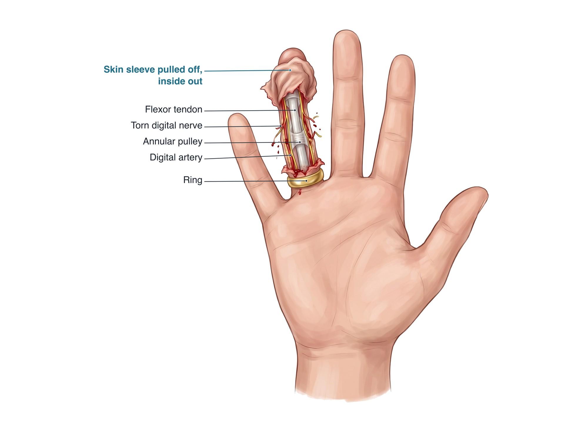
Degloved finger (ring avulsion)
A ring avulsion happens when a ring catches on a hook, fence, door or machine as the person falls or pulls away. The ring drags the skin and fat of the finger off toward the tip. The digital arteries and nerves that run along each side of the finger are stretched and torn, while the tendon and bone may stay intact. Surgeons sort these injuries by how badly blood flow is lost: from a finger that still has good circulation, to one that needs its arteries rejoined with microsurgery, to a completely degloved finger that may need amputation.
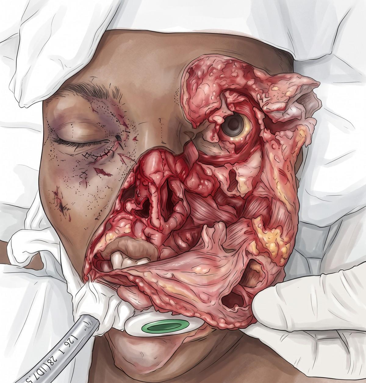
Degloved face
A degloved face is a facial degloving injury, where the skin and soft tissue of the face are torn off the facial bones. It usually comes from high-speed crashes, motorcycle crashes, falls onto the face and animal attacks, and it often comes with facial fractures. The face has a very rich blood supply, so a torn facial flap often survives when it is cleaned and stitched back into place in layers, unlike degloved skin on the legs. Damage to the facial nerve branches, the nerves coming out of the bone, the eyelids and the lips can leave numbness, weakness and visible scars.
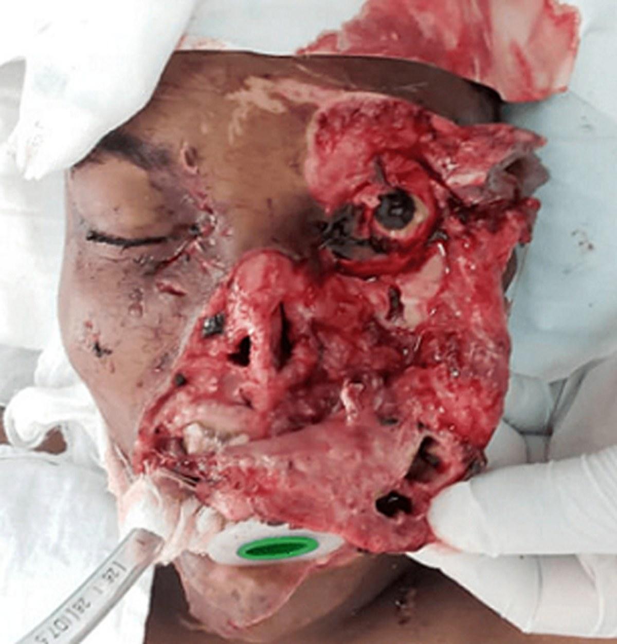
Surgeons also grade how deep the damage goes. Some injuries stop at the deep fascia. Others also involve muscle, tendon, nerves and bone, often with a fracture such as a tibia and fibula fracture.
Causes
A degloving injury needs a strong force that pulls the skin one way while the deep tissue stays still. Common causes are:
- A wheel rolling over a leg or foot in a car, truck or forklift accident
- A hand or arm pulled into rollers, belts or other industrial machines
- Motorcycle and bicycle crashes, with the body sliding across the road
- Falls where the hip or thigh hits the ground and slides
- Rings or jewelry catching on a fixed object
- Sports injuries, especially in football and wrestling, for closed lesions over the knee and thigh
Closed lesions are most common over the outer hip and upper thigh (the greater trochanter), the pelvis, the knee and the lower back.
Symptoms
Open injuries are obvious. The skin is torn and pulled back, and fat, muscle, tendon or bone may be visible. There is usually heavy bleeding and severe pain.
Closed injuries are easy to miss. Signs include:
- A soft, fluid-filled swelling under the skin, often over the hip or thigh
- Bruising and a squishy feeling when the area is pressed
- Skin that moves too freely over the deeper tissue
- Numbness of the skin above the swelling
- Swelling that grows over days or weeks after the injury
Diagnosis
Doctors examine the wound or swelling and check the blood flow, nerves and tendons of the limb. X-rays look for fractures underneath. For a closed lesion, ultrasound shows a fluid pocket between the fat and the deep fascia. MRI shows the size and age of the collection, and whether a capsule (a fibrous wall) has formed around it. A CT scan is often done after a major crash and may show the lesion next to pelvic fractures. In open injuries, the surgeon judges in the operating room which skin is still alive.
Treatment
Treatment depends on whether the skin is broken, how much tissue is damaged, and the blood supply to the skin.
Open degloving injuries are surgical emergencies:
- Control of bleeding, tetanus protection and antibiotics.
- Debridement, which means cutting away dead and dirty tissue, often repeated over several operations.
- Saving skin that still has a blood supply. Dead skin is removed, and some can be thinned and reused as a skin graft.
- Negative pressure wound therapy (a sealed vacuum dressing) to prepare the wound.
- Coverage with skin grafts or flaps (tissue moved with its own blood supply).
- In the worst cases, especially with crushed bone and nerves, amputation.
Closed degloving injuries (Morel-Lavallee lesions):
- Small lesions: compression wraps and rest.
- Larger lesions: draining the fluid with a needle, sometimes with an injection to make the walls stick together (sclerotherapy).
- Lesions that keep coming back or have a thick capsule: surgery to open, clean and close the space with drains.
If the limb swells badly, the pressure inside the muscle compartments must be watched. A fasciotomy may be needed for compartment syndrome.
Recovery
Recovery is often long. Open injuries may need several operations over weeks. Grafts and flaps take a few weeks to take hold, followed by months of wound care and therapy. Closed lesions treated with drainage may settle within a few weeks to months, but some come back.
Many people need physical or hand therapy to regain motion and strength. Scars, skin grafts and numbness are usually permanent to some degree.
Complications
- Skin necrosis, when the degloved skin dies from lost blood supply
- Infection, of the wound or of a closed fluid pocket
- Recurrent fluid collections and a thick capsule in closed lesions
- Hypertrophic scars and contractures (tight scars that limit motion)
- Nerve damage with numbness or chronic pain
- Loss of a finger or limb in the most severe injuries
- Delayed fracture surgery when a closed lesion sits over the surgical site
How a degloving injury is shown in a personal injury case
Degloving injuries usually come from truck, motorcycle, workplace machine and car crashes. They often lead to repeat surgeries, visible scarring and lasting loss of function. A closed degloving injury can be hard for a jury to picture, because the skin may look almost normal in photographs.
A layered medical illustration shows what happened beneath the skin. It shows the skin and fat torn off the deep fascia, the torn vessels, and the fluid that filled the space. A side by side panel of normal anatomy makes the change easy to follow. These exhibits are a form of demonstrative evidence. They can be tailored to the patient's records and imaging as a medical legal illustration.
Related soft tissue injury pages: avulsion injury, laceration, abrasion and contusion.
FAQ
Can you heal from a degloving injury?+
Yes, most people heal, but it often takes several surgeries and months of recovery. Small closed injuries may heal with drainage and compression. Large open injuries need surgery to remove dead tissue and cover the wound with the original skin, skin grafts or flaps. Scarring, numbness and stiffness are common long term.
What does a degloving injury look like?+
An open degloving injury looks like the skin has been peeled back like a glove, exposing fat, muscle, tendon or bone. A closed degloving injury can look like a soft, fluid-filled swelling under intact skin, often with bruising. It may feel squishy when pressed, and the skin over it can be numb.
What is the difference between an avulsion and a degloving injury?+
An avulsion is any injury where tissue is torn away from the body. A degloving injury is a specific type of avulsion where the skin and fat are stripped off the deeper layers over a wide area, usually from a shearing or rolling force. All degloving injuries are avulsions, but not all avulsions are degloving injuries.
How serious is a Morel-Lavallee lesion?+
It is often missed at first, because the skin looks intact. Left alone, a large one can keep filling with fluid, form a thick capsule, get infected, or kill the skin above it. It is often found with pelvic and hip fractures, and it can delay or complicate fracture surgery. Early drainage and compression usually give the best results.
How does a degloving injury happen?+
A strong force drags the skin in one direction while the deeper tissue stays still. Typical causes are a limb run over by a car wheel, a hand caught in a roller or conveyor belt, a ring snagging on an object, a motorcycle crash, or a fall with the body sliding across the ground.
Is a degloving injury painful?+
Usually yes, with severe pain at the time of injury. Some areas can be numb, because the small nerves running from the deep tissue to the skin are torn. Pain from scarring, nerve damage and skin grafts can last for months.
What is a degloved finger or ring avulsion?+
A ring avulsion happens when a ring catches on something and the skin of the finger is pulled off like a glove. It can damage the arteries and nerves of the finger. Treatment ranges from repair to microsurgery to rejoin vessels, and sometimes amputation of the finger.
How long does a degloving injury take to heal?+
Small closed injuries can settle in a few weeks to a few months. Open injuries that need grafts or flaps usually take several months, and full recovery of strength and movement can take a year or longer.
Need this illustration for a patient or a case?
Describe the injury or upload a scan and get a clear, labeled medical illustration for a consult, a chart or a case exhibit.
Try Natomy →